. 2002 Aug 15;30(16):3592-601.
doi: 10.1093/nar/gkf470.

Metallo-beta-lactamase fold within nucleic acids processing enzymes: the beta-CASP family

Affiliations

Metallo-beta-lactamase fold within nucleic acids processing enzymes: the beta-CASP family

Isabelle Callebaut et al. Nucleic Acids Res. .

Abstract

A separate family of enzymes within the metallo-beta-lactamase fold comprises several important proteins acting on nucleic acid substrates, involved in DNA repair (Artemis, SNM1 and PSO2) and RNA processing [cleavage and polyadenylation specificity factor (CPSF) subunit]. Proteins of this family, named beta-CASP after the names of its representative members, possess specific features relative to those of other metallo-beta-lactamases, that are concentrated in the C-terminal part of the domain. In this study, using sensitive methods of sequence analysis, we identified highly conserved amino acids specific to the beta-CASP family, some of which were unidentified to date, that are predicted to play critical roles in the enzymatic function. The identification and characterisation of all the extant, detectable beta-CASP members within sequence databases and genome data also allowed us to unravel particular sequence features which are likely to be involved in substrate specificity, as well as to describe new but as yet uncharacterised members which may play critical roles in DNA and RNA metabolism.


Figures
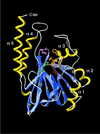
Figure 1
Figure 1
Three-dimensional representation of a metallo-β-lactamase domain. The structure of Stenotrophomonas maltophilia metallo-β-lactamase (; PDB identifier 1SML) is used for illustration purposes. The two β-sheets are formed by strands β1–β7 and β8–β12, respectively. Side chains of amino acids participating in zinc binding are coloured red (motif 1 after strand β5), yellow (motif 2 after strand β6), pink (motif 3 after strand β9/), violet (motif 4 after strand β11) and green (motif 5 after strand β12). Clear sequences similarities are found with members of the metallo-β-lactamase superfamily up to motif 4, whereas the region linking motif 4 to motif 5, comprising here helix α4, is proposed to be extremely large for members of the β-CASP family discussed here. This figure was prepared using Swiss-PdbViewer (27).
Figure 2
Figure 2
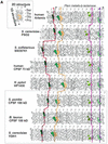
(Opposite and above) Comparison of the HCA plots of several members of the β-CASP family. (A) The first four metallo-β-lactamase conserved motifs; (B) the β-CASP region. The interest of using HCA for sequence comparison, especially at low levels of identity, lies in the possibility that such a representation offers to combine lexical analysis (1D structure) and secondary structure localisation. Briefly, the sequence is written on a duplicated α-helical net in which strong hydrophobic amino acids (V, I, L, F, M, Y, W) are contoured. These are not randomly distributed but instead form clusters that were shown to mainly correspond to the internal faces of regular secondary structures (α-helices and β-strands). A cluster thus includes hydrophobic but also non-hydrophobic amino acids lying between its first and last hydrophobic amino acids. Non-hydrophobic sequences separating clusters mainly correspond to loop regions. The observed good correspondence between hydrophobic clusters and regular secondary structures is linked to the use of an appropriate connectivity distance (i.e. the minimum distance separating two hydrophobic amino acids belonging to two different clusters) related to the α-helical pitch. The way to read the sequence and special symbols are indicated in the inset. As secondary structures are often much more stable than sequences, a good conservation of key hydrophobic clusters, whose hydrophobic amino acids participate in the protein core, is generally observed. Hence, it is therefore possible to appreciate the required fold conservation for remote sequence relationships. Conservation of key hydrophobic clusters is indicated in green, with vertical lines highlighting correspondences. Highly conserved amino acids of the four metallo-β-lactamase domain motifs are coloured red, yellow, pink and violet (A), according to Figure 1. The β-CASP ones are coloured red (B, motifs A–C). Amino acids that substitute these highly conserved amino acids in some sequences (e.g. CPSF 100 kDa) are contoured accordingly. Other conserved, non-hydrophobic amino acids are coloured grey and yellow. Local similarities between the CPSF 100 kDa proteins are indicated in orange. The C-terminal limit of the globular domain following the four metallo-β-lactamase motifs (β-CASP motif) is indicated with a star and the total number of amino acids within each protein is given within brackets. The three sequences belonging to the CPSF 100 kDa group are boxed. These have lost part or most of the highly conserved amino acids. Moreover, long intervening sequences separate motifs B and C from the rest of the domain.
Figure 2
Figure 2
(Opposite and above) Comparison of the HCA plots of several members of the β-CASP family. (A) The first four metallo-β-lactamase conserved motifs; (B) the β-CASP region. The interest of using HCA for sequence comparison, especially at low levels of identity, lies in the possibility that such a representation offers to combine lexical analysis (1D structure) and secondary structure localisation. Briefly, the sequence is written on a duplicated α-helical net in which strong hydrophobic amino acids (V, I, L, F, M, Y, W) are contoured. These are not randomly distributed but instead form clusters that were shown to mainly correspond to the internal faces of regular secondary structures (α-helices and β-strands). A cluster thus includes hydrophobic but also non-hydrophobic amino acids lying between its first and last hydrophobic amino acids. Non-hydrophobic sequences separating clusters mainly correspond to loop regions. The observed good correspondence between hydrophobic clusters and regular secondary structures is linked to the use of an appropriate connectivity distance (i.e. the minimum distance separating two hydrophobic amino acids belonging to two different clusters) related to the α-helical pitch. The way to read the sequence and special symbols are indicated in the inset. As secondary structures are often much more stable than sequences, a good conservation of key hydrophobic clusters, whose hydrophobic amino acids participate in the protein core, is generally observed. Hence, it is therefore possible to appreciate the required fold conservation for remote sequence relationships. Conservation of key hydrophobic clusters is indicated in green, with vertical lines highlighting correspondences. Highly conserved amino acids of the four metallo-β-lactamase domain motifs are coloured red, yellow, pink and violet (A), according to Figure 1. The β-CASP ones are coloured red (B, motifs A–C). Amino acids that substitute these highly conserved amino acids in some sequences (e.g. CPSF 100 kDa) are contoured accordingly. Other conserved, non-hydrophobic amino acids are coloured grey and yellow. Local similarities between the CPSF 100 kDa proteins are indicated in orange. The C-terminal limit of the globular domain following the four metallo-β-lactamase motifs (β-CASP motif) is indicated with a star and the total number of amino acids within each protein is given within brackets. The three sequences belonging to the CPSF 100 kDa group are boxed. These have lost part or most of the highly conserved amino acids. Moreover, long intervening sequences separate motifs B and C from the rest of the domain.
Figure 3
Figure 3
Multiple alignment of conserved motifs of representative members of the β-CASP family. The alignment is divided into two main blocks, the first one including metallo-β-lactamase motif 4 (with the conserved aspartic acid in red) and the β-CASP motif A (with the conserved aspartic or glutamic acid in red), the second one centred on the two β-CASP motifs B and C (conserved histidine residues in red). The identifiers of proteins that are involved, or are predicted to be involved, in DNA and RNA metabolism are coloured blue and red, respectively. Positions of the alignment N- and C-termini are indicated by the number of residues. Distances between conserved blocks, as well as the C-terminal end position of sequences (right), are indicated in brackets. Predicted secondary structures are shown up to the alignment [H and E stand for helix and strand (extended), respectively]. Conserved hydrophobic amino acids (V, I, L, F, M, Y, W) are boxed in green, conserved aromatic amino acids (F, Y, W) in violet, acidic and basic amino acids in pink and blue, respectively, small amino acids (G, A, T, S, C) in yellow. Amino acids that can substitute these residues in some circumstances are shown in bold (e.g. A, T, C and S can substitute hydrophobic positions). Underlined sequences correspond to human SMN1C and CPSF 73 kDa-related proteins.

References

    1. Aravind L. (1999) An evolutionary classification of the metallo-β-lactamase fold proteins. In Silico Biol., 1, 69–91. - PubMed
    1. Melino S., Capo,C., Dragani,B., Aceto,A. and Petruzzelli,R. (1998) A zinc-binding motif conserved in glyoxalase II, β-lactamase and arylsulfatases. Trends Biochem. Sci., 23, 381–382. - PubMed
    1. Daiyasu H., Osaka,K., Ishino,Y. and Toh,H. (2001) Expansion of the zinc metallo-hydrolase family of the β-lactamase fold. FEBS Lett., 503, 1–6. - PubMed
    1. Moshous D., Callebaut,I., de Chasseval,R., Corneo,B., Cavazzana-Calvo,M., Le Deist,F., Tezcan,I., Sanal,O., Bertrand,Y., Philippe,N. et al. (2001) ARTEMIS, a novel DNA double-strand break repair/V(D)J recombination protein, is mutated in human severe combined immune deficiency. Cell, 105, 177–186. - PubMed
    1. Ma Y., Pannicke,U., Schwarz,K. and Lieber,M.R. (2002) Hairpin opening and overhang processing by an Artemis/DNA-dependent complex in non-homologous end joining and V(D)J recombination. Cell, 108, 781–794. - PubMed

Publication types

MeSH terms