. 2002 Aug 15;30(16):3624-31.
doi: 10.1093/nar/gkf469.

The human XPC DNA repair gene: arrangement, splice site information content and influence of a single nucleotide polymorphism in a splice acceptor site on alternative splicing and function

Affiliations

The human XPC DNA repair gene: arrangement, splice site information content and influence of a single nucleotide polymorphism in a splice acceptor site on alternative splicing and function

Sikandar G Khan et al. Nucleic Acids Res. .

Abstract

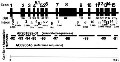
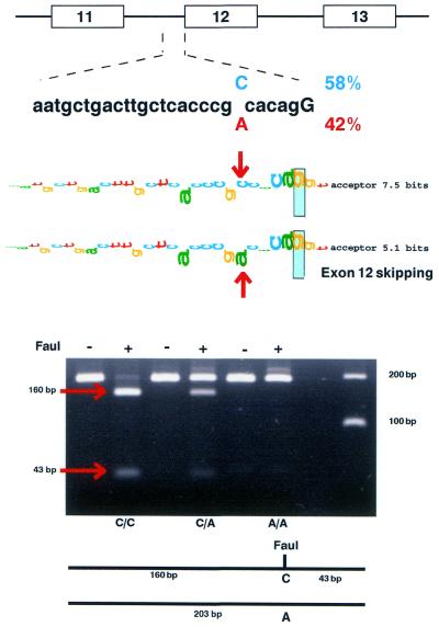
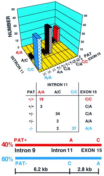
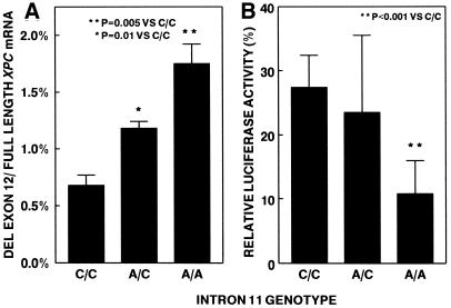
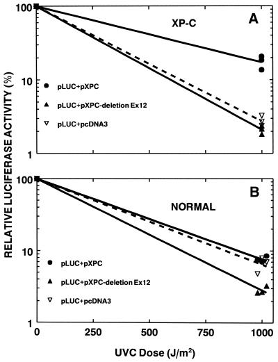
XPC DNA repair gene mutations result in the cancer-prone disorder xeroderma pigmentosum. The XPC gene spans 33 kb and has 16 exons (82-882 bp) and 15 introns (0.08-5.4 kb). A 1.6 kb intron was found within exon 5. Sensitive real- time quantitative reverse transcription-polymerase chain reaction methods were developed to measure full-length XPC mRNA (the predominant form) and isoforms that skipped exons 4, 7 or 12. Exon 7 was skipped in approximately 0.07% of XPC mRNAs, consistent with the high information content of the exon 7 splice acceptor and donor sites (12.3 and 10.4 bits). In contrast, exon 4 was skipped in approximately 0.7% of the XPC mRNAs, consistent with the low information content of the exon 4 splice acceptor (-0.1 bits). A new common C/A single nucleotide polymorphism in the XPC intron 11 splice acceptor site (58% C in 97 normals) decreased its information content from 7.5 to 5.1 bits. Fibroblasts homozygous for A/A had significantly higher levels (approximately 2.6-fold) of the XPC mRNA isoform that skipped exon 12 than those homozygous for C/C. This abnormally spliced XPC mRNA isoform has diminished DNA repair function and may contribute to cancer susceptibility.


Figures

Figure 1
Figure 1
Structural map of the human XPC gene. The 16 exons and 15 introns of the 33 kb XPC gene are numbered and their size in bp or kb is indicated below each segment. The parts of the genomic DNA sequence that we determined were submitted to GenBank (accession nos AF261892– AF261901). AC090645 is the chromosome 3 reference sequence that was later posted in GenBank.
Figure 2
Figure 2
XPC intron 11 splice acceptor polymorphism. (Top) A C/A SNP is located at the –5 position of the XPC intron 11 splice acceptor. (Middle) The walkers indicate the change in splice acceptor information content from 7.5 to 5.1 bits by the polymorphism. (Lower) RFLP assay for detection of the C/A polymorphism by use of digestion with FauI. The 203 bp sequence generated by PCR is cut into fragments of 160 and 43 bp by FauI if the C is present in the sequence, permitting detection of C/C, C/A and A/A genotypes.
Figure 3
Figure 3
Linkage disequilibrium among three XPC polymorphisms in 97 normal donors. (Upper) The number of donors with the XPC intron 9 PAT+/–, intron 11 A/C and exon 15 C/A genotypes is indicated on the bar graph. (Middle) The table shows the number of individuals homozygous for all three polymorphisms (in red or blue) or heterozygous for at least one of the polymorphisms (in black). (Lower) Schematic diagram of the physical location and haplotypes of three XPC polymorphisms. The XPC intron 11 splice acceptor A at position –5 is linked to C2920 in exon 15 and PAT+ in intron 9 in a haplotype covering ∼9 kb and occurring at a genotype frequency of ∼40% (in red). The XPC intron 11 splice acceptor C at position –5 is linked to A2920 in exon 15 and PAT in intron 9 in a haplotype covering ∼9 kb and occurring at a genotype frequency of ∼60% (in blue).
Figure 4
Figure 4
Relation of XPC intron 11 genotype to deletion of exon 12 in XPC mRNA and DNA repair function. (A) Real-time QRT–PCR was used to measure the amount of XPC mRNA deleting exon 12 and retaining exon 12. The proportion of deleted exon 12 XPC mRNA was significantly greater in cells with the A/A and A/C genotypes compared with the C/C genotype (the nine fibroblast lines assayed in duplicate are listed in Table 2). (B) Post-UV host cell reactivation was used to measure DNA repair function. The relative luciferase activity was significantly greater in the cells with the C/C genotype than in cells with the A/A genotype (the nine fibroblast lines assayed in triplicate are listed in Table 2).
Figure 5
Figure 5
Post-UV host cell reactivation assay assessment of DNA repair activity of XPC cDNA deleting exon 12. (A) Increased expression of luciferase activity in XPC cells [XP4PA (SV40)] by co-transfection of full-length XPC cDNA expression vector (pcDNA3/HA-XPC) (solid circle) was not seen on co-transfection with the XPC cDNA expression vector deleted of exon 12 (pcDNA3/HA-XPC-deleted-exon 12) (solid triangle) or with the empty vector (pcDNA3/HA) (open triangle). (B) Reduction in expression of luciferase in normal cells (AG13129) by co-transfection with XPC cDNA expression vector deleted for exon 12 (solid triangle). Each data point represents an independent transfection. Data points at 1000 J/m2 are displaced for clarity.

Similar articles

Cited by

References

    1. Van Steeg H. and Kraemer,K.H. (1999) Xeroderma pigmentosum and the role of UV-induced DNA damage in skin cancer. Mol. Med. Today, 5, 86–94. - PubMed
    1. Kraemer K.H. (1999) Heritable diseases with increased sensitivity to cellular injury. In Freedberg,I.M., Eisen,A.Z., Wolff,K., Austen,K.F., Goldsmith,L., Katz,S.I. and Fitzpatrick,T.B. (eds), Fitzpatrick’s Dermatology in General Medicine. McGraw Hill, New York, NY, pp. 1848–1862.
    1. Kraemer K.H., Lee,M.M. and Scotto,J. (1987) Xeroderma pigmentosum. Cutaneous, ocular and neurologic abnormalities in 830 published cases. Arch. Dermatol., 123, 241–250. - PubMed
    1. Kraemer K.H., Lee,M.-M., Andrews,A.D. and Lambert,W.C. (1994) The role of sunlight and DNA repair in melanoma and nonmelanoma skin cancer: the xeroderma pigmentosum paradigm. Arch. Dermatol., 130, 1018–1021. - PubMed
    1. Legerski R. and Peterson,C. (1992) Expression cloning of a human DNA repair gene involved in xeroderma pigmentosum group C. Nature, 359, 70–73. [Erratum (1992) Nature, 360, 610.] - PubMed