. 2002 Aug 20;99(17):11025-30.
doi: 10.1073/pnas.162172899. Epub 2002 Aug 12.

Caspase 3 activity is required for skeletal muscle differentiation

Affiliations

Caspase 3 activity is required for skeletal muscle differentiation

Pasan Fernando et al. Proc Natl Acad Sci U S A. .

Abstract

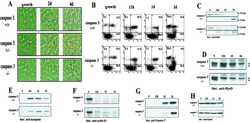
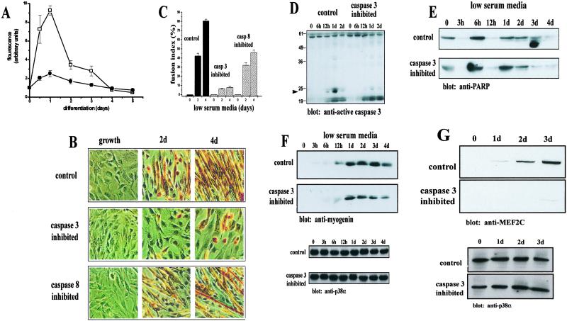
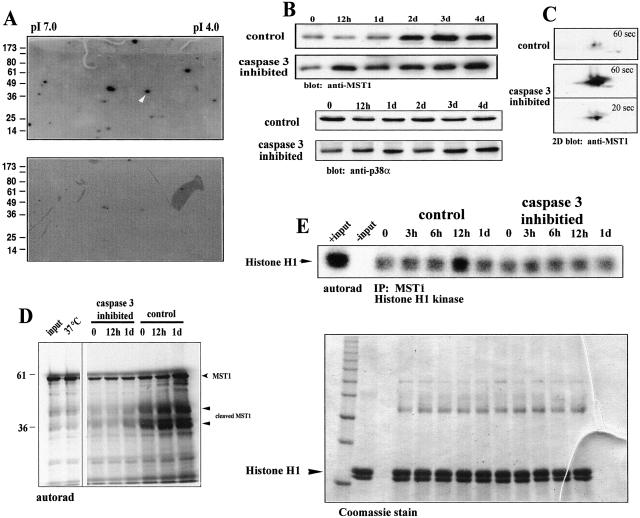
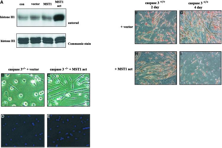
The cellular alterations associated with skeletal muscle differentiation share a high degree of similarity with key phenotypic changes usually ascribed to apoptosis. For example, actin fiber disassembly/reorganization is a conserved feature of both apoptosis and differentiating myoblasts and the conserved muscle contractile protein, myosin light chain kinase, is required for the apoptotic feature of membrane blebbing. As such, these observations suggest that the induction of differentiation and apoptosis in the myogenic lineage may use overlapping cellular mechanisms. Here, we report that skeletal muscle differentiation depends on the activity of the key apoptotic protease, caspase 3. Peptide inhibition of caspase 3 activity or homologous deletion of caspase 3 leads to dramatic reduction in both myotube/myofiber formation and expression of muscle-specific proteins. Subsequently, we have identified Mammalian Sterile Twenty-like kinase as a crucial caspase 3 effector in this cellular process. Mammalian Sterile Twenty-like kinase is cleavage-activated by caspase 3, and restoration of this truncated kinase in caspase 3 null myoblasts restores the differentiation phenotype. Taken together, these results confirm a unique and unanticipated role for a caspase 3-mediated signal cascade in the promotion of myogenesis.


Figures

Fig 1.
Fig 1.
Caspase 3 null myoblasts have deficient myotube formation. (A) Primary myoblast cultures were derived from wild-type and caspase 3−/− mice (n = 5 for each genotype). Cultures were fixed and stained for myosin heavy chain and counterstained with the nuclear marker hematoxylin (dark staining pattern). A severe lack of myogenesis is noted in caspase 3−/− cells. (B) Flow cytometry analysis of wild-type and caspase 3−/− myoblasts after incubation in low-serum medium. Cells were either left unstained (lower left quadrant), stained with Annexin V-FITC (lower right quadrant), propidium iodide (upper left quadrant), or both (upper right quadrant). A comparable proportion of apoptotic cells was observed for both wild-type and caspase 3 null myoblasts after 2 days of low-serum treatment. (C) PARP cleavage is equivalent in both wild-type and caspase 3 null myoblasts. Both the 118-kDa PARP protein and the 85-kDa cleavage fragment were detected. A reduction of muscle differentiation-specific proteins in caspase 3 null myoblasts was observed by using Western blot analyses for hypophosphorylated MyoD (D) (top and bottom arrows indicate the position of phosphorylated and hypophosphorylated MyoD, respectively), myogenin (E), and Troponin-T (G). The amount of myogenin protein was ≈2-fold higher in wild-type cells after 12 h. Comparable levels of Troponin-T were found after 2 days of low-serum treatment. (F) Caspase 3 null myoblasts displayed a prolonged accumulation of cyclin D1 in the presence of low-serum (differentiation) media. (H) Equal loading for Western blot analyses was assessed by using an anti-tubulin antibody. Band intensity was assessed by using SCION IMAGE.
Fig 2.
Fig 2.
Caspase 3 activity is required for skeletal muscle differentiation. (A) A sharp increase in caspase 3 activity (□) was found in C2C12 myoblasts after 1 day of low-serum treatment. A much smaller increase in caspase 8 activity (•) was noted, although a similar trend in activation was found (mean ± SEM., n = 5). (B) Morphology of C2C12 cells after inhibition of caspase 3 or caspase 8 activity. Cells were incubated in low-serum media with inhibitors specific for either caspase 3 (Z-DEVD.fmk) or caspase 8 (Z-LETD.fmk) or DMSO (control) and stained with MF20. Myotube formation is drastically attenuated in caspase 3-inhibited cells after a 4-day treatment. A minor attenuation of myotube formation is observed with caspase 8 inhibition. (C) Caspase 3-inhibited cells lack fusion capacity. Calculation of myoblast fusion indices as a percentage of cells containing two or more nuclei within a differentiated myotube (MF20 positive cell). Values were determined as mean ± SEM from three to four independently derived cultures. (D) Western blot analysis of active caspase 3 from control and caspase 3-inhibited cells. The arrow indicates a 21-kDa caspase 3 fragment, indicative of caspase 3 activation abundant in control cells and at very low levels in caspase 3-inhibited samples. (E) Comparable levels of PARP cleavage in both control and caspase 3-inhibited cells. PARP cleavage was accessed by Western blot. Shown is the 118-kDa PARP species. (F) Accumulation of myogenin is delayed and is in lower abundance in caspase 3-inhibited C2C12 myoblasts. The level of myogenin in control cells is approximately twice that of in caspase 3-inhibited cells by day 4. Equal loading was accessed by Western blot with anti-p38α (shown below). (G) Caspase 3-inhibited C2C12 myoblasts express very low levels of MEF2C relative to control cells. Equal loading (shown below) was accessed as in F. Band intensity was assessed by using SCION IMAGE.
Fig 3.
Fig 3.
Activated caspase 3 induces differentiation in growing myoblasts. (A) Subconfluent C2C12 myoblasts were transfected with 20 pg of active caspase 3 protein and examined after 12 h for the presence of myosin heavy chain (anti-MF20) detected by using a FITC-conjugated secondary antibody. A confluent population of cells were incubated in low-serum media (differentiation media) to induce myoblast differentiation and examined after 12 h. In all cells examined, nuclei were localized with DAPI staining. Figure is representative of seven trials. (B) Caspase 3 activity results in a withdrawal of cells from the cell cycle. A subconfluent population of cells maintained in growth media was transfected with 20 pg of active caspase 3 protein. Cells were fixed after 24–28 h and stained for the cell proliferation marker cyclin D1 (FITC-labeled) and active caspase 3 (Alexa 594). Cells containing active caspase 3 do not express cyclin D1. Total nuclei are shown stained with DAPI. Figure is representative of five trials.
Fig 4.
Fig 4.
MST1 is activated by caspase 3 during skeletal muscle differentiation. (A) Two-dimensional in-gel kinase assay. Caspase 3 inhibition results in a significant loss of MBP-directed cellular kinase activities. The putative location of active MST1 kinase, based on predicted pI (5.2) and molecular weight (36 kDa), is indicated by the white arrowhead. (B) Western blot analysis of C2C12 cells by using an anti-MST1 antibody. Control-treated cells show a varied amount of MST1 during 0 and 1 day of differentiation and exemplify a proteolytic-associated event. The level of MST1 protein does not change on the addition of low-serum (differentiation) media in caspase 3-inhibited myoblasts. Shown below is loading control by using p38α. (C) Two-dimensional isoelectric focusing/Western blot of control and caspase 3-treated cells after 12 h in low-serum media. Subsequent immunoblot analysis was performed by using an anti-MST1 antibody. Shown are 1-min exposures for both control and caspase 3-inhibited samples. A 20-s exposure for the caspase 3 sample is included as a comparison of signal intensity. (D) [35S]methionine shows that labeling MST1 is cleaved in differentiating myoblasts. The right-hand side shows the position of [35S]Met-MST1. The arrows below indicate MST1 cleavage products of approximately 36 and ≈45 kDa. (E) MST1 kinase activity increases during myoblast differentiation. [γ-32P]ATP-labeled Histone H1 after immunoprecipitation of MST1 from C2C12 myoblasts under differentiation conditions. A substantial activation of MST1 in the control sample after 12 h of differentiation coincides with the cleavage of MST1 as shown in A and C. Sample loading was indexed by staining the gel with Coomassie blue before drying and autoradiography (E Bottom). The arrow indicates the position of histone H1.
Fig 5.
Fig 5.
Activated MST1 rescues the caspase 3−/− myoblast phenotype. (A) Kinase activity of MST1 protein constructs. MST1-act-transfected cells display a high level of kinase activity. Accuracy of sample loading is indicated by the Coomassie-stained gel shown below (n = 4). (B) Caspase 3 knockout myoblasts transfected with vector alone failed to differentiate. (C) Caspase 3 knockout myoblasts containing MST1-act differentiated to form fused, multinucleated myotubes. The efficiency of differentiation in MST1-act myoblasts was >85%. Total nuclei were visualized by using DAPI staining (D and E). (FI) Wild-type myoblasts were transfected with vector or with MST1-act and allowed to differentiate for up to 4 days. After 2 days of differentiation, cells containing MST1-act (H) share a similar phenotype with vector-transfected cultures differentiated for 4 days (G). Extensive cell death is observed when cells are transfected with MST1-act and differentiated for 4 days (I).

References

    1. Megeney L. A. & Rudnicki, M. A. (1995) Biochem. Cell. Biol. 73, 723-732. - PubMed
    1. Black B. L. & Olson, E. N. (1998) Annu. Rev. Cell Dev. Biol. 14, 167-196. - PubMed
    1. Black B. L., Martin, J. F. & Olson, E. N. (1995) J. Biol. Chem. 270, 2889-2892. - PubMed
    1. Molkentin J. D., Black, B. L., Martin, J. F. & Olson, E. N. (1995) Cell 83, 1125-1136. - PubMed
    1. Yee S. P. & Rigby, P. W. (1993) Genes Dev. 7, 1277-1289. - PubMed

Publication types