. 2002 Aug 20;99(17):11375-80.
doi: 10.1073/pnas.172360799. Epub 2002 Aug 12.

Defects in caveolin-1 cause dilated cardiomyopathy and pulmonary hypertension in knockout mice

Affiliations

Defects in caveolin-1 cause dilated cardiomyopathy and pulmonary hypertension in knockout mice

You-Yang Zhao et al. Proc Natl Acad Sci U S A. .

Abstract

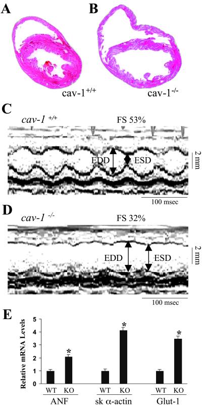
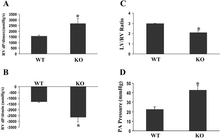
Caveolins are important components of caveolae, which have been implicated in vesicular trafficking and signal transduction. To investigate the in vivo significance of Caveolins in mammals, we generated mice deficient in the caveolin-1 (cav-1) gene and have shown that, in the absence of Cav-1, no caveolae structures were observed in several nonmuscle cell types. Although cav-1(-/-) mice are viable, histological examination and echocardiography identified a spectrum of characteristics of dilated cardiomyopathy in the left ventricular chamber of the cav-1-deficient hearts, including an enlarged ventricular chamber diameter, thin posterior wall, and decreased contractility. These animals also have marked right ventricular hypertrophy, suggesting a chronic increase in pulmonary artery pressure. Direct measurement of pulmonary artery pressure and histological analysis revealed that the cav-1(-/-) mice exhibit pulmonary hypertension, which may contribute to the right ventricle hypertrophy. In addition, the loss of Cav-1 leads to a dramatic increase in systemic NO levels. Our studies provided in vivo evidence that cav-1 is essential for the control of systemic NO levels and normal cardiopulmonary function.


Figures

Fig 1.
Fig 1.
Generation of cav-1 null mutation mice. (A) Schematic representation of the wild-type and mutant loci of the cav-1 gene together with the targeting vector. Exons for the gene encoding cav-1 are represented by a black box. The size of intron 2 is approximately 10 kb. The targeting vector carries the lacZ gene and the neomycin resistance gene (neo). (B) A representative Southern blot analysis of XbaI-digested DNA isolated from ES clones. Hybridization was carried out with a 0.4-kb DNA fragment located 5 kb downstream of exon 2. (C) PCR analysis of tail genomic DNA from littermates generated by heterozygous pairs. Lane M, molecular size marker DNA. The locations of PCR primers (P1 to P4) are shown in A. (D). Western blotting analysis of cav-1 and cav-3 expression in the hearts from littermates. Ventricle lysates were immunoblotted with a polyclonal antibody against Cav-1. The same blot was reprobed with a monoclonal antibody against Cav-3. Equal loading amounts were confirmed by β-actin immunoblotting. The experiment was repeated twice with similar results.
Fig 2.
Fig 2.
Transmission electron microscopy analysis. (A) Representative micrograph of heart ventricular endothelial and muscle cells from cav-1 wild-type mice. Caveolae are indicated in both endothelial and muscle cells by arrows. (B) Micrograph of heart ventricular endothelial and muscle cells from cav-1 knockout mice. Caveolae were only found in the muscle cells not in the endothelial cells. (C and D) Micrographs of the endothelial cells from larger blood vessels in the cav-1 knockout mice. Clathrin coated vesicles were indicated (arrowheads) for size comparison. Interestingly, in both the heart and diaphragm muscle of cav-1−/− mice, larger caveolae-like structures could be occasionally observed in the vessels with diameters larger than capillaries (>10 μm) not surrounded by pericytes or smooth muscle cells. These structures appear to be provided with stomatal diaphragms, and were also reported by Drab et al. (35). e, endothelial cells; mf, cardiac muscle cells.
Fig 3.
Fig 3.
Pathological analysis of the cav-1 knockout hearts. (A and B) Histological sections of hearts from 5-month-old littermates. Hearts were fixed in paraformaldehyde and stained with hematoxylin and eosin. Data are representative of three independent experiments with nearly identical results. (C and D) Transthoracic M-mode echocardiographic tracings in a wild-type mouse (C) and cav-1 knockout mouse (D). LV dimensions are indicated by the double-sided arrows. EDD, end diastolic dimension; ESD, end systolic dimension. Cav-1 knockout mice have chamber dilation with reduced wall motion, indicating depressed cardiac function and increased wall stress. (E) Quantitative analysis of the expression levels of ANF, skeletal α-actin and glut-1 genes in the heart by real time RT-PCR. The data were expressed as mean ± SEM. *, P < 0.05. ANF, proatrial natriuretic factor; sk α-actin, skeletal α-actin; Glut-1, glucose transporter-1.
Fig 4.
Fig 4.
Pulmonary hypertension in cav-1-deficient mice. (A and B) Maximal in vivo cardiac contractility. Cardiac catheterization was performed at basal conditions in intact, anesthetized mice of wild-type (WT) (n = 8) and cav-1 knockout (KO) (n = 9) background. (A) Maximal first derivative of RV pressure, RV dP/dt max; (B) minimal first derivative of RV pressure, RV dP/dt min. (C) Comparison of LV/RV ratios between age- and gender-matched WT (n = 6) and cav-1 KO (n = 6) mice. (D) Direct measurements of pulmonary artery (PA) pressure. Values are mean ± SEM. *, P < 0.01. WT, cav-1+/+; KO, cav-1−/−.

References

    1. Palade G. E. (1953) J. Appl. Phys. 24, 1424.
    1. Rothberg K. G., Heuser, J. E., Donzell, W. C., Ying, Y. S., Glenney, J. R. & Anderson, R. G. (1992) Cell 68, 673-682. - PubMed
    1. Palade G. E. (1960) Anat. Rec. 136, 254.
    1. Anderson R. G., Kamen, B. A., Rothberg, K. G. & Lacey, S. W. (1992) Science 255, 410-411. - PubMed
    1. Anderson R. G. (1998) Annu. Rev. Biochem. 67, 199-225. - PubMed

Publication types