

Neural processing of emotional faces requires attention
- PMID: 12177449
- PMCID: PMC123278
- DOI: 10.1073/pnas.172403899
Neural processing of emotional faces requires attention
Abstract
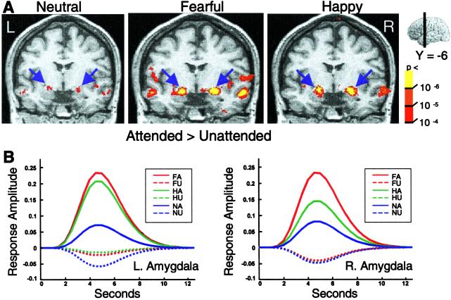
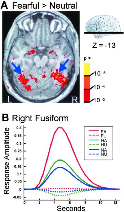
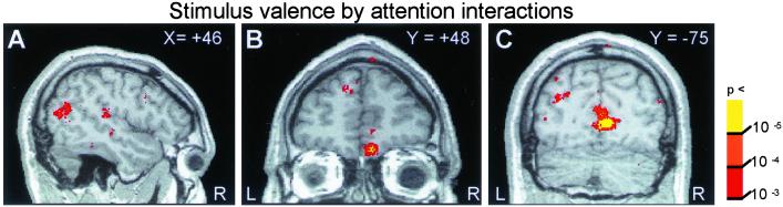
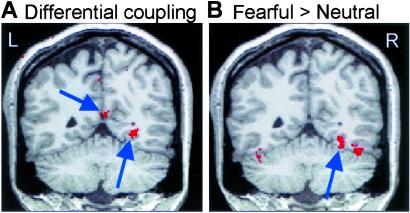
Attention gates the processing of stimuli relatively early in visual cortex. Yet, existing data suggest that emotional stimuli activate brain regions automatically, largely immune from attentional control. To resolve this puzzle, we used functional magnetic resonance imaging to first measure activation in regions that responded differentially to faces with emotional expressions (fearful and happy) compared with neutral faces. We then measured the modulation of these responses by attention, using a competing task with a high attentional load. Contrary to the prevailing view, all brain regions responding differentially to emotional faces, including the amygdala, did so only when sufficient attentional resources were available to process the faces. Thus, the processing of facial expression appears to be under top-down control.
Figures
Similar articles
-
Attentional control of the processing of neural and emotional stimuli.Brain Res Cogn Brain Res. 2002 Dec;15(1):31-45. doi: 10.1016/s0926-6410(02)00214-8. Brain Res Cogn Brain Res. 2002. PMID: 12433381 Review.
-
The influence of directed covert attention on emotional face processing.Neuroimage. 2010 Apr 1;50(2):545-51. doi: 10.1016/j.neuroimage.2009.12.073. Epub 2009 Dec 24. Neuroimage. 2010. PMID: 20035882
-
Attention inhibition of early cortical activation to fearful faces.Brain Res. 2010 Feb 8;1313:113-23. doi: 10.1016/j.brainres.2009.11.060. Epub 2009 Dec 11. Brain Res. 2010. PMID: 20004181
-
Dissociable effects of bottom-up and top-down factors on the processing of unattended fearful faces.Neuropsychologia. 2007 Oct 1;45(13):3075-86. doi: 10.1016/j.neuropsychologia.2007.05.019. Epub 2007 Jun 13. Neuropsychologia. 2007. PMID: 17631362 Free PMC article.
-
Functional atlas of emotional faces processing: a voxel-based meta-analysis of 105 functional magnetic resonance imaging studies.J Psychiatry Neurosci. 2009 Nov;34(6):418-32. J Psychiatry Neurosci. 2009. PMID: 19949718 Free PMC article. Review.
Cited by
-
Self-regulatory depletion increases emotional reactivity in the amygdala.Soc Cogn Affect Neurosci. 2013 Apr;8(4):410-7. doi: 10.1093/scan/nss082. Epub 2012 Jul 25. Soc Cogn Affect Neurosci. 2013. PMID: 22842815 Free PMC article.
-
Emotion and attention interaction: a trade-off between stimuli relevance, motivation and individual differences.Front Hum Neurosci. 2013 Jul 12;7:364. doi: 10.3389/fnhum.2013.00364. eCollection 2013. Front Hum Neurosci. 2013. PMID: 23874284 Free PMC article.
-
Functional neural correlates of emotional expression processing deficits in behavioural variant frontotemporal dementia.J Psychiatry Neurosci. 2013 May;38(3):174-82. doi: 10.1503/jpn.120008. J Psychiatry Neurosci. 2013. PMID: 23031250 Free PMC article.
-
Curvilinear relationship between phonological working memory load and social-emotional modulation.Cogn Emot. 2013;27(2):283-304. doi: 10.1080/02699931.2012.712948. Epub 2012 Aug 28. Cogn Emot. 2013. PMID: 22928750 Free PMC article.
-
Reward learning and negative emotion during rapid attentional competition.Front Psychol. 2015 Mar 12;6:269. doi: 10.3389/fpsyg.2015.00269. eCollection 2015. Front Psychol. 2015. PMID: 25814971 Free PMC article.
References
-
- Lavie N. (1995) J. Exp. Psychol. Hum. Percept. Perform. 21, 451-468. - PubMed
-
- Simons D. J. & Levin, D. T. (1997) Trends Cognit. Sci. 1, 261-267. - PubMed
-
- Joseph J. S., Chun, M. M. & Nakayama, K. (1997) Nature (London) 387, 805-807. - PubMed
-
- Rees G., Frith, C. D. & Lavie, N. (1997) Science 278, 1616-1619. - PubMed
-
- Ohman A., Esteves, F. & Soares, J. J. F. (1995) J. Psychophysiol. 9, 99-108.
Publication types
MeSH terms
LinkOut - more resources
Full Text Sources
