. 2002 Aug 20;99(17):11464-9.
doi: 10.1073/pnas.172403999. Epub 2002 Aug 12.

Newly generated neurons in the amygdala and adjoining cortex of adult primates

Affiliations

Newly generated neurons in the amygdala and adjoining cortex of adult primates

Patrick J Bernier et al. Proc Natl Acad Sci U S A. .

Abstract

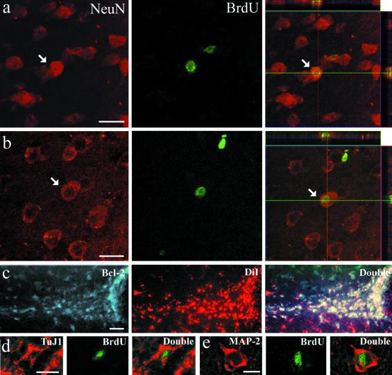
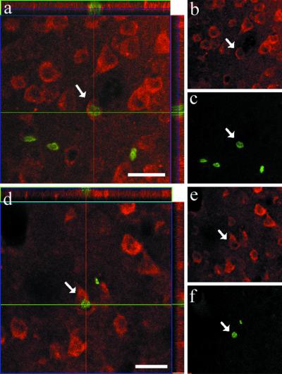
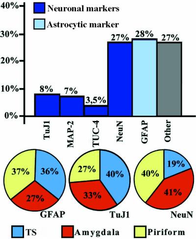
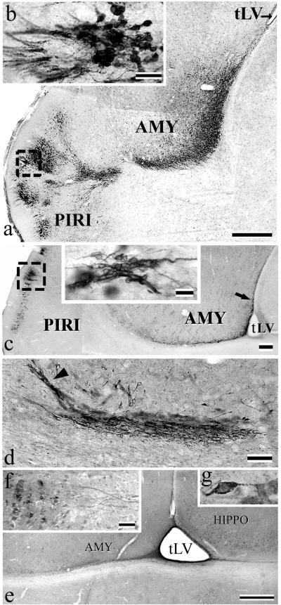
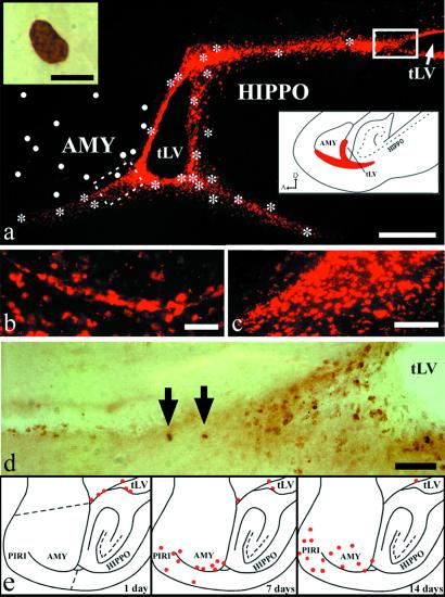
The subventricular zone remains mitotically active throughout life in rodents. Studies with tritiated thymidine, which is incorporated into the DNA of mitotic cells, have revealed that the rodent subventricular zone produces neuroblasts that migrate toward the olfactory bulb along the rostral migratory stream. A similar migratory stream has been documented in monkeys by using the thymidine analogue BrdUrd. The same approach showed that neurogenesis occurred in the dentate gyrus of adult primates, including humans. In the present study, experiments combining injections of BrdUrd and the dye 1,1'-dioctadecyl-3,3,3',3'-tetramethylindo-carbocyanine, with the immunostaining for molecular markers of neurogenesis (polysialylated neural cell adhesion molecule, beta-tubulin-III, collapsin response mediator protein-4, neuronal nuclear protein) in New World (Saimiri sciureus) and Old World (Macaca fascicularis) monkeys have revealed that new neurons are produced in the amygdala, piriform cortex, and adjoining inferior temporal cortex in adult primates. These newborn neurons expressed the antiapoptotic protein Bcl-2 and formed a more-or-less continuous pathway that extended from the tip of the temporal ventricular horn to the deep portion of the temporal lobe. The production of newborn neurons in the amygdala, piriform cortex, and inferior temporal cortex seems to parallel the continuing addition of neurons in the olfactory bulb. These two concomitant phenomena may ensure structural stability and functional plasticity to the primate olfactory system and temporal lobe.


Figures

Fig 1.
Fig 1.
Colocalization of neurogenesis markers in the rostral portion of the temporal lobe of squirrel monkeys that were killed 21 and 28 days after the last BrdUrd injection. Examples of a cell in the amygdala (a) and piriform cortex (b) that have incorporated BrdUrd (green) and also express NeuN (red). The right side shows reconstructed orthogonal images of the same NeuN+/BrdUrd+ cells, which are viewed from the sides in both x–z (top) and y–z (right) planes. (c) Cells in TS are colabeled for DiI (red) and for Bcl-2 (green). (d) TuJ1+/BrdUrd+ cell in TS and (e) MAP-2+/BrdUrd+ cell in amygdala. The various neuronal markers are stained in red, whereas BrdUrd is in green. [Bars = 25 μm (a and b), 100 μm (c), 15 μm (d and e).]
Fig 2.
Fig 2.
(af) Two BrdUrd+/NeuN+ cells encountered in the inferior temporal cortex of an adult squirrel monkey that have incorporated BrdUrd (green) and also express NeuN (red). This animal was killed 28 days after the last BrdUrd injection. (Left) Orthogonal views. (Right) Separate labeling views. (Bars in a and d = 25 μm.)
Fig 3.
Fig 3.
Bar and circle histograms depicting the relative proportion of the different types of cells that have incorporated BrdUrd in the TS and the rostral sector of the temporal lobe (see Fig. 5e for exact delimitation) of adult squirrel monkeys that were killed 28 days after the last BrdUrd injection. The bar histogram shows the percentage of neuronal (MAP-2, NeuN), immature neuronal (TuJ1, TUC-4) or astrocytic (GFAP) markers relative to the total number of BrdUrd-labeled nuclei. The column named ‘Other’ refers to the percentage of cells that do not express NeuN or GFAP. The circle diagrams illustrate the proportion of BrdUrd+ cells that were also labeled for GFAP, TuJ1, and NeuN in the TS (blue), amygdala (yellow), and piriform cortex (red).
Fig 4.
Fig 4.
The squirrel monkey TS as seen on parasagittal sections immunostained with various neurogenesis markers. (a) Photomontage showing typical Bcl-2 immunostaining that extends from the tip of the tLV to the amygdala (AMY) and piriform cortex (PIRI). (b) High-power view of a cluster of Bcl-2+ cells (box in a) in superficial layers of piriform cortex. (c) Chains of PSA-NCAM+ cells beneath the amygdala. (Inset) A high power view of PSA-NCAM+ chains in piriform cortex (PIRI) (box). (d) Chains of β-tubulin-III+ cells in amygdala (see arrow in c). Some chains are oriented toward amygdala (arrowhead in d). (e) Overview of TUC-4 immunoreactivity in the lower part of amygdala (AMY). Note the labeling that emerges form the tip of tLV. (f) A typical cluster of TUC-4+ neurons in piriform cortex, from which emerges an array of thick immunopositive fibers. (g) High-power view of a TUC-4+ neuron in piriform cortex. [Bars = 500 μm (a), 20 μm (b and f), 250 μm (c), 25 μm (Inset in c), 50 μm (d), 1 mm (e), and 15 μm (g).]
Fig 5.
Fig 5.
(ad) DiI-labeling and distribution of BrdUrd+ cells, as seen on a parasagittal section of the temporal lobe of an adult squirrel monkey. (a) General view of the typical DiI staining; dots represent BrdUrd+ cells found in amygdala (AMY), whereas asterisks indicate BrdUrd+ cells lying along the DiI+ pathway and its branches. Inset (Upper Left) displays a high-power view of one typical BrdUrd+ nucleus. For the sake of clarity the numerous BrdUrd+ cells in the hippocampal formation (HIPPO) have not been indicated here. Inset (Lower Right) depicts the germinal matrix extensions under amygdala (AMY) and hippocampus (HIPPO), as they can be seen on a sagittal section of the temporal lobe of a 26-day-old human embryo (25). (b and c) A typical chain of DiI+ cells en route to the amygdala (b) (dotted line box in a) and a pool of DiI+ cells at the tip of the temporal horn of the lateral ventricle (tLV) (c). (d) BrdUrd+ cells (arrows) that are part of a pool of similar cells at the tip of the tLV (uninterrupted-line box in a). (e) Plotting of BrdUrd+ nuclei on sagittal sections of adult macaques killed at three different times after the last BrdUrd injection. The red circles indicate BrdUrd+ nuclei that appear at day 1, 7, and 14 after the last BrdUrd injection. For the sake of clarity the numerous BrdUrd+ cells in the hippocampal formation have not been indicated here. The dotted lines in the upper drawing delineate the sector in which quantification was made (see Fig. 3). [Bars = 250 μm (a), 5 μm (Inset in a), 50 μm (b and e), and 100 μm (c).]

References

    1. Doetsch F., Garcia-Verdugo, J. M. & Alvarez-Buylla, A. (1997) J. Neurosci. 17, 5046-5061. - PMC - PubMed
    1. Gould E., Reeves, A. J., Fallah, M., Tanapat, P. & Gross, C. G. (1999) Proc. Natl. Acad. Sci. USA 96, 5263-5267. - PMC - PubMed
    1. Lois C. & Alvarez-Buylla, A. (1994) Science 26, 1145-1148. - PubMed
    1. Kornack D. R. & Rakic, P. (2001) Proc. Natl. Acad. Sci. USA 98, 4752-4757. - PMC - PubMed
    1. O'Rourke N. A., Chenn, A. & McConnell, S. K. (1997) Development (Cambridge, U.K.) 124, 997-1005. - PubMed

Publication types