. 2002 Aug;129(4):1581-91.
doi: 10.1104/pp.003798.

Expression and molecular analysis of the Arabidopsis DXR gene encoding 1-deoxy-D-xylulose 5-phosphate reductoisomerase, the first committed enzyme of the 2-C-methyl-D-erythritol 4-phosphate pathway

Affiliations

Expression and molecular analysis of the Arabidopsis DXR gene encoding 1-deoxy-D-xylulose 5-phosphate reductoisomerase, the first committed enzyme of the 2-C-methyl-D-erythritol 4-phosphate pathway

Lorenzo Carretero-Paulet et al. Plant Physiol. 2002 Aug.

Abstract

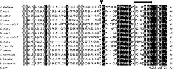
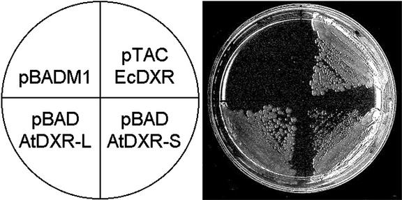
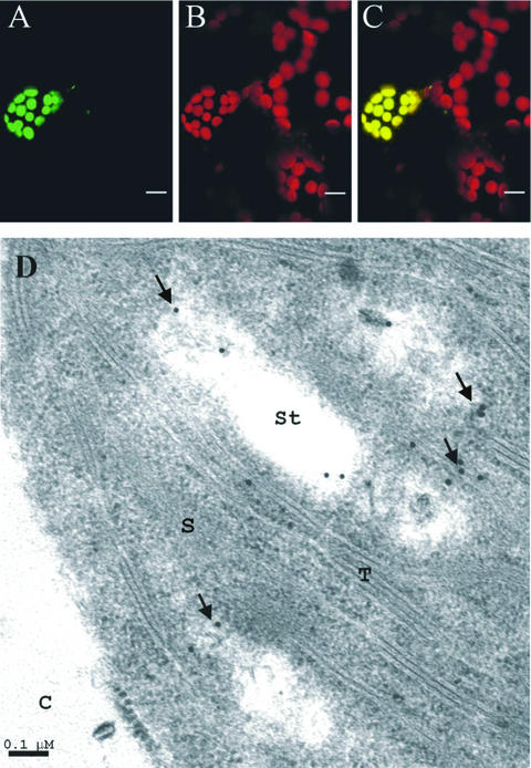
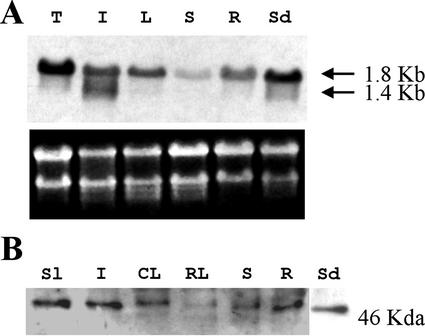
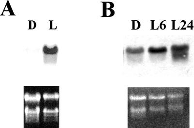
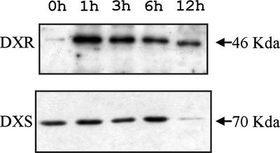
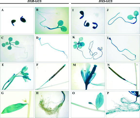
1-Deoxy-D-xylulose 5-phosphate reductoisomerase (DXR) catalyzes the first committed step of the 2-C-methyl-D-erythritol 4-phosphate pathway for isoprenoid biosynthesis. In Arabidopsis, DXR is encoded by a single-copy gene. We have cloned a full-length cDNA corresponding to this gene. A comparative analysis of all plant DXR sequences known to date predicted an N-terminal transit peptide for plastids, with a conserved cleavage site, and a conserved proline-rich region at the N terminus of the mature protein, which is not present in the prokaryotic DXR homologs. We demonstrate that Arabidopsis DXR is targeted to plastids and localizes into chloroplasts of leaf cells. The presence of the proline-rich region in the mature Arabidopsis DXR was confirmed by detection with a specific antibody. A proof of the enzymatic function of this protein was obtained by complementation of an Escherichia coli mutant defective in DXR activity. The expression pattern of beta-glucuronidase, driven by the DXR promoter in Arabidopsis transgenic plants, together with the tissue distribution of DXR transcript and protein, revealed developmental and environmental regulation of the DXR gene. The expression pattern of the DXR gene parallels that of the Arabidopsis 1-deoxy-D-xylulose 5-phosphate synthase gene, but the former is slightly more restricted. These genes are expressed in most organs of the plant including roots, with higher levels in seedlings and inflorescences. The block of the 2-C-methyl-D-erythritol 4-phosphate pathway in Arabidopsis seedlings with fosmidomycin led to a rapid accumulation of DXR protein, whereas the 1-deoxy-D-xylulose 5-phosphate synthase protein level was not altered. Our results are consistent with the participation of the Arabidopsis DXR gene in the control of the 2-C-methyl-D-erythritol 4-phosphate pathway.


Figures

Figure 1
Figure 1
Multiple sequence alignment of the N-terminal region of plant DXR. The sequence of Arabidopsis DXR was aligned to the other plant DXR sequences known to date, deduced either from complete cDNA clones or expressed sequence tag (EST) entries. Only those EST sequences confirmed by at least two independent entries were considered. Sequence alignment was performed with the ClustalW 1.8 program (http://dot.imgen.bcm.tmc.edu:9331/multi-align/multi-align.html) and optimized by visual inspection. The origin of the DXR sequence is indicated on the left and the number of amino acid residues on the right. Residues are written in white inside black boxes if they are identical in all plant sequences, in white inside gray boxes if two alternative residues are found in equivalent positions, or in black over white background if a lower conservation is observed. Gaps in the sequence are represented with a dash. The last residue of the transit peptide predicted by the ChloroP program (Emanuelsson et al., 1999) in the different sequences is indicated by an asterisk above the corresponding letter. The putative cleavage site deduced from the collective analysis of all plant DXR sequences is indicated with an arrowhead. The Arabidopsis peptide used for antibody production is marked with a line on the top. The cDNA and EST sequences are accessible at the GenBank with the following accession numbers: Arabidopsis (AF148852), Artemisia annua (AF182287), periwinkle (AF250235), Glycine max (EST 1, BE804032; EST 2, BE211397; and EST 3, BG839054), tomato (AF331705), Lycopersicon hirsutum (EST, AW617386), Medicago truncatula (EST 1, BG456710; and EST 2, BG450566), peppermint (AF116825), Oryza sativa (AF367205), Solanum tuberosum (EST, BE924278), and Zea mays (AJ297566). For reference, the N-terminal sequence of the DXR from E. coli is represented at the bottom.
Figure 2
Figure 2
Complementation of E. coli dxr mutant with Arabidopsis DXR. The E. coli dxr::TET mutant EcAB1–2 (Rodríguez-Concepción et al., 2000) was transformed with a control expression plasmid without DXR insert (pBADM1), a pBADM1 derivative coding for a long form of Arabidopsis DXR (pBAD-AtDXR-L), a pBADM1 derivative coding for a short form of Arabidopsis DXR (pBAD-AtDXR-S), or a expression plasmid coding for the DXR from E. coli (pTAC-EcDXR). Transformants were plated in Luria-Bertani medium containing 6 μg mL−1 tetracycline to select for the dxr::TET mutant, 100 μg mL−1 ampicillin to select for the plasmid, 100 μm isopropyl-β-d-thiogalactoside to induce expression of EcDXR, and 0.02% (w/v) l-Ara to induce expression of AtDXR-S and AtDXR-L. After 17 h at 37°C, the colony size was 2 to 3 mm for the mutant transformed with pTAC-EcDXR or pBAD-AtDXR-L and 0.3 to 0.4 mm for the mutant transformed with pBAD-AtDXR-S.
Figure 3
Figure 3
Targeting and subcellular localization of plant DXR. Leaves of 15-d-old Arabidopsis seedlings were microbombarded with a construct encoding Arabidopsis DXR fused to the N terminus of GFP. Cells expressing the fusion protein were studied by laser confocal microscopy. The images show green fluorescence of DXR-GFP (A), red autofluorescence of chlorophyll (B), and the superimposed green and red fluorescence (C). Bars in A through C indicate 10 μm. Arabidopsis DXR was immunolocalized in leaves of 15-d-old seedlings with the Ab-AtDXR1 polyclonal antibody. The electron micrograph (D) shows localization of 15-nm gold particles in chloroplast. C, Cytoplasm; S, stroma; St, starch granule; T, thylakoid. Bar in D indicates 0.1 μm.
Figure 4
Figure 4
Distribution of DXR transcripts and protein in Arabidopsis plants. A, Fifteen micrograms of total RNA from Arabidopsis tissues was analyzed by northern blot as described in “Materials and Methods.” Exposure time was 48 h. Ethidium bromide staining of the gel before transfer is also shown. B, Crude extracts (30 μg of protein) from Arabidopsis tissues were analyzed by western blot as described in “Materials and Methods.” DXR protein was detected with the Ab-AtDXR1 polyclonal antibody. Developing time was 1 min. CL, Cauline leaves, Sl, siliques; I, inflorescences; L, rosette and cauline leaves; R, roots of 15-d-old seedlings grown either in Murashige and Skoog plates exposed to light (A) or in soil (B); RL, rosette leaves; S, stems from adult plant; Sd, 15-d-old seedlings grown in 16-h light/8-h dark regime; T, Arabidopsis cell suspension line T87.
Figure 5
Figure 5
Accumulation of Arabidopsis DXR transcript induced by light. Samples were analyzed by northern blot as described in “Materials and Methods.” A, Twenty micrograms of total RNA from light-grown (L) and dark-grown (D) 12-d-old seedlings. B, Ten micrograms of total RNA from dark-grown 15-d-old seedlings (D) or 14-d-old dark-grown seedlings exposed to light for 6 h (L6) or 24 h (L24). Exposure time was 40 h for the filter of A and 72 h for the filter of B.
Figure 6
Figure 6
Accumulation of Arabidopsis DXR protein induced by fosmidomycin. One hundred micromolar fosmidomycin was added to liquid cultures containing 7-d-old Arabidopsis seedlings grown under continuous light with agitation. Samples were collected just before treatment or at different times after treatment and analyzed by western blot. Arabidopsis DXR protein was detected with the Ab-AtDXR1 antibody. Arabidopsis DXS protein was detected with a specific polyclonal antibody (Estévez et al., 2000). Collection times are indicated at the top.
Figure 7
Figure 7
Histochemical analysis of GUS activity in transgenic Arabidopsis plants expressing the GUS gene under control of the DXR (A–H) or the DXS (I–P) promoter. A and I, Germinating seeds imbibed for 24 h in Murashige and Skoog medium; B and J, 6-d-old light-grown seedlings; C and K, 15-d-old light-grown seedlings; D and L, 9-d-old dark-grown seedlings; E and M, flowers; F and N, mature siliques; G and O, cauline leaf with emerging axillary inflorescence; H and P, roots of adult plant.

References

    1. Araus JL, Bort J, Brown RH, Bassett CL, Cortadellas N. Immunocytochemical localization of phosphoenolpyruvte carboxylase and photosynthetic gas-exchange characteristics in ears of Triticum durum Desf. Planta. 1993;191:507–514.
    1. Ausubel FM, Brent R, Kingston RE, Moore DD, Seidman JG, Smith JA, Struhl K, editors. Current Protocols in Molecular Biology. New York: John Wiley and Sons, Inc.; 1987.
    1. Axelos M, Curei C, Mazzolini L, Bardet C, Lescure B. A protocol for transient gene expression in Arabidopsis thaliana protoplasts isolated from cell suspension cultures. Plant Physiol Biochem. 1992;30:123–128.
    1. Bouvier F, d'Harlingue A, Suire C, Backhaus RA, Camara B. Dedicated roles of plastid transketolases during the early onset of isoprenoid biogenesis in pepper fruits. Plant Physiol. 1998;117:1423–1431. - PMC - PubMed
    1. Bradford M. A rapid and sensitive method for the quantitation of microgram quantities of protein utilizing the principle of protein-dye binding. Anal Biochem. 1976;72:248–254. - PubMed

Publication types

MeSH terms