

High resolution chromosome 3p, 8p, 9q and 22q allelotyping analysis in the pathogenesis of gallbladder carcinoma
- PMID: 12177780
- PMCID: PMC2376134
- DOI: 10.1038/sj.bjc.6600490
High resolution chromosome 3p, 8p, 9q and 22q allelotyping analysis in the pathogenesis of gallbladder carcinoma
Abstract
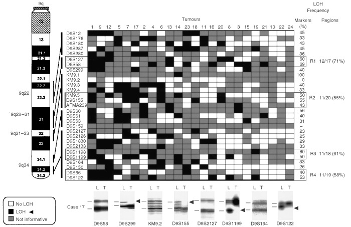
Our recent genome-wide allelotyping analysis of gallbladder carcinoma identified 3p, 8p, 9q and 22q as chromosomal regions with frequent loss of heterozygosity. The present study was undertaken to more precisely identify the presence and location of regions of frequent allele loss involving those chromosomes in gallbladder carcinoma. Microdissected tissue from 24 gallbladder carcinoma were analysed for PCR-based loss of heterozygosity using 81 microsatellite markers spanning chromosome 3p (n=26), 8p (n=14), 9q (n=29) and 22q (n=12) regions. We also studied the role of those allele losses in gallbladder carcinoma pathogenesis by examining 45 microdissected normal and dysplastic gallbladder epithelia accompanying gallbladder carcinoma, using 17 microsatellite markers. Overall frequencies of loss of heterozygosity at 3p (100%), 8p (100%), 9q (88%), and 22q (92%) sites were very high in gallbladder carcinoma, and we identified 13 distinct regions undergoing frequent loss of heterozygosity in tumours. Allele losses were frequently detected in normal and dysplastic gallbladder epithelia. There was a progressive increase of the overall loss of heterozygosity frequency with increasing severity of histopathological changes. Allele losses were not random and followed a sequence. This study refines several distinct chromosome 3p, 8p, 9q and 22q regions undergoing frequent allele loss in gallbladder carcinoma that will aid in the positional identification of tumour suppressor genes involved in gallbladder carcinoma pathogenesis.
Figures


Similar articles
-
Genome-wide allelotyping analysis reveals multiple sites of allelic loss in gallbladder carcinoma.Cancer Res. 2001 May 1;61(9):3795-800. Cancer Res. 2001. PMID: 11325854
-
Fragile histidine triad gene abnormalities in the pathogenesis of gallbladder carcinoma.Am J Pathol. 2002 Jun;160(6):2073-9. doi: 10.1016/S0002-9440(10)61157-1. Am J Pathol. 2002. PMID: 12057912 Free PMC article.
-
Loss of heterozygosity in dysplasia and carcinoma of the gallbladder.Mod Pathol. 1999 Aug;12(8):763-9. Mod Pathol. 1999. PMID: 10463477
-
High resolution chromosome 3p allelotyping of human lung cancer and preneoplastic/preinvasive bronchial epithelium reveals multiple, discontinuous sites of 3p allele loss and three regions of frequent breakpoints.Cancer Res. 2000 Apr 1;60(7):1949-60. Cancer Res. 2000. PMID: 10766185
-
Genetic alterations in gallbladder carcinoma.Surg Today. 2005;35(2):101-5. doi: 10.1007/s00595-004-2906-2. Surg Today. 2005. PMID: 15674488 Review.
Cited by
-
Gallbladder cancer epidemiology, pathogenesis and molecular genetics: Recent update.World J Gastroenterol. 2017 Jun 14;23(22):3978-3998. doi: 10.3748/wjg.v23.i22.3978. World J Gastroenterol. 2017. PMID: 28652652 Free PMC article. Review.
-
Frequent Downregulation and Promoter Hypermethylation of DLC1: Relationship with Clinical Outcome in Gallbladder Cancer.J Gastrointest Cancer. 2022 Jun;53(2):237-244. doi: 10.1007/s12029-020-00560-3. Epub 2021 Jan 8. J Gastrointest Cancer. 2022. PMID: 33417200
-
Pathologies of Precursor Lesions of Biliary Tract Carcinoma.Cancers (Basel). 2022 Oct 30;14(21):5358. doi: 10.3390/cancers14215358. Cancers (Basel). 2022. PMID: 36358777 Free PMC article. Review.
-
Pathology of gallbladder carcinoma: current understanding and new perspectives.Pathol Oncol Res. 2015 Jul;21(3):509-25. doi: 10.1007/s12253-014-9886-3. Epub 2015 Jan 25. Pathol Oncol Res. 2015. PMID: 25618479 Review.
-
Hypomethylation of the protein gene product 9.5 promoter region in gallbladder cancer and its relationship with clinicopathological features.Cancer Sci. 2006 Nov;97(11):1205-10. doi: 10.1111/j.1349-7006.2006.00320.x. Epub 2006 Sep 12. Cancer Sci. 2006. PMID: 16965602 Free PMC article.
References
-
- Albores-SaavedraJHensonDE2000Tumours of gallbladder and extrahepatic bile ducts Fascicle 233rd ednWashington, DC: Armed Forces Institute of Pathology
-
- AlbrechtSvon DeimlingAPietschTGiangasperoFBrandnerSKleihuesPWiestlerOD1994Microsatellite analysis of loss of heterozygosity on chromosomes 9q, 11p and 17p in medulloblastomas Neuropathol Appl Neurobiol 207481 - PubMed
-
- AllioneFEisingerFParcPNoguchiTSobolHBirnbaumD1998Loss of heterozygosity at loci from chromosome arm 22Q in human sporadic breast carcinomas Int J Cancer 75181186 - PubMed
-
- BaffaRSantoroRBullrichFMandesBIshiiHCroceCM2000Definition and refinement of chromosome 8p regions of loss of heterozygosity in gastric cancer Clin Cancer Res 613721377 - PubMed
-
- BooksteinRBovaGSMacGroganDLevyAIsaacsWB1997Tumour-suppressor genes in prostatic oncogenesis: a positional approach Br J Urol 79Suppl 12836 - PubMed
Publication types
MeSH terms
Grants and funding
LinkOut - more resources
Full Text Sources
Medical
