

Disinhibition in the human motor cortex is enhanced by synchronous upper limb movements
- PMID: 12181301
- PMCID: PMC2290478
- DOI: 10.1113/jphysiol.2002.023986
Disinhibition in the human motor cortex is enhanced by synchronous upper limb movements
Abstract
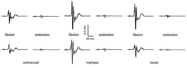
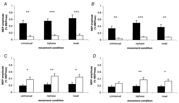
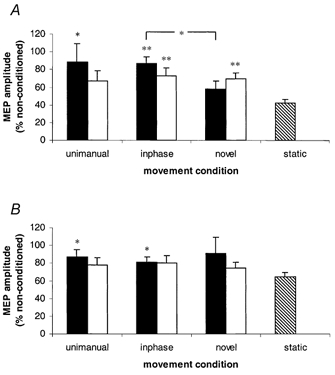
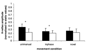
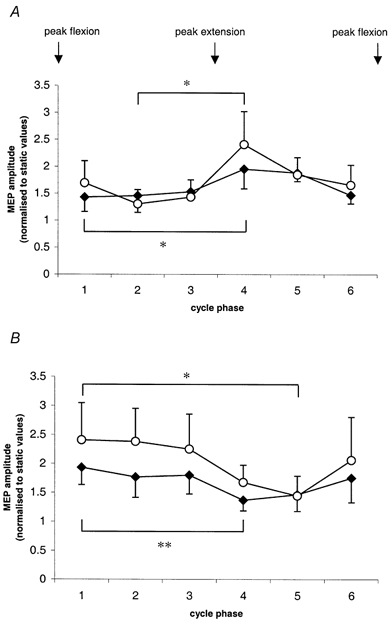
The phasic modulation of wrist flexor corticomotor disinhibition has previously been demonstrated during the flexion phase of rhythmical passive flexion-extension of the human wrist. Here we ask if rhythmical bimanual flexion-extension movements of the wrists of neurologically intact subjects, modulate inhibitory activity in the motor cortex. In the first experiment intracortical inhibition was assessed when one wrist was passively flexed and extended on its own, with the addition of the opposite limb voluntarily moving synchronously in a mirror symmetric pattern, and also in a near-symmetric asynchronous pattern. Two subsequent experiments investigated firstly the modulation of spinal reflex pathway activity during the same three movement conditions, and secondly the effect of contralateral wrist movement alone on the excitability of corticomotoneuronal pathways to a static test limb. When the wrist flexors of both upper limbs were shortening simultaneously (i.e. synchronously), intracortical inhibition associated with flexor representations was suppressed to a greater extent than when the two muscles were shortening asynchronously. The results of the three experiments indicate that modulation of inhibitory activity was taking place at the cortical level. These findings may have further application in the study of rehabilitation procedures where the effects of simultaneous activation of affected and unaffected upper limbs in hemiparetic patients are to be investigated.
Figures
References
-
- Carson R, Riek S, Smethurst C, Parraga J, Byblow W. Neuromuscular-skeletal constraints upon the dynamics of unimanual and bimanual coordination. Experimental Brain Research. 2000;131:196–214. - PubMed
-
- Carson RG. The dynamics of isometric bimanual coordination. Experimental Brain Research. 1995;105:465–476. - PubMed
-
- Carson RG, Riek S, Bawa P. Electromyographic activity, H-reflex modulation and corticospinal input to forearm during active and passive rhythmic movements. Human Movement Science. 1999;18:307–343.
-
- Cheney P, FetZ E, Mewes K. Neural mechanisms underlying corticospinal and rubrospinal control of limb movements. Progress in Brain Research. 1991;87:213–252. - PubMed
Publication types
MeSH terms
LinkOut - more resources
Full Text Sources