. 2002 Aug;13(8):2795-809.
doi: 10.1091/mbc.01-07-0348.

Function of dynein and dynactin in herpes simplex virus capsid transport

Affiliations

Function of dynein and dynactin in herpes simplex virus capsid transport

Katinka Döhner et al. Mol Biol Cell. 2002 Aug.

Abstract

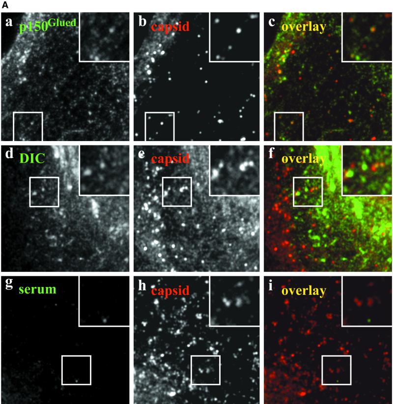
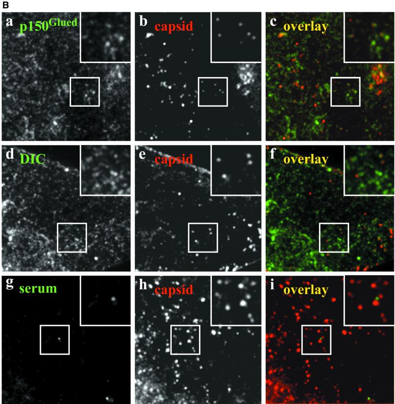
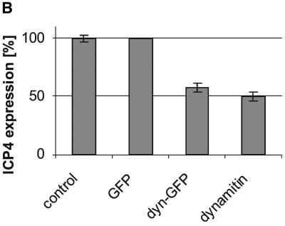
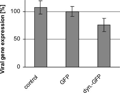
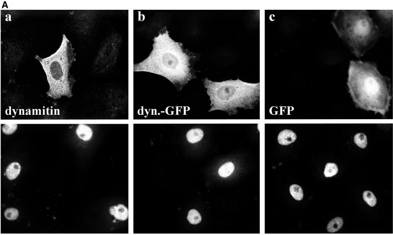
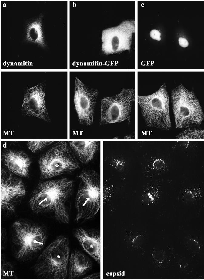
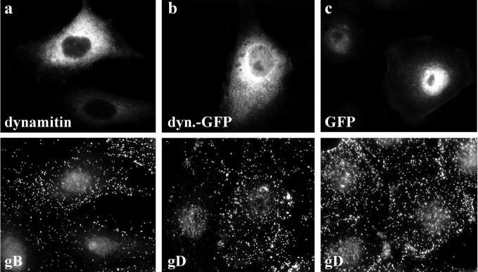
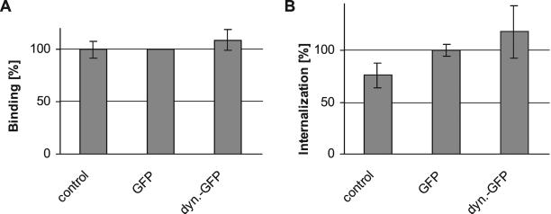
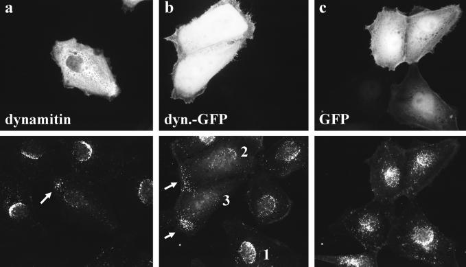
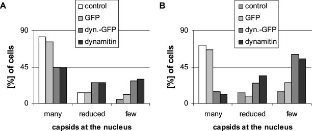
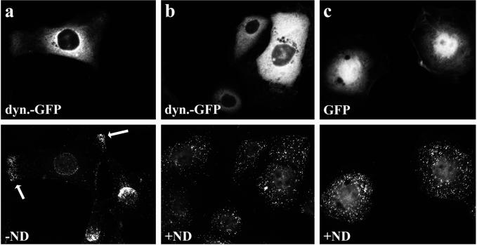
After fusion of the viral envelope with the plasma membrane, herpes simplex virus type 1 (HSV1) capsids are transported along microtubules (MTs) from the cell periphery to the nucleus. The motor ATPase cytoplasmic dynein and its multisubunit cofactor dynactin mediate most transport processes directed toward the minus-ends of MTs. Immunofluorescence microscopy experiments demonstrated that HSV1 capsids colocalized with cytoplasmic dynein and dynactin. We blocked the function of dynein by overexpressing the dynactin subunit dynamitin, which leads to the disruption of the dynactin complex. We then infected such cells with HSV1 and measured the efficiency of particle binding, virus entry, capsid transport to the nucleus, and the expression of immediate-early viral genes. High concentrations of dynamitin and dynamitin-GFP reduced the number of viral capsids transported to the nucleus. Moreover, viral protein synthesis was inhibited, whereas virus binding to the plasma membrane, its internalization, and the organization of the MT network were not affected. We concluded that incoming HSV1 capsids are propelled along MTs by dynein and that dynein and dynactin are required for efficient viral capsid transport to the nucleus.


Figures

Figure 1
Figure 1
(A) Incoming wild-type HSV1 capsids colocalize with p150Glued and DIC. A significant fraction of incoming HSV1 capsids colocalizes with p150Glued and DIC (yellow dots in the overlays, c and f). This apparent colocalization is not due to binding of rabbit IgG to the herpes virus Fc receptor on the surface of infected cells because there is no signal from a nonimmune rabbit serum (i). Moreover, immunoblots confirmed that there is no unspecific cross-reactivity of the anti-dynactin or anti-dynein antibodies to structural viral proteins (our unpublished results). Immunofluorescence microscopy of PtK2 cells infected with wild-type HSV1 for 1 h in the presence of 0.5 mM cycloheximide. Cells were fixed with PHEMO-fix at room temperature, blocked with 0.2 mg/ml pooled human IgG and 5 mg/ml BSA and double-labeled with anti-p150Glued (pAb Portos; a), anti-DIC (pAb L5, d) or a nonimmune rabbit serum (serum, g) and anti-VP5 (mAb 5C10; b, e, and h). Note, that the anti-VP5 antibodies cross-reacted in PtK2 cells weakly with the cortical actin and stress fibers. (B) Incoming HSV1 capsids of a gE deletion mutant colocalize with p150Glued and DIC. Incoming HSV1 capsids of the mutant strain R7202 that does not express a viral Fc receptor colocalize with p150Glued and DIC (yellow dots in the overlays, c and f). Moreover, there is no colocalization (i) after double labeling with a nonimmune rabbit serum (g) and capsid antibodies (h). Immunofluorescence microscopy of PtK2 cells infected with HSV1 deleted for gE (R7202) for 1 h in the presence of 0.5 mM cycloheximide. Cells were fixed with PHEMO-fix at room temperature, blocked with 0.2 mg/ml pooled human IgG and 5 mg/ml BSA and double-labeled with anti-p150Glued (pAb Portos; a), anti-DIC (pAb L5, d), or a nonimmune rabbit serum (g) and anti-VP5 (mAb 5C10; b, e, and h).
Figure 1
Figure 1
(A) Incoming wild-type HSV1 capsids colocalize with p150Glued and DIC. A significant fraction of incoming HSV1 capsids colocalizes with p150Glued and DIC (yellow dots in the overlays, c and f). This apparent colocalization is not due to binding of rabbit IgG to the herpes virus Fc receptor on the surface of infected cells because there is no signal from a nonimmune rabbit serum (i). Moreover, immunoblots confirmed that there is no unspecific cross-reactivity of the anti-dynactin or anti-dynein antibodies to structural viral proteins (our unpublished results). Immunofluorescence microscopy of PtK2 cells infected with wild-type HSV1 for 1 h in the presence of 0.5 mM cycloheximide. Cells were fixed with PHEMO-fix at room temperature, blocked with 0.2 mg/ml pooled human IgG and 5 mg/ml BSA and double-labeled with anti-p150Glued (pAb Portos; a), anti-DIC (pAb L5, d) or a nonimmune rabbit serum (serum, g) and anti-VP5 (mAb 5C10; b, e, and h). Note, that the anti-VP5 antibodies cross-reacted in PtK2 cells weakly with the cortical actin and stress fibers. (B) Incoming HSV1 capsids of a gE deletion mutant colocalize with p150Glued and DIC. Incoming HSV1 capsids of the mutant strain R7202 that does not express a viral Fc receptor colocalize with p150Glued and DIC (yellow dots in the overlays, c and f). Moreover, there is no colocalization (i) after double labeling with a nonimmune rabbit serum (g) and capsid antibodies (h). Immunofluorescence microscopy of PtK2 cells infected with HSV1 deleted for gE (R7202) for 1 h in the presence of 0.5 mM cycloheximide. Cells were fixed with PHEMO-fix at room temperature, blocked with 0.2 mg/ml pooled human IgG and 5 mg/ml BSA and double-labeled with anti-p150Glued (pAb Portos; a), anti-DIC (pAb L5, d), or a nonimmune rabbit serum (g) and anti-VP5 (mAb 5C10; b, e, and h).
Figure 2
Figure 2
Quantification of immediate-early viral gene expression in transfected cells. β-Galactosidase activity per cell was reduced in dynamitin-GFP–overexpressing cells compared with untransfected or GFP-transfected cells. Two-sided Student's t test confirmed that viral protein synthesis is significantly lower in dynamitin-GFP–transfected cells compared with either GFP-transfected cells (p = 1.13 × 10⁻⁴) or to untransfected control cells (p = 2.1 × 10⁻⁵). There was no significant difference in β-galactosidase expression between control and GFP-transfected cells (p = 1.58 × 10⁻¹). The mean values for five independent experiments each performed in duplicates are shown.
Figure 3
Figure 3
Overexpression of dynamitin reduces immediate-early viral gene expression. (A) Overexpression of dynamitin (a) or dynamitin-GFP (b) reduced the immediate-early viral gene expression compared with control (a–c) or GFP-transfected cells (c). Immunofluorescence microscopy of PtK2 cells transfected with dynamitin or dynamitin-GFP and 30 h later infected with HSV1 for 3 h. Cells were fixed with PFA and either double-labeled with anti-myc (a; top panel) to detect transfected cells and anti-ICP4, an immediate-early protein of HSV1 (a–c; bottom panels) or single-labeled with anti-ICP4 (b and c; bottom panels), and the transfected proteins were detected by their intrinsic GFP fluorescence (b and c; top panels). (B) Quantification of viral ICP4 synthesis after transfection. The overexpression of dynamitin reduced immediate-early viral gene expression. Two-sided Student's t test confirmed that ICP4 expression is significantly lower in dynamitin (p = 2.52 × 10⁻³) or dynamitin-GFP (p = 3.57 × 10⁻³) transfected cells compared with GFP-transfected cells. There was no significant difference in ICP4 expression between untransfected and GFP-transfected cells (p = 1). The experiment described in A was quantitated (three independent experiments; altogether >500 cells analyzed for each condition). Cells overexpressing dynamitin, dyna-mitin-GFP, or GFP and untransfected cells were divided into two classes: cells displaying a nuclear ICP4 signal and cells not displaying a nuclear ICP4 signal.
Figure 4
Figure 4
Organization of the microtubule network in transfected PtK2 cells. Immunofluorescence microscopy showing the MT network of PtK2 cells after overexpression of dynamitin or dynamitin-GFP. In almost all cells the MTs emanated from the perinuclear region of the cell. After overexpression of dynamitin (a) or dynamitin-GFP (b), there seemed to be fewer cells with a well-defined MTOC. In untransfected cells (b and d) and in cells overexpressing GFP (c), the MTs were focused at the MTOC in many cells. In untransfected cells with an apparent MTOC (arrows in d) and in cells with unfocussed MTs (asterisks in d) numerous capsids reached the nucleus (d, capsids). Twenty-eight hours after transfection, cells were fixed with PHEMO-fix and double-labeled with anti-myc (pAb, a; top panel) and anti-tubulin (mAb 1A2, a; bottom panel). Cells transfected with GFP proteins were only labeled with anti-tubulin (mAb 1A2, b and c; bottom panels). Note that because of the PHEMO-fixation, GFP was relocalized to the nucleus. In unfixed cells, most of the transiently expressed GFP was localized to the cytoplasm. In d, untransfected PtK2 cells were infected with HSV1 for 3 h and fixed with PHEMO-fix at 37°C. Capsids were labeled with anti-LC (d; right panel) and MTs with 1A2 (d; left panel).
Figure 5
Figure 5
HSV1 binding and internalization in transfected cells. In cells overexpressing dynamitin (a) or dynamitin-GFP (b) virus binding was not reduced compared with untransfected cells or cells overexpressing GFP (c). Immunofluorescence microscopy of PtK2 cells transfected with dynamitin or dynamitin-GFP and 20 h later inoculated with HSV1 for 2 h at 4°C. After 2 h at 4°C cells were washed and shifted to 37°C for 15 min to reduce unspecific binding, fixed in PFA and permeabilized with TX-100. They were double-labeled with anti-dynamitin (a; top panel) and anti-gB (a; bottom panel), and cells transfected with GFP proteins were only labeled with anti-gD (mAb DL6, b and c; bottom panels).
Figure 6
Figure 6
Quantification of HSV1 virus binding and internalization in transfected cells. (A) Virus binding after overexpression of dynamitin-GFP or GFP was identical to virus binding to mock-transfected cells (n = 2). Two-sided Student's t test confirmed that viral binding was not significantly different in dynamitin-GFP–transfected cells compared with either GFP-transfected cells (p = 4.21 × 10⁻¹) or to untransfected control cells (p = 4.03 × 10⁻¹). There was also no significant difference in viral binding between control and GFP-transfected cells (p = 9.42 × 10⁻¹). Virus binding was calculated as radioactivity per cell and expressed as percent of radioactivity per cell compared with GFP-transfected cells. (B) Virus internalization after overexpression of dynamitin-GFP was not reduced compared with GFP-transfected cells (n = 6). Two-sided Student's t test confirmed that viral internalization was not significantly different in dynamitin-GFP–transfected cells compared with GFP-transfected cells (p = 1.45 × 10⁻¹). However, compared with untransfected cells, virus internalization was higher in cells transfected with GFP (p = 2.46 × 10⁻³) or dynamitin-GFP (p = 7.11 × 10⁻³). Virus internalization was calculated as radioactivity per cell and expressed as percent of radioactivity per cell compared with GFP-transfected cells.
Figure 7
Figure 7
Overexpression of dynamitin reduces HSV1 capsid transport to the nucleus. Three hours after infection most capsids were bound to the nucleus in untransfected cells (a–c) and in cells overexpressing GFP (c). In cells overexpressing dynamitin (a) or dynamitin-GFP (b), only a few capsids reached the nucleus, and several capsids were seen in the periphery of the cells (arrows in a and b, bottom panels). Immunofluorescence microscopy of PtK2 cells transfected with dynamitin or dynamitin-GFP and 46 h later infected with HSV1 for 3 h in the presence of cycloheximide. Cells were fixed with PFA, permeabilized with TX-100, and double-labeled with anti-dynamitin (mAb p50; a, top panel) and anti-HC (pAb; a, bottom panel). Cells transfected with GFP proteins were labeled with anti-HC (pAb; b and c, bottom panels).
Figure 8
Figure 8
Quantification of viral capsid transport in transfected cells. For quantification, untransfected and transfected cells were randomly selected and classified into three different groups: cells with many capsids at the nucleus, typically forming a nuclear rim (Figure 7b, 1), cells with a reduced amount of capsids at the nucleus (Figure 7b, 2), and cells with <10 capsids at the nucleus (Figure 7b, 3). Either all (A) or only high expressing (B) cells were analyzed. Expression levels in the cytoplasm were determined using a digital camera. One can directly compare the expression levels for GFP with dynamitin-GFP (cf. MATERIALS AND METHODS). Dynamitin and dynamitin-GFP strongly inhibited HSV1 entry, whereas GFP had no effect on viral infection. Forty-five or 46 h after transfection with dynamitin, dynamitin-GFP, or GFP, PtK2 cells were infected with HSV1 for 3 or 2 h (A and B, respectively), in the presence of cycloheximide. Cells were fixed with PFA, permeabilized with TX-100, and double-labeled with anti-capsid (anti-HC or anti-LC) and anti-dynamitin (cf. Figure 8). Cells with aggregated protein or abnormal morphology were excluded from analysis.
Figure 9
Figure 9
The peripheral accumulation in dynamitin expressing cells requires an intact MT network. In cells overexpressing dynamitin-GFP (a) or dynamitin, few incoming capsids reach the nucleus, and many accumulate in the peripheral cytoplasm 3 h after infection. If such cells are infected in the absence of MTs, incoming capsids are distributed randomly over the entire cytoplasm, and there is no peripheral accumulation (b). Immunofluorescence microscopy of PtK2 cells transfected with dynamitin-GFP or GFP and 46 h later infected with HSV1 for 3 h in the presence of cycloheximide and in the absence (a) or presence (b and c) of nocodazole (ND). Cells were fixed with PFA, permeabilized with TX-100, and labeled with anti-VP5 (mAb 5C10; bottom panels).

References

    1. Baines JD, Roizman B. The UL10 gene of herpes simplex virus 1 encodes a novel viral glycoprotein, gM, which is present in the virion and in the plasma membrane of infected cells. J Virol. 1993;67:1441–1452. - PMC - PubMed
    1. Bearer EL, Breakefield XO, Schuback D, Reese TS, LaVail JH. Retrograde axonal transport of herpes simplex virus: evidence for a single mechanism and a role for tegument. Proc Natl Acad Sci USA. 2000;97:8146–8150. - PMC - PubMed
    1. Burkhardt JK, Echeverri CJ, Nilsson T, Vallee RB. Overexpression of the dynamitin (p50) subunit of the dynactin complex disrupts dynein-dependent maintenance of membrane organelle distribution. J Cell Biol. 1997;139:469–484. - PMC - PubMed
    1. Cohen GH, Ponce de Leon M, Diggelmann H, Lawrence WC, Vernon SK, Eisenberg RJ. Structural analysis of the capsid polypeptides of herpes simplex virus types 1 and 2. J Virol. 1980;34:521–531. - PMC - PubMed
    1. Diefenbach RJ, Miranda-Saksena M, Diefenbach E, Holland DJ, Boadle RA, Armati PJ, Cunningham AL. Herpes simplex virus tegument Protein US11 interacts with conventional kinesin heavy chain. J Virol. 2002;76:3282–3291. - PMC - PubMed

Publication types