. 2002 Sep 3;99(18):11712-7.
doi: 10.1073/pnas.182412399. Epub 2002 Aug 14.

TCL1 participates in early embryonic development and is overexpressed in human seminomas

Affiliations

TCL1 participates in early embryonic development and is overexpressed in human seminomas

Maria Grazia Narducci et al. Proc Natl Acad Sci U S A. .

Abstract

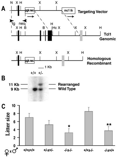
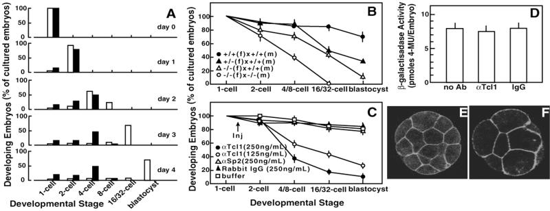
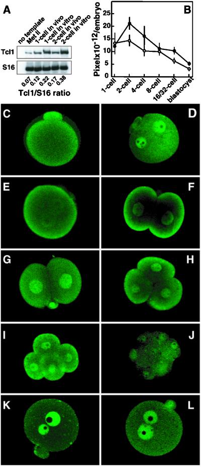
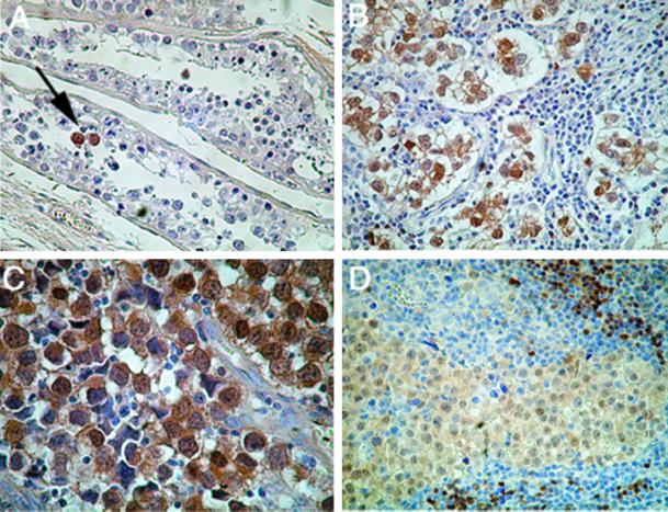
Overexpression of the TCL1 oncogene has been shown to play a causative role in T cell leukemias of humans and mice. The characterization of Tcl1-deficient mice in these studies indicates an important developmental role for Tcl1 in early embryogenesis. In wild-type embryos, Tcl1 is abundant in the first three mitotic cycles, during which it shuttles between nuclei and the embryo cortical regions in a cell-cycle-dependent fashion. The absence of this protein in early embryogenesis results in reduced fertility of female mice. The present studies elucidate the mechanism responsible for the reduced female fertility through analysis of the oogenesis stages and early embryo development in Tcl1-deficient mice. Even though Tcl1(-/-) females display normal oogenesis and rates of oocyte maturation/ovulation and fertilization, the lack of maternally derived Tcl1 impairs the embryo's ability to undergo normal cleavage and develop to the morula stage, especially under in vitro culture conditions. Beyond this crisis point, differentiative traits of zygotic genome activation and embryo compaction can take place normally. In contrast with this unanticipated role in early embryogenesis, we observed an overexpression of TCL1 in human seminomas. This finding suggests that TCL1 dysregulation could contribute to the development of this germinal cell cancer as well as lymphoid malignancies.


Figures

Figure 1
Figure 1
Gene targeting of the Tcl1 locus and reduced female fertility. (A) Configuration of the targeted genomic Tcl1 gene and the PGK-neo targeting vector. PGK-neo cassette replaces a BamHI (B) to HincII (Hc) fragment containing exons 2, 3, and 4. Arrows show transcriptional orientation of neo and TK genes. Restriction endonuclease cleavage sites are indicated. X, XbaI; H, HindIII; N, NotI; PGK, phosphoglycerate kinase; MC1, polyoma enhancer fragment; TK, herpes simplex thymidine kinase. (B) Representative genomic blot probed with an external probe recognizing a 9-kb wild type HindIII (H) fragment. The 11-kb fragment shows the rearranged homologous recombinant allele. (C) Litter size of wild-type and Tcl1 knockout female mice. The number of pups (mean ± SEM) in the litters produced by various mating pair was determined the day of birth. Mice (2–3 months old) were bred for a period of 3 months. Mating pairs analyzed in each group were (from left to right): 11, 12, 11, 5, and 4. Difference from wild type litters: *, P = 0.0001; **, P = 0.006, calculated by ANOVA.
Figure 2
Figure 2
Preimplantation development of in vitro cultured Tcl1-deficient embryos. (A) Comparison of developmental abilities of wild-type and Tcl1 knockout embryos. One-cell embryos derived from the wild-type (open bars) and knockout (solid bars) were cultured in vitro and scored daily for 5 days in three independent experiments having 20–40 embryos in each experimental group. (B and C) Developmental abilities of Tcl1-defective embryos (B) and of wild-type embryos made defective in Tcl1 by cytoplasmic injection of anti-Tcl1 antibody (C). The arrow indicates the time of injection. Data represent the mean ± SEM of percentages obtained in at least three independent experiments having 20–40 embryos in each experimental group. Symbols indicate the embryos obtained in different crosses that progressed to the appropriate developmental stage at the time of the score. (D) Effect of anti-Tcl1 antibody on the expression of the DNA construct phsplacZ in wild-type embryos at ZGA. Histograms represents the mean β-galactosidase activity ± SEM of 25–35 single injected embryos, pooled from three independent experiments. Differences between treatments: P > 0.75, calculated by ANOVA. (E and F) Immunofluorescence detection of β-catenin by confocal microscopy in compacted Tcl1+/+ (E) and Tcl1−/− (F) embryos. One-cell embryos were in vitro cultured for 3 days. Note the reduced number of blastomeres in the presence of normal blastomere flattening in Tcl1−/− embryos.
Figure 3
Figure 3
Expression of Tcl1 protein in wild-type preimplantation embryos. (A) Semiquantitative reverse transcription–PCR amplification of Tcl1 and ribosomal protein S16 mRNAs of oocytes and early wild-type embryos. Numbers below lanes indicate Tcl1/S16 incorporation ratios. (B) Quantitative immunofluorescence analysis of Tcl1 protein in preimplantation wild-type embryos using anti-Tcl1 antibodies. (○) One-cell embryos collected 15 hr after hCG and allowed to develop in vitro to the appropriate developmental stage; (●) embryos collected at the appropriate developmental stage directly from the tubes of pregnant animals and immediately processed for the assay. (CL) Tcl1 immunolocalization in Met II oocytes and preimplantation embryos by confocal microscopy. Representative embryos are shown in the panel. (C) Met II oocyte. (D) Mid 1-cell embryo at the S phase. (E) Late 1-cell embryo at the late G2 phase. (F) Early 2-cell embryo at the early G1 phase. (G) Mid 2-cell embryo at the S phase. (H) Three- to 4-cell embryo at the early G1 phase. (I) Four-cell embryo at the late G2 phase. (J) Eight-cell embryo. (K) Late 1-cell embryo cultured in vitro in the presence of 2 μg/ml aphidicolin. (L) Late 1-cell embryo cultured in vitro in the presence of 5 μg/ml cytochalasin D.
Figure 4
Figure 4
TCL1 expression in human germ cell neoplasia. (A) Testicular tubules showing normal spermatogenesis in adult testis with focal Tcl1 positive cells (arrow) of intratubular germ cell neoplasia. Nonneoplastic germ cells are Tcl1 negative (original magnification, ×250). (B) Testicular seminoma, classical type of neoplastic testis cells show strong nuclear immunoreactivity (original magnification, ×400). (C) Testicular seminoma: neoplastic cells show strong nuclear and cytoplasmic immunoreactivity (original magnification, ×630). (D) Mediastinal (thymic) dysgerminoma: neoplastic cells show weak to medium level positivity for Tcl1, both in the nucleus and in the cytoplasm. Clusters of B lymphocytes display the normal Tcl1 pattern of immunoreactivity (Upper Right) (original magnification, ×400).

References

    1. Russo G, Isobe M, Pegoraro L, Finan J, Nowell P C, Croce C M. Cell. 1988;53:137–144. - PubMed
    1. Virgilio L, Narducci M G, Isobe M, Billips L G, Cooper M D, Croce C M, Russo G. Proc Natl Acad Sci USA. 1994;91:12530–12534. - PMC - PubMed
    1. Virgilio L, Lazzeri C, Bichi R, Nibu K, Narducci M G, Russo G, Rothstein J L, Croce C M. Proc Natl Acad Sci USA. 1998;95:3885–3889. - PMC - PubMed
    1. Pekarsky Y, Koval A, Hallas C, Bichi R, Tresini M, Malstrom S, Russo G, Tsichlis P, Croce C M. Proc Natl Acad Sci USA. 2000;97:3028–3033. - PMC - PubMed
    1. Laine J, Kunstle G, Obata T, Sha M, Noguchi M. Mol Cell. 2000;6:395–407. - PubMed

Publication types