. 2002 Sep;46(9):2735-46.
doi: 10.1128/AAC.46.9.2735-2746.2002.

Importance of the fourth alpha-helix within the CAP homology domain of type II topoisomerase for DNA cleavage site recognition and quinolone action

Affiliations

Importance of the fourth alpha-helix within the CAP homology domain of type II topoisomerase for DNA cleavage site recognition and quinolone action

Dirk Strumberg et al. Antimicrob Agents Chemother. 2002 Sep.

Abstract

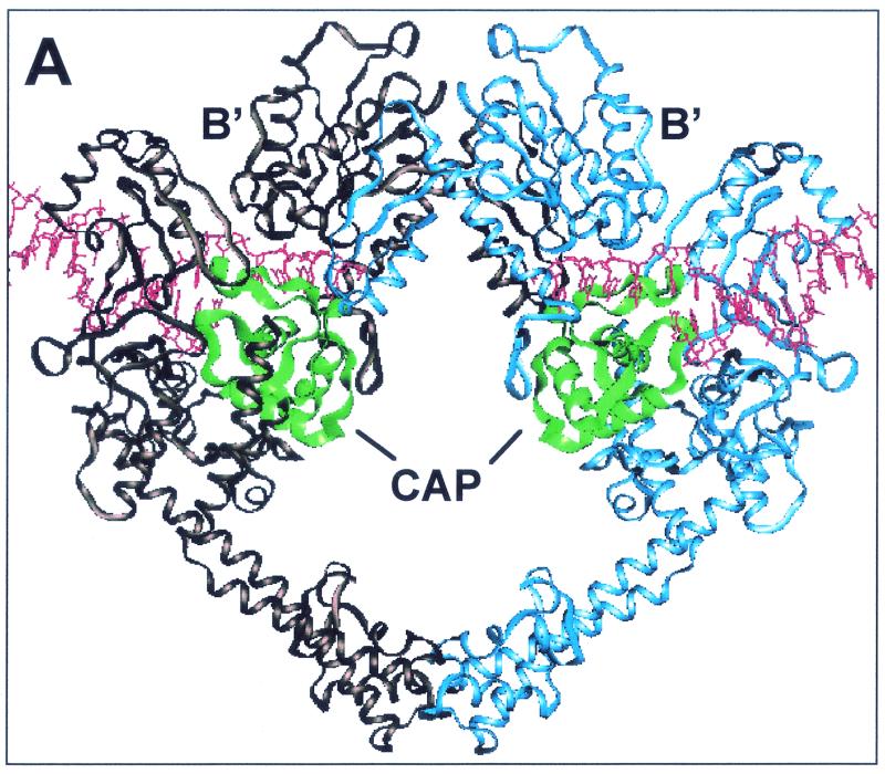
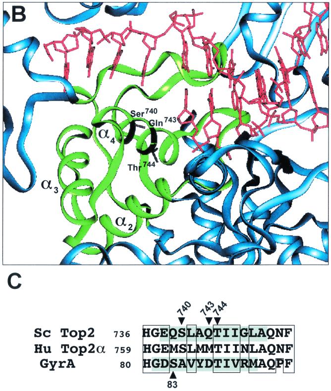
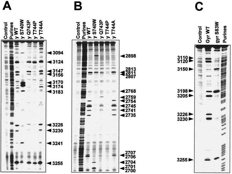
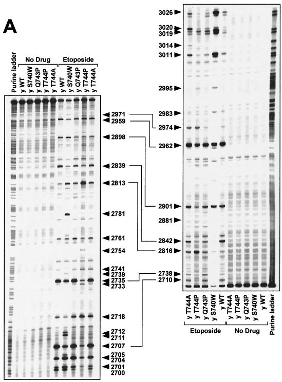
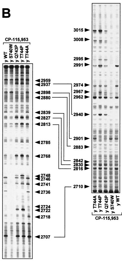
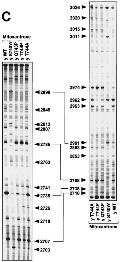
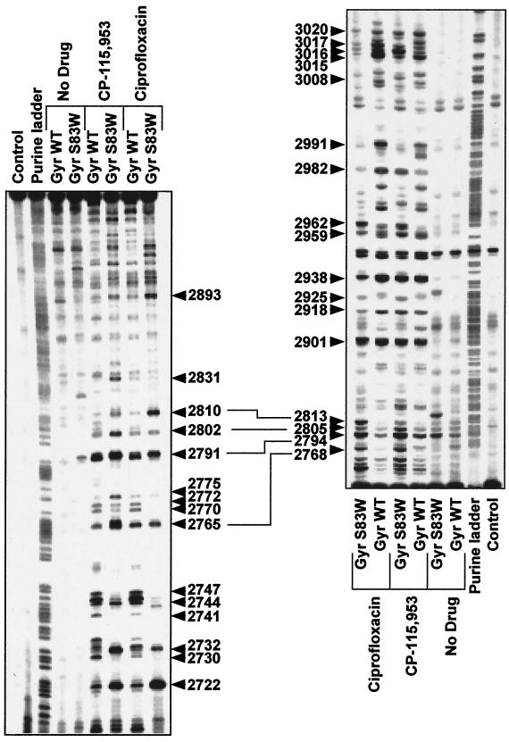
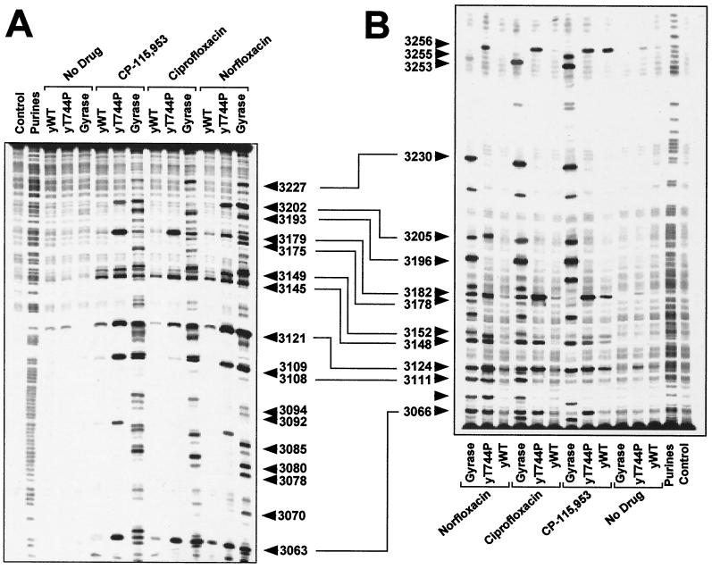
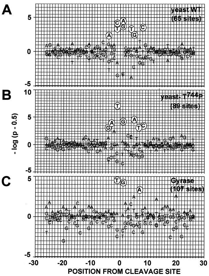
We report that point mutations causing alteration of the fourth alpha-helix (alpha4-helix) of the CAP homology domain of eukaryotic (Saccharomyces cerevisiae) type II topoisomerases (Ser(740)Trp, Gln(743)Pro, and Thr(744)Pro) change the selection of type II topoisomerase-mediated DNA cleavage sites promoted by Ca(2+) or produced by etoposide, the fluoroquinolone CP-115,953, or mitoxantrone. By contrast, Thr(744)Ala substitution had minimal effect on Ca(2+)- and drug-stimulated DNA cleavage sites, indicating the selectivity of single amino acid substitutions within the alpha4-helix on type II topoisomerase-mediated DNA cleavage. The equivalent mutation in the gene for Escherichia coli gyrase causing Ser(83)Trp also changed the DNA cleavage pattern generated by Ca(2+) or quinolones. Finally, Thr(744)Pro substitution in the yeast type II topoisomerase rendered the enzyme sensitive to antibacterial quinolones. This study shows that the alpha4-helix within the conserved CAP homology domain of type II topoisomerases is critical for selecting the sites of DNA cleavage. It also demonstrates that selective amino acid residues in the alpha4-helix are important in determining the activity and possibly the binding of quinolones to the topoisomerase II-DNA complexes.


Figures

FIG. 1.
FIG. 1.
CAP homology domain and positions of Ser740, Gln743, and Thr744 in yeast Top2p. (A) Illustration of dimeric S. cerevisiae topoisomerase II structure (Protein Data Bank accession number 1bgw). Drawings were generated using the program QUANTA (version 97). The helical ribbon representation shows the 92-kDa fragment of the yeast enzyme with a DNA fragment modeled into each of the putative DNA-binding sites (6). The CAP homology domain of each protomer is highlighted in green. (B) Close view of the putative DNA-binding region, presenting the proposed proximity of the α4-helix within the CAP homology domain, including Ser 740, Gln 743, and Thr744 (in a stick model), to DNA. (C) Alignment of protein sequence for the yeast (Sc Top2p), human Top2pα (Hu Top2pα) and E. coli gyrase. The mutated residues studied in the present report are indicated by arrowheads. Shaded residues correspond to the α4-helix (5). No shading is shown for human Top2p because of lack of structural data. Boxed regions indicate similarity between the amino acid residues. Ser763 in the human Top2pα sequence corresponds to Ser740 in yeast Top2p.
FIG. 1.
FIG. 1.
CAP homology domain and positions of Ser740, Gln743, and Thr744 in yeast Top2p. (A) Illustration of dimeric S. cerevisiae topoisomerase II structure (Protein Data Bank accession number 1bgw). Drawings were generated using the program QUANTA (version 97). The helical ribbon representation shows the 92-kDa fragment of the yeast enzyme with a DNA fragment modeled into each of the putative DNA-binding sites (6). The CAP homology domain of each protomer is highlighted in green. (B) Close view of the putative DNA-binding region, presenting the proposed proximity of the α4-helix within the CAP homology domain, including Ser 740, Gln 743, and Thr744 (in a stick model), to DNA. (C) Alignment of protein sequence for the yeast (Sc Top2p), human Top2pα (Hu Top2pα) and E. coli gyrase. The mutated residues studied in the present report are indicated by arrowheads. Shaded residues correspond to the α4-helix (5). No shading is shown for human Top2p because of lack of structural data. Boxed regions indicate similarity between the amino acid residues. Ser763 in the human Top2pα sequence corresponds to Ser740 in yeast Top2p.
FIG. 2.
FIG. 2.
Comparison of Ca2+-promoted DNA cleavage patterns of yeast Top2pWT, Top2pS740W, Top2pQ743P, Top2pT744P, and Top2pT744A as well as gyraseWT and GyrAS83W. DNA fragments from the human c-myc first intron (A) and from the junction between the c-myc first intron and first exon (B) were prepared by PCR. For each fragment, 5′-end-labeling was performed with 32P. Yeast Top2p reactions were performed at 37°C for 30 min (A and B), and gyrase reactions were performed at 25°C for 2 h (C). In all reactions, the CaCl2 concentration was 5 mM. Lanes: Control, without Top2 enzyme; Purine, ladder obtained after formic acid reaction; y WT, yeast wild-type Top2p; y S74 0W, yeast Top2pS740W; y Q743P, yeast Top2pQ743P; y T744P, yeast Top2pT744P; y T744A, yeast Top2pT744A; gyrWT, gyraseWT from E. coli; GyrAS83W, mutant Ser83Trp gyrase. Numbers correspond to genomic positions of the nucleotide covalently linked to the enzyme via the 5′ phosphate.
FIG. 3.
FIG. 3.
DNA cleavage patterns generated by yeast Top2pWT, Top2pS740W, Top2pQ743P, Top2pT744P, and Top2pT744A enzymes in the presence of etoposide, the fluoroquinolone CP-115,953, and the intercalator mitoxantrone. DNA fragments from the junction between the c-myc first intron and first exon between positions 2671 and 3072 were prepared by PCR using one primer labeled with 32P at the 5′ terminus. The left-hand panels show labeling of the upper DNA strand at position 2671. The right-hand panels show that labeling of the lower DNA strand was at position 3072. Top2 enzymes are indicated above each lane. Lanes: y WT, yeast wild-type Top2p; y S740W, yeast Top2pS740W; y Q743P, yeast Top2pQ743P; y T744P, yeast Top2pT744P; y T744A, yeast Top2pT744A. Concentrations were 100 μM for etoposide (A) and CP-115,953 (B) and 1 μM for mitoxantrone (C). Reactions were performed at 37°C for 30 min in the presence of 5 mM MgCl2. Purine ladders were obtained after formic acid reaction. Numbers correspond to genomic positions of the nucleotide covalently linked to Top2p via the 5′ phosphate. Double-headed arrows correspond to DNA cleavage sites with a 4-bp stagger that represent potential DNA double-strand breaks.
FIG. 3.
FIG. 3.
DNA cleavage patterns generated by yeast Top2pWT, Top2pS740W, Top2pQ743P, Top2pT744P, and Top2pT744A enzymes in the presence of etoposide, the fluoroquinolone CP-115,953, and the intercalator mitoxantrone. DNA fragments from the junction between the c-myc first intron and first exon between positions 2671 and 3072 were prepared by PCR using one primer labeled with 32P at the 5′ terminus. The left-hand panels show labeling of the upper DNA strand at position 2671. The right-hand panels show that labeling of the lower DNA strand was at position 3072. Top2 enzymes are indicated above each lane. Lanes: y WT, yeast wild-type Top2p; y S740W, yeast Top2pS740W; y Q743P, yeast Top2pQ743P; y T744P, yeast Top2pT744P; y T744A, yeast Top2pT744A. Concentrations were 100 μM for etoposide (A) and CP-115,953 (B) and 1 μM for mitoxantrone (C). Reactions were performed at 37°C for 30 min in the presence of 5 mM MgCl2. Purine ladders were obtained after formic acid reaction. Numbers correspond to genomic positions of the nucleotide covalently linked to Top2p via the 5′ phosphate. Double-headed arrows correspond to DNA cleavage sites with a 4-bp stagger that represent potential DNA double-strand breaks.
FIG. 4.
FIG. 4.
DNA cleavage patterns generated by gyraseWT and mutant GyrAS83W in the presence of the quinolones CP-115,953 and ciprofloxacin. The same DNA fragments presented in Fig. 3 were used. The left-hand panel shows labeling of the upper DNA strand; the right-hand panel shows labeling of the lower DNA strand. Drugs and enzymes are indicated above each lane. Concentrations were 100 μM for CP-115,953 and ciprofloxacin, respectively. Cleavage reactions were performed at 25°C for 2 h in the presence of 5 mM MgCl2. Purine ladders were obtained after formic acid reaction. Lanes: Control, no Top2p, no drug treatment; Gyr WT: gyraseWT from E. coli; GyrAS83W, mutant Ser83Trp gyrase. Numbers correspond to genomic positions of the nucleotide covalently linked to Top2p. Double-headed arrows correspond to DNA cleavage sites with a 4-bp stagger that represent potential DNA double-strand breaks.
FIG. 5.
FIG. 5.
Generation of DNA cleavage by the yeast Top2pT744P mutant in the presence of antibacterial quinolones. A 254-bp DNA fragment from the c-myc first intron was prepared. (A) Labeling of the upper DNA strand at position 3035. (B) Labeling of the lower DNA strand at position 3288. Drugs (100 μM each) and enzymes are indicated above the lanes. Lanes: yWT, yeast wild-type Top2p; Y T744P, Top2pT744P. Numbers correspond to genomic positions of the nucleotide covalently linked to Top 2p. Double-headed arrows correspond to DNA cleavage sites with a 4-bp stagger that represent potential DNA double-strand breaks.
FIG. 6.
FIG. 6.
Probability of the observed base frequency deviations at Top2p cleavage sites for the yeast wild-type enzyme, for Top2pT744P and for E. coli gyrase in the presence of CP-115,953. Position 0 corresponds to the cleavage site, and positions −1 and +1 to the bases immediately 3′ and 5′ from the cleavage site, respectively. The panels present the probability of the observed base frequency deviations from expectation for the indicated enzyme. On the y axis, P is the probability of observing that deviation or more, either as excess (above baseline) or deficiency (below baseline), relative to the expected frequency of each individual base (48). Cleavage sites for the yeast wild-type Top2p (A), the yeast Top2pT744P (B), and E. coli gyrase (C) were analyzed after treatment with 100 μM CP-115,953.

Similar articles

Cited by

References

    1. Anderson, V. E., T. D. Gootz, and N. Osheroff. 1998. Topoisomerase IV catalysis and the mechanism of quinolone action. J. Biol. Chem. 273:17879-17885. - PubMed
    1. Andoh, T., and R. Ishida. 1998. Catalytic inhibitors of DNA topoisomerase II. Biochim. Biophys. Acta 1400:155-171. - PubMed
    1. Beck, W. T., M. K. Danks, J. S. Wolverton, R. Kim, and M. Chen. 1993. Drug resistance associated with altered DNA topoisomerase II. Adv. Enzyme Regul. 33:113-127. - PubMed
    1. Berger, J. 1998. Type II DNA topoisomerases. Curr. Opin. Struct. Biol. 8:26-32. - PubMed
    1. Berger, J. M., D. Fass, J. C. Wang, and S. C. Harrison. 1998. Structural similarities between topoisomerases that cleave one or both DNA strands. Proc. Natl. Acad. Sci. USA 95:7876-7881. - PMC - PubMed

Publication types

MeSH terms

LinkOut - more resources