. 2002 Sep;46(9):3001-12.
doi: 10.1128/AAC.46.9.3001-3012.2002.

The antifungal echinocandin caspofungin acetate kills growing cells of Aspergillus fumigatus in vitro

Affiliations

The antifungal echinocandin caspofungin acetate kills growing cells of Aspergillus fumigatus in vitro

J C Bowman et al. Antimicrob Agents Chemother. 2002 Sep.

Abstract

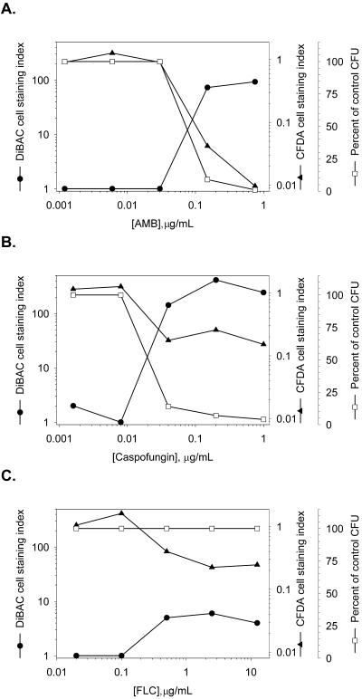
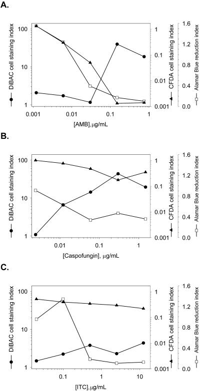
Caspofungin acetate is an antifungal antibiotic that inhibits synthesis of 1,3-beta-D-glucan, an essential component of the fungal cell wall. While caspofungin causes cell death in yeasts and dimorphic fungi such as Candida albicans, its effect on Aspergillus fumigatus is less well understood. We used the fluorescent dyes 5,(6)-carboxyfluorescein diacetate (CFDA) and bis-(1,3-dibutylbarbituric acid) trimethine oxonol (DiBAC), which stain live and dead cells, respectively, to further characterize the antifungal activity of caspofungin. For comparison, compounds whose mode of action was either fungistatic (fluconazole, itraconazole) or fungicidal (amphotericin B) were also evaluated. A correlation between caspofungin-induced loss of viability, decreased CFDA staining, and increased DiBAC staining was established first with C. albicans. For A. fumigatus, caspofungin caused similar dye-staining changes, which were quantified by fluorimetric analysis of stained hyphae grown in a medium that promoted dispersed growth. The minimum concentration of caspofungin required to produce these changes also decreased the level of growth-dependent reduction of the indicator dye Alamar Blue. We observed a differential effect of caspofungin as a function of cell position: 88% of apical cells and 61% of subapical branching cells failed to stain with the viable dye CFDA, but only 24% of subapical cells were unstained. Complementary results were seen with germlings from DiBAC-stained, caspofungin-treated cultures. Extended incubation of A. fumigatus with a single dose of caspofungin affected the same proportion of apical and subapical branching cells for up to 72 h. The dye-staining patterns illustrate that the cells at the active centers for new cell wall synthesis within A. fumigatus hyphae are killed when they are exposed to caspofungin.


Figures

FIG. 1.
FIG. 1.
Photomicrographs of untreated and drug-treated C. albicans stained with the fluorescent dyes CFDA and DiBAC. C. albicans cells from an untreated culture or from cultures treated with AMB (0.15 μg/ml), caspofungin (0.2 μg/ml), or FLC (2.5 μg/ml) were harvested after 15 h, stained with the dye CFDA, which stains viable cells, or the dye DiBAC, which stains nonviable cells, and photographed at ×1,000 magnification. Exposure times for epifluorescence of untreated samples stained with DiBAC, AMB-treated samples stained with CFDA, FLC-treated samples stained with CFDA, and FLC-treated samples stained with DiBAC were 20 s; all others were less than 1 s (as determined by the camera controller). Left panels, Nomarski optics; right panels, epifluorescence with an FITC filter set.
FIG. 2.
FIG. 2.
Dose-dependent changes in normalized CFDA or DiBAC fluorescence of C. albicans. Cultures were challenged with serial dilutions of AMB (A), caspofungin (B), or FLC (C). Aliquots of these cultures were stained with CFDA or DiBAC or were spread onto agar plates to determine the number of CFU. Fluorescence values were normalized per 106 cells (CFDA) or 107 cells (DiBAC), as described in Materials and Methods.
FIG. 3.
FIG. 3.
In vitro growth of A. fumigatus in RPMI-Junlon. (A) Tubes containing RPMI 1640 medium (the two leftmost tubes [tubes 1 and 2]) or RPMI-Junlon (the two rightmost tubes [tubes 3 and 4]) were either inoculated with 105 A. fumigatus conidia (tubes 2 and 4) or left uninoculated (tubes 1 and 3) and were incubated for 15 h at 37°C. The tubes were swirled briefly prior to being photographed. (B) A. fumigatus conidia (105) were grown in either RPMI-Junlon (○) or RPMI 1640 medium alone (▴), incubated for 24 h at 37°C, and stained with CFDA. Stained samples were serially diluted twofold across a 96-well plate, and the fluorescence was read (excitation wavelength = 485 nm, emission wavelength = 538 nm).
FIG.4.
FIG.4.
Photomicrographs of untreated and drug-treated A. fumigatus stained with the fluorescent dyes CFDA and DiBAC. A. fumigatus germlings incubated for 6 h with 0.15 μg of AMB per ml, 0.3 μg of caspofungin per ml, or 2.5 μg of ITC per ml or without drug were stained with the dye CFDA, which stains viable cells, or the dye DiBAC, which stains nonviable cells, and photographed at ×400 magnification. Fluorescent micrograph exposure times for untreated samples stained with DiBAC, AMB-treated samples stained with CFDA, and ITC-treated samples stained with CFDA or DiBAC were 20 s; all others were less than 1 s (as determined by the camera controller). Left panels, Nomarski optics; right panels, epifluorescence with an FITC filter set.
FIG. 5.
FIG. 5.
High-magnification photomicrographs of caspofungin-treated, DiBAC-stained A. fumigatus. A. fumigatus germlings incubated for 6 h with 0.3 μg of caspofungin per ml were stained with DiBAC and photographed at ×2,000 magnification. Images from two separate germlings from the same culture are shown. Left panels, Nomarski optics; right panels, epifluorescence with an FITC filter set.
FIG. 6.
FIG. 6.
A. fumigatus killing curves. A. fumigatus conidia (105) were inoculated into RPMI-Junlon and incubated at 37°C for 14 h before 0.3 μg of drug per ml (AMB [▵], caspofungin [▾], or ITC [□]) or vehicle (○) was added. At 3-h intervals after drug addition, aliquots from each culture were spread onto agar plates for determination of the number of CFU.
FIG. 7.
FIG. 7.
Quantitative fluorescence of dye-stained A. fumigatus. A. fumigatus cultures were grown as described in the text and incubated with serial dilutions of AMB (A), caspofungin (B), or ITC (C) for 6 h. Aliquots of these cultures were stained with CFDA or DiBAC; parallel titrations were performed with cultures containing 10% (vol/vol) Alamar Blue. The values for CFDA and DiBAC staining were normalized to the total amount of protein, and the fold change relative to the amount for the untreated control (staining index) was determined as described in Materials and Methods.
FIG. 8.
FIG. 8.
Classification scheme for cells within A. fumigatus germlings. An A. fumigatus culture was treated with 0.3 μg of caspofungin per ml, stained with the viable dye CFDA, and photographed at ×1,600 magnification. Left panel, Nomarski optics; right panel, epifluorescence with an FITC filter set plus bright-field illumination. Septa (indicated by arrows) were identified as described in the text, and the cells were classified as apical (e.g., cells 1 and 6), subapical (e.g., cells 3 and 5), or subapical branching (e.g., cells 2 and 4).
FIG. 9.
FIG. 9.
Effect of extended drug treatment on A. fumigatus fluorescent dye staining. (A) Untreated, DiBAC-stained germlings harvested 24 h after vehicle addition. Magnification, ×400. Arrows indicate conidiophores. Caspofungin-treated germlings (0.3 μg/ml) harvested 72 h after drug addition were stained with CFDA (B) or DiBAC (C), and photographed at ×800 magnification. In panels B and C, arrows with single tails indicate lysed apical cells and arrows with double tails indicate lysed conidiophores. The fluorescent micrograph in panel A required a 20-s exposure. Left panels, Nomarski optics; right panels, epifluorescence with an FITC filter set.

References

    1. Abruzzo, G. K., A. M. Flattery, C. J. Gill, L. Kong, J. G. Smith, V. B. Pikounis, J. M. Balkovec, A. F. Bouffard, J. F. Dropinski, H. Rosen, H. Kropp, and K. Bartizal. 1997. Evaluation of the echinocandin antifungal MK-0991 (L-743,872): efficacies in mouse models of disseminated aspergillosis, candidiasis, and cryptococcosis. Antimicrob. Agents Chemother. 41:2333-2338. - PMC - PubMed
    1. Abruzzo, G. K., C. J. Gill, A. M. Flattery, L. Kong, C. Leighton, J. G. Smith, V. B. Pikounis, K. Bartizal, and H. Rosen. 2000. Efficacy of the echinocandin caspofungin against disseminated aspergillosis and candidiasis in cyclophosphamide-induced immunosuppressed mice. Antimicrob. Agents Chemother. 44:2310-2318. - PMC - PubMed
    1. Anderson, J. G., and J. E. Smith. 1971. The production of conidiophores and conidia by newly germinated conidia of Aspergillus niger (microcycle conidiation). J. Gen. Microbiol. 69:185-197. - PubMed
    1. Archer, D. B. 1977. Chitin biosynthesis in protoplasts and subcellular fractions of Aspergillus fumigatus. Biochem. J. 164:653-658. - PMC - PubMed
    1. Archer, D. B., D. A. MacKenzie, and M. J. Ridout. 1995. Heterologous protein secretion by Aspergillus niger growing in submerged culture as dispersed or aggregated mycelia. Appl. Microbiol. Biotechnol. 44:157-160. - PubMed