. 2002 Aug 15;16(16):2085-96.
doi: 10.1101/gad.994902.

Saccharomyces forkhead protein Fkh1 regulates donor preference during mating-type switching through the recombination enhancer

Affiliations

Saccharomyces forkhead protein Fkh1 regulates donor preference during mating-type switching through the recombination enhancer

Kaiming Sun et al. Genes Dev. .

Abstract

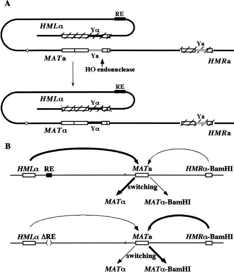
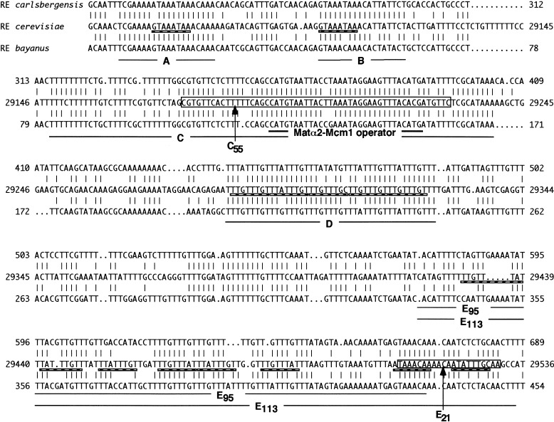
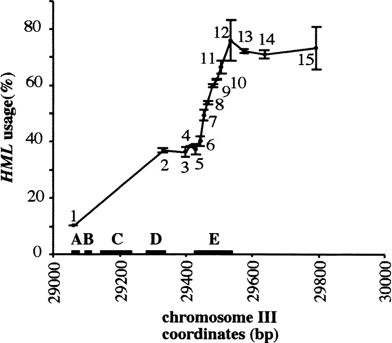
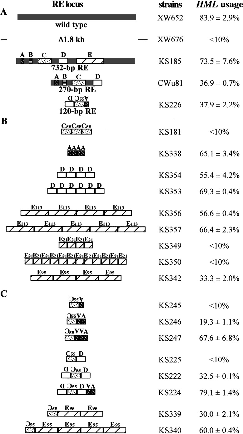
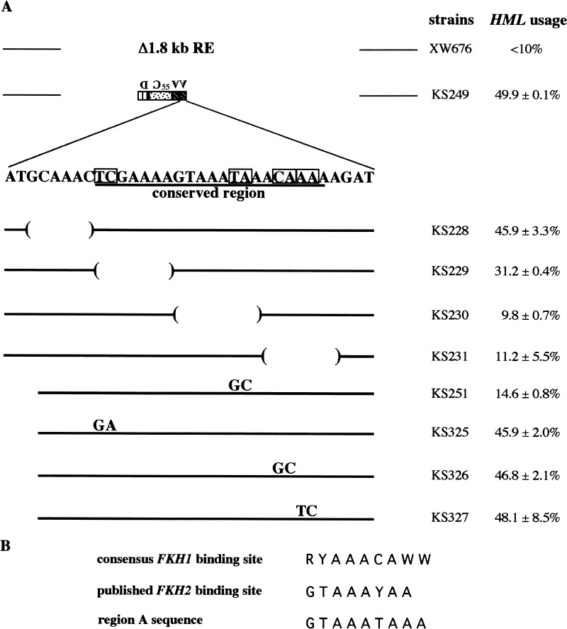
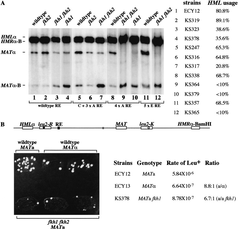
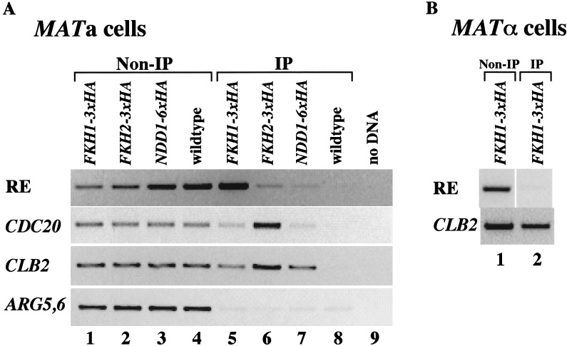
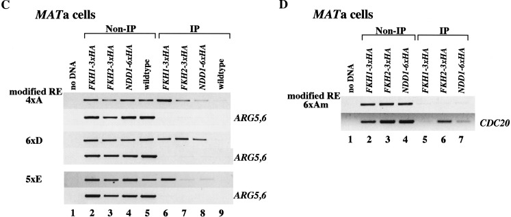
Saccharomyces mating-type switching results from replacement by gene conversion of the MAT locus with sequences copied from one of two unexpressed donor loci, HML or HMR. MATa cells recombine with HMLalpha approximately 90% of the time, whereas MATalpha cells choose HMRa 80%-90% of the time. HML preference in MATa is controlled by the cis-acting recombination enhancer (RE) that regulates recombination along the entire left arm of chromosome III. Comparison of RE sequences between S. cerevisiae, S. carlsbergensis, and S. bayanus defines four highly conserved regions (A, B, C, and D) within a 270-bp minimum RE. An adjacent E region enhances RE activity. Multimers of region A, D, or E are sufficient to promote selective use of HML. Regions A, D, and E each bind in vivo the transcription activator forkhead proteins Fkh1p and Fkh2p and their associated Ndd1p, although there are no adjacent open reading frames (ORFs). Deletion of FKH1 significantly reduces MATa's use of HML, as does mutation of the Fkh1/Fkh2-binding sites in a multimer of region A. We conclude that Fkh1p regulates MATa donor preference through direct interaction with RE.


Figures

Figure 1
Figure 1
Mating-type switching at the MATa locus. (A) Expression of HO endonuclease creates a double-strand break (DSB) at the Y/Z1 border of the MAT locus and initiates repair by using the silent donor HML and leads to gene conversion. The two donor loci (HML and HMR) are maintained in a transcriptionally inactive chromatin structure. Other shared regions of homology are indicated. The preferential use of HML donor in MATa cells is regulated by a cis-acting element, recombination enhancer (RE), located ∼17 kb centromere-proximal to HML. The positions of HML, RE, centromere, MAT and HMR are indicated. (B) HMRa is replaced by HMRα modified to carry a point mutation that creates a BamHI site, so that it is possible to distinguish which sequences have been gene converted into the MAT locus. Donor preference is assayed in populations of cells induced to switch from MATa to either MATα or MATα-B (Wu and Haber 1995). MATa cells recombine with HML ∼90% of the time in the presence of RE; when RE is deleted, MATa cells prefer to choose HMR 80%–90% of the time.
Figure 2
Figure 2
Comparison of RE among three species: Saccharomyces carlsbergensis, S. cerevisiae, and S. bayanus (analyzed in “LASERGENE navigator”). Identical residues in the alignment of the DNA sequences of RE carlsbergensis (top), RE cerevisiae (middle), and RE bayanus (bottom) are indicated by vertical lines. The four most conserved subdomains (designated as A, B, C, and D) and a second near-perfect array of TTT(G/A) repeats (designated as E113) are underlined. Region C contains the Matα2-Mcm1 consensus binding site (double underlined). Regions A, B, D, and E contain a total of 15 Fkh1p and Fkh2p consensus binding sites (hatched line). The fragments used to replace the wild-type RE in donor preference assay—designated as C55 (boxed), E21 (boxed), E95 (solid line), and E113 (solid line)—are indicated.
Figure 3
Figure 3
Identification of region E, which, in combination with A, B, C, and D, can provide full RE activity in MATa cells through a series of deletions from centromere-proximal end of RE region. The different RE fragments were introduced into the chromosome III to replace a 1.8-kb sequence containing RE. The strains used here (XW676, CWu81, KS210, KS209, KS208, KS207, KS206, KS205, KS204, KS203, KS202, KS198, KS199, KS184, and KS185) are indicated as 1–15, respectively. Number 1 is the strain that has no RE fragment inserted into a 1.8-kb deletion. Number 2 carries the chromosome III region from 29059 bp to 29330 bp, which was reported previously as the 270-bp RE (Wu et al. 1998). Numbers 3–14 each represent strains that carry a fragment starting from 29059 bp of chromosome III to centromere-proximal end. The numbers of TTT(G/A) repeats are #3 (0), #4 (0), #5 (0), #6 (3), #7 (5), #8 (7), #9 (11), #10 (14), #11 (16), #12 (20), #13 (20), and #14 (20). Number 15 carries the 732-bp RE starting from 29059 bp to 29790 bp. Donor preference is shown as the percentage of HML usage in MATa cells. Important region E is from ∼29423 to ∼29535 bp as indicated above.
Figure 4
Figure 4
Effect of different combinations of regions A, C, D, and E on the percentage of HML usage in MATa cells. The different synthetic RE fragments were introduced into the chromosome III to replace the 1.8-kb sequence containing RE. C55, E21, E95, and E113 are indicated in Fig. 2. All other regions, A, C, and D, used here contain full length of consensus sequences.
Figure 5
Figure 5
Identification of functional region A-binding site. (A) Identification of essential sequences within region A by introduction of the A + A + C55 + D synthetic REs carrying different deletions or 2 bp mutations on each A into the chromosome III to replace a 1.8-kb sequence containing RE. The 2-bp changes are boxed. The conserved region is underlined. Donor preference is shown as the percentage of HML usage in MATa cells. (B) Comparison of consensus Fkh1p- and Fkh2p-binding sites reported previously (Zhu et al. 2000) with region A sequence. R: A or G; Y: T or C; W: A or T.
Figure 6
Figure 6
The effects of Fkh1p and Fkh2p on donor preference and leu2 heteroallelic recombination in MATa cells. (A) Analysis of HMLα usage in different strains carrying single or double forkhead deletions in either wild-type RE or synthetic RE with C + 3 × A, 4 × A, and 5 × E. HO-induced switching of MATa to MATα (from HMLα) or MATα-Bam (from HMRα-Bam) can be assessed densitometrically on a Southern blot of DNA digested with BamHI and HindIII restriction endonucleases. Yα-specific probe was used to give rise to four specific bands indicated above. Donor preference is shown as the percentage of HML usage in MATa cells. (B) Spontaneous recombination between leu2-R inserted at kb 22 of chromosome III and leu2-K located at kb 233 of the right arm of MAT produces Leu+ prototrophs. The wild-type MATa and MATα and mutant fkh1 fkh2 MATa were patched on a YEPD plate and then replica-plated to synthetic medium lacking leucine. The rate of LEU2 prototroph formation was determined by a fluctuation test (Lea and Coulson 1949).
Figure 7
Figure 7
Fkh1p, Fkh2p, and Ndd1p are recruited to the RE in vivo. (A,B) Chromatin immunoprecipitation (ChIP) assays with a different pair of primers that can specifically amplify the wild-type RE region, the promoter regions of CDC20 and CLB2 (positive controls), and the coding region of ARG5,6 (negative control). Strains used in this assay are CFY480 (A, lanes 1,5) and CFY854 (A, lanes 2,6) (Hollenhorst et al. 2000), 155 (A, lanes 3,7) (Koranda et al. 2000), W303 (A, lanes 4,8), and KS369 (MATα; B, lanes 1,2). (C,D) ChIP assays with pairs of primers that can specifically amplify either the modified RE region, the promoter of CDC20 (positive controls), or the coding region of ARG5,6 (negative control). The wild-type RE is replaced by either four copies of region A, six copies of D, five copies of E, or six copies of mutant region A, indicated as 4 × A, 6 × D, 5 × E, or 6 × Am at left. Controls that have fragments amplified from ARG5,6coding region and CDC20 promoter are indicated at right. All strains used here are MATa. Strains used in this assay are KS358 (4 × A), KS359 (6 × D), and KS360 (5 × E) (C, lanes 2,6); KS370 (4 × A), KS371 (6 × D), and KS372 (5 × E) (C, lanes 3,7); KS374 (4 × A), KS375 (6 × D), and KS376 (5 × E) (C, lanes 4,8); KS361 (4 × A), 362 (6 × D), and 363 (5 × E) (C, lanes 5,9); KS365 (6 × Am) (D, lanes 2,5); KS373 (6 × Am) (D, lanes 3,6); and KS377 (6 × Am) (D, lanes 4,7).
Figure 7
Figure 7
Fkh1p, Fkh2p, and Ndd1p are recruited to the RE in vivo. (A,B) Chromatin immunoprecipitation (ChIP) assays with a different pair of primers that can specifically amplify the wild-type RE region, the promoter regions of CDC20 and CLB2 (positive controls), and the coding region of ARG5,6 (negative control). Strains used in this assay are CFY480 (A, lanes 1,5) and CFY854 (A, lanes 2,6) (Hollenhorst et al. 2000), 155 (A, lanes 3,7) (Koranda et al. 2000), W303 (A, lanes 4,8), and KS369 (MATα; B, lanes 1,2). (C,D) ChIP assays with pairs of primers that can specifically amplify either the modified RE region, the promoter of CDC20 (positive controls), or the coding region of ARG5,6 (negative control). The wild-type RE is replaced by either four copies of region A, six copies of D, five copies of E, or six copies of mutant region A, indicated as 4 × A, 6 × D, 5 × E, or 6 × Am at left. Controls that have fragments amplified from ARG5,6coding region and CDC20 promoter are indicated at right. All strains used here are MATa. Strains used in this assay are KS358 (4 × A), KS359 (6 × D), and KS360 (5 × E) (C, lanes 2,6); KS370 (4 × A), KS371 (6 × D), and KS372 (5 × E) (C, lanes 3,7); KS374 (4 × A), KS375 (6 × D), and KS376 (5 × E) (C, lanes 4,8); KS361 (4 × A), 362 (6 × D), and 363 (5 × E) (C, lanes 5,9); KS365 (6 × Am) (D, lanes 2,5); KS373 (6 × Am) (D, lanes 3,6); and KS377 (6 × Am) (D, lanes 4,7).

References

    1. Bon E, Neuveglise C, Casaregola S, Artiguenave F, Wincker P, Aigle M, Durrens P. Genomic exploration of the hemiascomycetous yeasts: 5. Saccharomyces bayanus var. uvarum. FEBS Lett. 2000;487:37–41. - PubMed
    1. Chen DC, Yang BC, Kuo TT. One-step transformation of yeast in stationary phase. Curr Genet. 1992;21:83–84. - PubMed
    1. Galitski T, Saldanha AJ, Styles CA, Lander ES, Fink GR. Ploidy regulation of gene expression. Science. 1999;285:251–254. - PubMed
    1. Gietz RD, Schiestl RH, Willems AR, Woods RA. Studies on the transformation of intact yeast cells by the LiAc/SS-DNA/PEG procedure. Yeast. 1995;11:355–360. - PubMed
    1. Gietz RD, Triggs-Raine B, Robbins A, Graham KC, Woods RA. Identification of proteins that interact with a protein of interest: Applications of the yeast two-hybrid system. Mol Cell Biochem. 1997;172:67–79. - PubMed

Publication types

MeSH terms

Associated data