. 2002 Aug 15;16(16):2108-19.
doi: 10.1101/gad.1003902.

The Drosophila heterochromatic gene encoding poly(ADP-ribose) polymerase (PARP) is required to modulate chromatin structure during development

Affiliations

The Drosophila heterochromatic gene encoding poly(ADP-ribose) polymerase (PARP) is required to modulate chromatin structure during development

Alexei Tulin et al. Genes Dev. .

Abstract

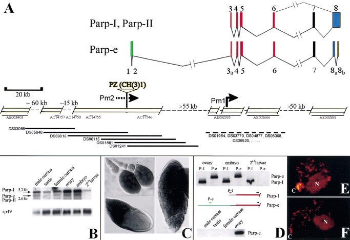
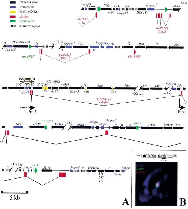
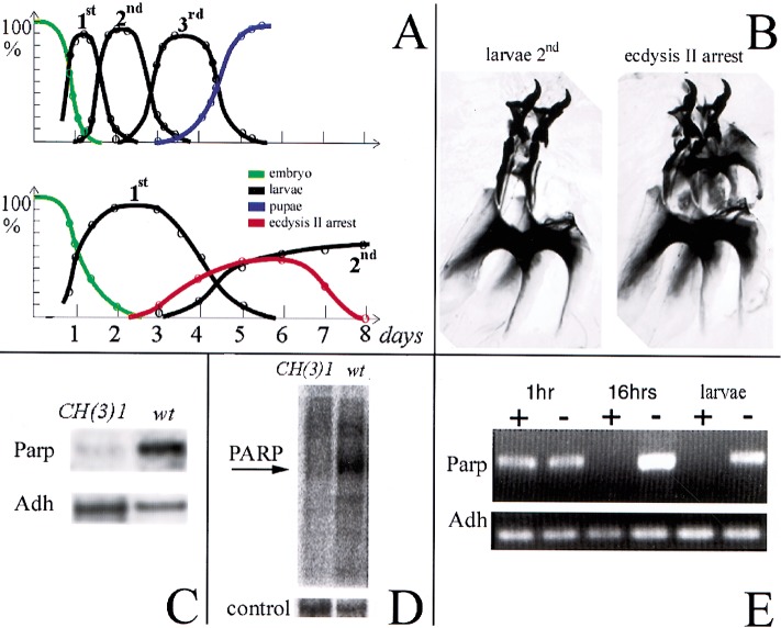
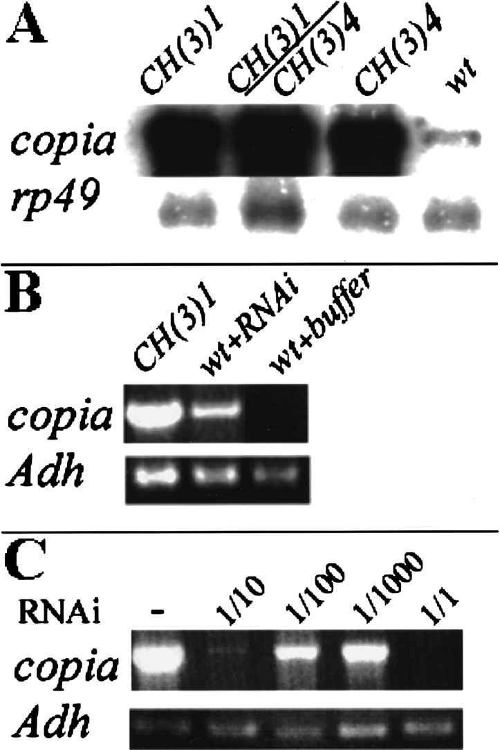
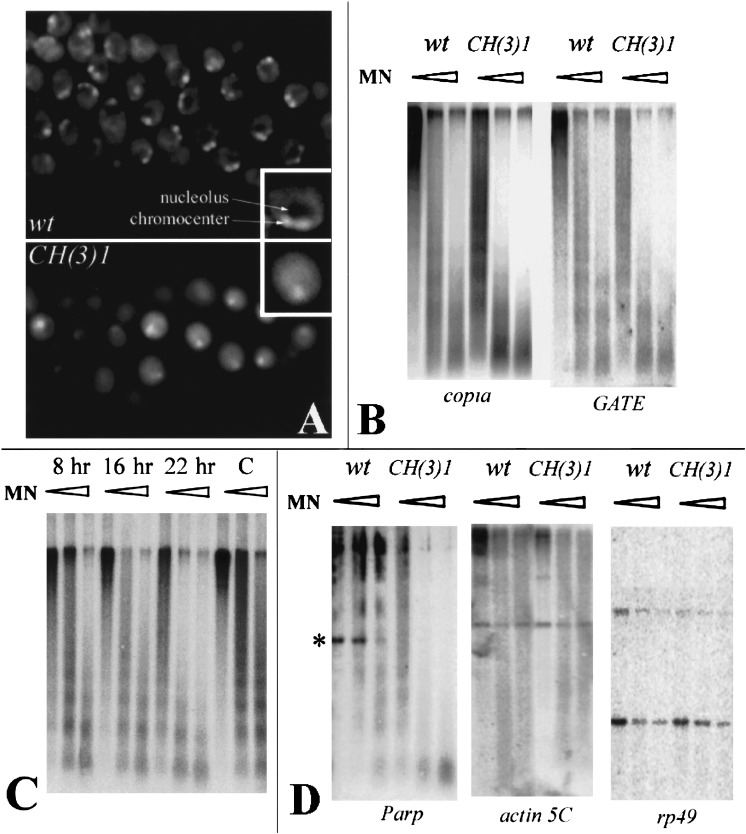
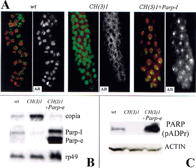
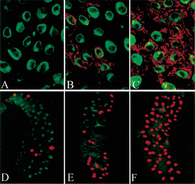
Poly(ADP-ribose) polymerase (PARP) is a major NAD-dependent modifying enzyme that mediates important steps in DNA repair, transcription, and apoptosis, but its role during development is poorly understood. We found that a single Drosophila Parp gene spans more than 150 kb of transposon-rich centromeric heterochromatin and produces several differentially spliced transcripts, including a novel isoform, PARP-e, predicted to encode a protein lacking enzymatic activity. An insertion mutation near the upstream promoter for Parp-e disrupts all Parp expression. Heterochromatic but not euchromatic sequences become hypersensitive to micrococcal nuclease, nucleoli fail to form, and transcript levels of the copia retrotransposon are elevated more than 50-fold; the variegated expression of certain transgenes is dominantly enhanced. Larval lethality can be rescued and PARP activity restored by expressing a cDNA encoding PARP-e. We propose that PARP-e autoregulates Parp transcription by influencing the chromatin structure of its heterochromatic environment. Our results indicate that Parp plays a fundamental role organizing the structure of Drosophila chromatin.


Figures

Figure 1
Figure 1
Structure and expression of the Drosophila Parp locus. (A) Deduced genomic structure of the 300-kb Parp region; open boxes are sequenced. The arrangement of the exons encoding Parp-I is shown at top (Uchida et al. 1993; Hanai et al. 1998). (Bottom) The positions of three unlinked Drosophila genomic contigs (thin black lines: AE002935, AE002666, and AE002892) homologous to Parp-I exons are shown at right (Adams et al. 2000). Pm1 indicates the Parp-I promoter deduced from 5′ cDNA sequences (Hanai et al. 1998). A single cDNA isolated from early ovarian stages, GM10715, defines the alternatively spliced Parp-e transcript. The 5′-most 273 bp of GM10715 matches the genomic sequence flanking a P element insertion, CH(3)1 (Zhang and Spradling 1994). A map of this region (left portion of figure) was constructed by chromosome walking using a P1 genomic library (Kimmerly et al. 1996) (bottom, thick black lines). The two resulting scaffolds were sequenced and found to span four small preexisting genomic sequence contigs (thin black lines) and to link to a fifth (AE003403). The color code indicates which portion of PARP is encoded: DNA binding (red), automodification (purple), catalytic (blue), noncoding (green and yellow). (B) Multiple Parp transcripts. A Northern blot of poly(A)-containing RNA from the indicated developmental stages reveals both a 3.2-kb RNA, the size predicted for Parp-I, and a 2.6-kb RNA, the approximate size expected for Parp-II and Parp-e. Parp-homologous RNAs are abundant in both ovaries and embryos, and are reduced but still detectable in second instar larvae and adults. (C) Whole mount in situ hybridization using a 1.4-kb cDNA probe from the DNA-binding domain common to all isoforms labels Parp RNA in nurse cells and in oocytes from stage 9–14 follicles. (D) RT-PCR using isoform-specific primers (see diagrams) that distinguish between Parp-I (or Parp-II) and Parp-e demonstrate that Parp-e is produced in ovaries and embryos, but not at detectable levels in second instar larvae, or in adults outside the ovary. (E) Nuclei are shown from brains of third instar larvae expressing a Parp-I-DsRed fusion gene (see Materials and Methods). Protein is abundant in the chromocenter (C), the nucleolus (N), and at sites within euchromatin. (F) Third instar larval brain nuclei stained with anti-poly(ADP-ribose) antibody 10H show that protein-associated ADP-ribose is found in the same regions as PARP-DsRed.
Figure 2
Figure 2
DNA sequence of the heterochromatic region containing Parp. (A) A diagram summarizing the sequence organization of the region as determined from this study (see Materials and Methods) and from Adams et al. (2000) is shown. Genes defined by cDNAs sequenced as part of this study are shown in red (boxes are exons). The names of retrotransposons (black) and of transposons (blue) are given above the region of homology represented by an arrow (arrowhead: 3′ end). Regions containing only small sequence blocks related to a particular transposon are indicated by parallel bars. The position of the CH(3)1 insertion and the location of the putative Parp promoters Pm1 and Pm2 are indicated. Gaps in the sequence of known or estimated size are represented by hash marks. (B) An ideogram of chromosome 3 heterochromatin shows the cytological region of CH(3)1 insertion (Zhang and Spradling 1994). (Bottom) A chromosome set from a CH(3)1/TM3 third instar larval neuroblast is shown that has been hybridized in situ with Parp cDNA (green) and transposon-specific sequences (red). The partial overlap of the Parp and CH(3)1 sequences indicates that Parp and CH(3)1 are located near each other in 3R heterochromatin. (Note: the TOTO-3 used for this confocal micrograph does not reveal full morphological detail, but chromosomes were also scored using DAPI; CH(3)1 was localized previously to h55 (Zhang and Spradling 1994).
Figure 3
Figure 3
The CH(3)1 complementation group disrupts Parp expression and activity. (A) Timelines of development of wild-type (top) and CH(3)1 homozygotes (bottom) are shown. The fraction of animals at each developmental stage are plotted as a function of time, revealing the strong developmental delay caused by CH(3)1. (B) Preparation of larval mouth hooks, which distinguish larval instars, are illustrated showing the characteristic appearance of the normal l2 mouthhooks (left) and of mouthhooks from CH(3)1 mutants arrested at the onset of ecdysis 2 (right). (C) Northern blot of poly(A)-containing RNA from wild-type larvae and 4-d-old CH(3)1 larvae showing reduced levels of Parp 3.2-kb mRNA. (D) Proteins labeled by ADP-ribosylation in wild-type (wt) and CH(3)1 mutant larvae. An autoradiogram of a gel of 32P-labeled protein is shown (see Materials and Methods). The prominent band at 117 kD in wild-type has the expected molecular weight of Parp itself. Stained protein in a segment of the same gel is shown as a loading control. (E) RNAi treatment of embryos eliminates detectable Parp mRNA in 16-h embryos and larvae. An RT-PCR assay recognizing all forms of the Parp transcripts is shown; primers specific for the alcohol dehydrogenase gene (Adh) gene serve as a loading control.
Figure 4
Figure 4
Parp mutations or Parp (RNAi) elevate copia transcript levels. (A) A Northern blot of total RNA from second instar larvae of the indicated genotypes was probed with copia sequences. The 5.5-kb copia transcript is overproduced up to 50-fold in CH(3)1 or CH(3)4 homozygotes, and in CH(3)1/CH(3)4 trans-heterozygotes compared to wild-type. An rp49 probe was used as a loading control. (B) Quantitative RT-PCR shows that injection of Parp-specific RNAi, but not buffer, causes copia RNA to be overproduced. Primers specific to Adh served as a loading control. (C) Copia RNA accumulation does not cause lethality. Injection of mutant CH(3)1 embryos with RNAi specific to copia suppressed the accumulation of excess copia RNA and resulted in the elimination of all copia transcripts detectable by RT-PCR within 16 h. Sequential dilutions of the RNAi gave a graded response. However, the treatment did not rescue larval lethality.
Figure 5
Figure 5
Parp mutations alter nuclear morphology and chromatin accessibility to nuclease. (A) DAPI-stained nuclei from second instar larval salivary glands of wild-type (top) or CH(3)1 mutants (bottom). A single nucleus is presented at higher magnification in the insets. Nuclei in the mutant appear more diffuse, have a less distinct chromocenter, and lack the region of low DNA density caused by the presence of a normal nucleolus. (B) Nuclei from CH(3)1 mutant larvae were treated with increasing concentrations of micrococcal nuclease (triangles) prior to DNA extraction, digestion with PstI, and analysis on Southern blots probed with a copia or GATE probe. Pst digestion produces no small internal fragment of copia or GATE resolvable within the molecular weight range of the gel. At all concentrations, retrotransposon-specific sequences were far more sensitive to digestion in the mutant. (C) The same analysis as in B was carried out using nuclei at the indicated times after injection of Parp-specific RNAi. Copia sequences from RNAi-injected animals become increasingly sensitive to micrococcal nuclease digestion with increasing time after RNAi injection, compared to buffer-injected controls (C). (D) Micrococcal nuclease assays were carried out as in B and analyzed with a probe from the Parp gene region encoding exons 3, 4, and 5, and with probes specific for the single-copy euchromatic genes actin 5C and rp49. Parp sequences are much more accessible to digestion in the mutant, including a band containing exon 3 and Pm1 (asterisk). To ensure that experiments with heterochromatic and single-copy probes were comparable, the same blot was used for copia, GATE, actin 5C, and rp49. The blot assayed with Parp in D was reprobed with copia as a control and showed the same differential digestion as in B.
Figure 6
Figure 6
Expression of Parp-I or Parp-e cDNA rescues defects in CH(3)1 mutants. (A) Partial restoration of normal nuclear morphology by expression of Parp-I. Immunofluorescent detection of the nucleolar antigen AJ1 (red) and DNA (green) is shown in larval salivary glands of the indicated genotypes. AJ1 staining alone is shown on the right. In CH(3)1 mutants (middle), AJ1 is cytoplasmic rather than in nucleoli as in wild-type (left). Expression of Parp-I cDNA (right) restores nucleoli and nuclear AJ1 staining in a mosaic manner; note cells at the top of the figure with normal localization, but cells near the bottom still show a mostly cytoplasmic distribution of AJ1 reactivity. (B) A Northern blot of RNA from larvae of the indicated genotypes shows that Parp-e cDNA expression greatly elevates the level of 2.6-kb Parp-e mRNA and also of the 3.2-kb Parp-I mRNA. Note that copia-specific RNA accumulation is greatly reduced in CH(3)1 mutant larvae that express Parp-e cDNA. rp49 hybridization serves as a loading control. (C) A Western blot of proteins isolated from larvae of the same genotypes as in C, and probed with an antibody specific for poly(ADP-ribosyl) moieties. Expression of Parp-e cDNA in a CH(3)1 homozygous background increases the amount of poly(ADP)-ribose-modified proteins to levels greater than in wild-type. As in wild-type, diverse proteins are affected, the most prominent of which is the size of PARP-I itself (shown). An actin antibody was used as a loading control.
Figure 7
Figure 7
ParpCH1 and Sir205327 have opposite dominant effects on the variegated expression of GAL4/UAS constructs. The variegated expression of an Arm-Gal4-driven UAS-Tim17B -DsRed construct (A,B) or a UAS-Sir2-DsRed construct (DE) is modified by background genotype. In a ParpCH1/+ background (A,D), expression is strongly reduced compared to expression in a wild-type background (B,E). Similar variegated expression of the same constructs driven by 69B-GAL4 is almost completely suppressed in a Sir205327/+ background (C,F). Green: DNA.

References

    1. Adams MD, Celniker SE, Holt RA, Evans CA, Gocayne JD, Amanatides PG, Scherer SE, Li PW, Hoskins RA, Galle RF, et al. The genome sequence of Drosophila melanogaster. Science. 2000;287:2185–2195. - PubMed
    1. Althaus FR. Poly ADP-ribosylation: a histone shuttle mechanism in DNA excision repair. J Cell Sci. 1992;102:663–670. - PubMed
    1. Althaus FR, Bachmann S, Hofferer L, Kleczkowska HE, Malanga M, Panzeter PL, Realini C, Zweifel B. Interactions of poly(ADPribose) with nuclear proteins. Biochimie. 1995;77:423–432. - PubMed
    1. Amé JC, Rolli V, Scureiber V, Niedergang C, Apiou F, Decker P, Muller S, Hoger T, Ménissier-de Murica J, de Murica G. PARP-2, a novel mammalian DNA damage-dependent poly(ADP-ribose) polymerase. J Biol Chem. 1999;274:17860–17868. - PubMed
    1. Baker BS, Boyd JB, Carpenter AT, Green MM, Nguyen TD, Ripoll P, Smith PD. Genetic controls of meiotic recombination and somatic DNA metabolism in Drosophila melanogaster. Proc Natl Acad Sci. 1976;73:4140–4144. - PMC - PubMed

Publication types

MeSH terms