. 2002 Aug 15;16(16):2135-46.
doi: 10.1101/gad.999002.

CDK-9/cyclin T (P-TEFb) is required in two postinitiation pathways for transcription in the C. elegans embryo

Affiliations

CDK-9/cyclin T (P-TEFb) is required in two postinitiation pathways for transcription in the C. elegans embryo

Eun Yong Shim et al. Genes Dev. .

Abstract

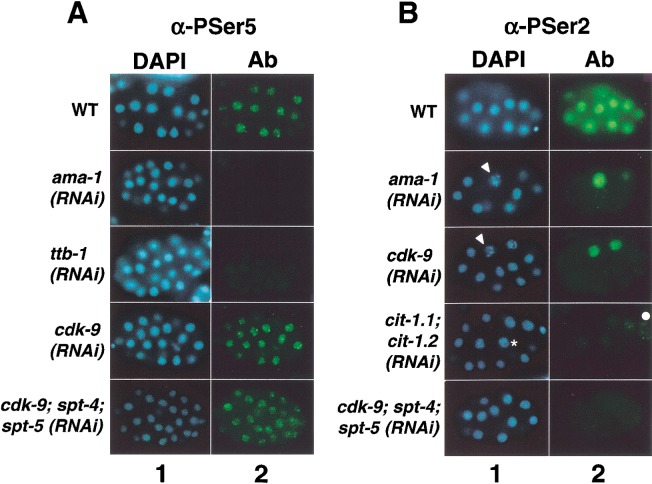
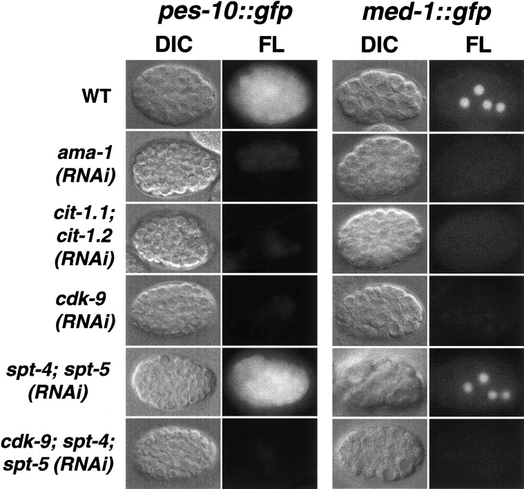
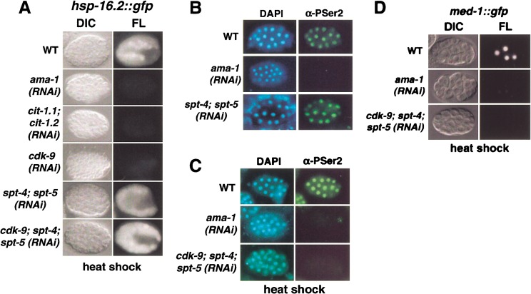
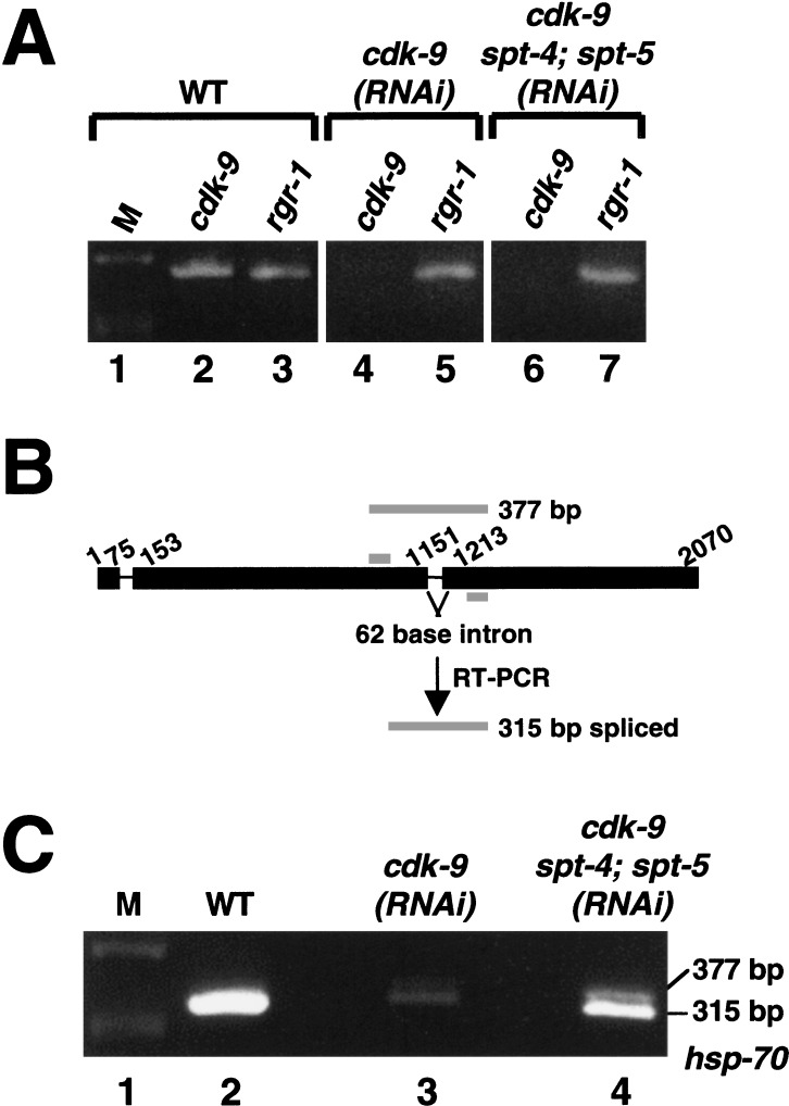
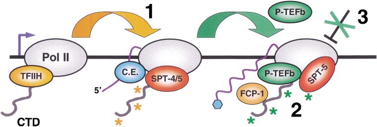
The metazoan transcription elongation factor P-TEFb (CDK-9/cyclin T) is essential for HIV transcription, and is recruited by some cellular activators. P-TEFb promotes elongation in vitro by overcoming pausing that requires the SPT-4/SPT-5 complex, but considerable evidence indicates that SPT-4/SPT-5 facilitates elongation in vivo. Here we used RNA interference to investigate P-TEFb functions in vivo, in the Caenorhabditis elegans embryo. We found that P-TEFb is broadly essential for expression of early embryonic genes. P-TEFb is required for phosphorylation of Ser 2 of the RNA Polymerase II C-terminal domain (CTD) repeat, but not for most CTD Ser 5 phosphorylation, supporting the model that P-TEFb phosphorylates CTD Ser 2 during elongation. Remarkably, although heat shock genes are cdk-9-dependent, they can be activated when spt-4 and spt-5 expression is inhibited along with cdk-9. This observation suggests that SPT-4/SPT-5 has an inhibitory function in vivo, and that mutually opposing influences of P-TEFb and SPT-4/SPT-5 may combine to facilitate elongation, or insure fidelity of mRNA production. Other genes are not expressed when cdk-9, spt-4, and spt-5 are inhibited simultaneously, suggesting that these genes require P-TEFb in an additional mechanism, and that they and heat shock genes are regulated through different P-TEFb-dependent elongation pathways.


Figures

Figure 1
Figure 1
Predicted C. elegans (ce) CDK-9, cyclin T, and SPT-4 proteins. (A) ceCDK-9, with similarity/identity to human (h) CDK-9 within the kinase domain indicated. Expressed cDNA sequences predict existence of two CDK-9 isoforms derived from alternative splicing of C-terminal exons (in black). (B) ceCIT-1.1 and ceCIT-1.2, compared with hcyclin T1, with similarity/identity to hcyclin T1 indicated. A phylogenic tree based on sequence comparison is shown at the bottom of the panel. (C) ceSPT-4 and ySPT-4 are compared with hSPT-4 as in (A).
Figure 2
Figure 2
Phenotypic analysis of RNAi embryos. (A) Expression of CDK-9 and RNA Pol II. Representative wild-type and RNAi embryos (indicated in rows) were stained with DAPI to visualize DNA, or with the indicated antibodies. The CDK-9 protein was not detected in any cdk-9(RNAi) or cdk-9; spt-4; spt-5(RNAi) embryos. The Pol II large subunit AMA-1 was detected with an antibody against the unphosphorylated CTD (8WG16). (B) Terminal and early cell division phenotypes of RNAi embryos. Embryos produced by N2 (wild-type) or pie-1∷gfp mothers were examined by differential interference (DIC) or fluorescence (FL) microscopy. Typical wild-type or RNAi embryos are shown in rows, as indicated to the left. In the leftcolumn, terminally arrested RNAi embryos are compared with a wild-type embryo that is continuing to develop. The right two columns show four-cell WT and RNAi pie-1∷gfp embryos. These RNAi embryos were indistinguishable from wild-type with respect to each aspect of PIE-1∷GFP germline and subcellular localization, including the presence of PIE-1 in germline RNA-protein P granules. Anterior is to the left.
Figure 3
Figure 3
CDK-9 and cyclin T are required for phosphorylation of Ser 2 of the Pol II CTD repeat, but not for most Ser 5 phosphorylation. In A and B, representative wild-type or RNAi embryos that are actively undergoing cell division are shown in rows, as indicated. Embryos were stained with DAPI to visualize DNA (column 1), and with α-P-Ser5 (A) or α-P-Ser2 (B) antibodies (Ab; column 2). (A) CTD Ser5 phosphorylation levels are not detectably reduced when CDK-9 is depleted by RNAi. α-P-Ser5 staining was comparable to wild-type at each stage in 100% of cdk-9(RNAi) embryos, but was dramatically reduced when transcription initiation was inhibited by depletion of the essential factor TFIIB [ttb-1(RNAi) embryos]. (B) CTD Ser2 phosphorylation is not detectable in embryos lacking P-TEFb. Throughout their development until terminal arrest, in 100% of cdk-9(RNAi), cit-1.1; cit-1.2(RNAi), and cdk-9; spt-4; spt-5(RNAi) embryos, α-P-Ser2 staining in interphase nuclei was not detectable above the background seen in ama-1(RNAi) embryos. Within each embryo set, in mitotic cells α-P-Ser2 also detected a cross-reactive epitope that does not derive from Pol II (see text). Examples of nuclei in early and late stages of mitotic chromosome condensation are indicated by an asterisk and arrowheads, respectively. No nuclear α-P-Ser2 staining is detectable in the wild-type germline precursor, which is not visible in the focal plane shown. Some α-P-Ser2-stained germline cells have weak perinuclear staining deriving from cross-reactivity of the secondary antibody with P granules (Walker et al. 2001). A white dot denotes a germline cell within the focal plane.
Figure 4
Figure 4
P-TEFb is required for expression of early embryonic genes. Wild-type (WT) and RNAi embryos (designated in rows) that were derived from transgenic GFP reporter strains were analyzed by DIC and fluorescence (FL) microscopy (as indicated above columns). These embryos are representative of the entire population analyzed in each of multiple independent experiments, in which >40 embryos were scored.
Figure 5
Figure 5
Requirement for P-TEFb is relieved at a heat shock gene by inhibition of spt-4 and spt-5. (A) Wild-type (WT) and RNAi hsp-16.2∷gfp embryos (designated in rows) were analyzed as in Fig. 4. Levels of HSP-16.2∷GFP expression varied within sets of wild-type, spt-4; spt-5(RNAi), and cdk-9; spt-4; spt-5(RNAi) embryos, but those shown correspond to representative differences between WT and RNAi embryos. (B) α-P-Ser2 staining in spt-4; spt-5(RNAi) embryos, analyzed as in Fig. 3. No mitotic cells are in focus in the ama-1(RNAi) (Pol II large subunit) embryo, which is shown to indicate background levels. (C) α-P-Ser2 staining is undetectable in heat shocked cdk-9; spt-4; spt-5(RNAi) embryos, which were analyzed as in (B). The α-P-Ser2 staining levels in heat shocked WT embryos were not detectably different from controls that were not heat shocked (data not shown). Some cross-reactive germline P granule staining is apparent in the ama-1(RNAi) embryo. (D) Heat shock did not induce MED-1∷GFP expression in cdk-9; spt-4; spt-5(RNAi) embryos, which were analyzed in parallel to the controls shown as in Fig. 4.
Figure 6
Figure 6
Restoration of endogenous hsp-70 transcription in cdk-9(RNAi) embryos by depletion of SPT-4/SPT-5. (A) Specific depletion of cdk-9 mRNA in RNAi embryos. Total RNA from the indicated embryo sets was assayed by RT-PCR to detect expression of cdk-9 (lanes 2,4,6), and a control mRNA (rgr-1; lanes 3,5,7). Each PCR primer set spanned an intron (data not shown). Products were analyzed on an agarose gel stained with ethidium bromide. DNA size markers are designated as M. (B) The hsp-70 gene, with exons indicated by a thick line. Primers used for RT-PCR (gray bars) flank intron 2. Sizes of predicted spliced and unspliced products are shown. (C) Endogenous hsp-70 expression. The samples analyzed in B were assayed by RT-PCR for hsp-70 RNA, using the primers shown in A. The hsp-70 mRNA (315-bp fragment) was present at markedly reduced levels in cdk-9(RNAi) embryos, but was significantly restored in cdk-9; spt-4; spt-5(RNAi) embryos. The 377-bp species in lane 4 corresponds to the unspliced hsp-70 sequence. It could not be established whether this product derived from incompletely processed RNA, because some samples contained trace amounts of genomic DNA (data not shown).
Figure 7
Figure 7
P-TEFb-dependent elongation mechanisms. Details and references are given in the text. (1) The TFIIH kinase (CDK-7; Kin28) phosphorylates CTD Ser 5 (orange asterisks) upon promoter clearance, and independently of P-TEFb. Pol II is then susceptible to DSIF (SPT-4/5)-dependent pausing. This phosphorylation also recruits capping enzyme (C.E.) to the CTD. (2) P-TEFb phosphorylates CTD Ser 2 and SPT-5 (green asterisks), and relieves inhibition by SPT-4/5. A speculative possibility is that SPT-4/5 may thereby be converted into a positively acting form. This mechanism may enhance the efficiency of elongation or mRNA processing by monitoring the presence or activity of factors required for these events. P-TEFb, the phosphatase FCP-1, and SPT-5 may remain associated with active elongating polymerase complexes, either continuously or intermittently, suggesting that SPT-5 may be regulated by opposing effects of P-TEFb and FCP-1. It is not known whether SPT-4 is then present. Heat shock genes can be expressed independently of cdk-9 when SPT-4 and SPT-5 are depleted by RNAi. (3) At most other genes, P-TEFb appears to be required in an additional mechanism. This mechanism may involve the release of a distinct, possibly chromatin-associated barrier to elongation, or an enhancement of cotranscriptional mRNA processing.

References

    1. Akhtar AX, Faye G, Bentley DL. Distinct activated and non-activated RNA polymerase II complexes in yeast. EMBO J. 1996;15:4654–4664. - PMC - PubMed
    1. Andrulis ED, Guzman E, Doring P, Werner J, Lis JT. High-resolution localization of Drosophila Spt5 and Spt6 at heat shock genes in vivo: Roles in promoter proximal pausing and transcription elongation. Genes & Dev. 2000;14:2635–2649. - PMC - PubMed
    1. Barboric M, Nissen RM, Kanazawa S, Jabrane-Ferrat N, Peterlin BM. NF-kappaB binds P-TEFb to stimulate transcriptional elongation by RNA polymerase II. Mol Cell. 2001;8:327–337. - PubMed
    1. Bellier S, Dubois MF, Nishida E, Almouzni G, Bensaude O. Phosphorylation of the RNA polymerase II largest subunit during Xenopus laevis oocyte maturation. Mol Cell Biol. 1997;17:1434–1440. - PMC - PubMed
    1. Bourgeois CF, Kim YK, Churcher MJ, West MJ, Karn J. Spt5 cooperates with human immunodeficiency virus type 1 tat by preventing premature RNA release at terminator sequences. Mol Cell Biol. 2002;22:1079–1093. - PMC - PubMed

Publication types

MeSH terms

Substances

LinkOut - more resources