. 2002 Sep;70(9):4841-50.
doi: 10.1128/IAI.70.9.4841-4850.2002.

Induction of the gene encoding macrophage chemoattractant protein 1 by Orientia tsutsugamushi in human endothelial cells involves activation of transcription factor activator protein 1

Affiliations

Induction of the gene encoding macrophage chemoattractant protein 1 by Orientia tsutsugamushi in human endothelial cells involves activation of transcription factor activator protein 1

Nam-Hyuk Cho et al. Infect Immun. 2002 Sep.

Abstract

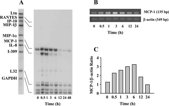
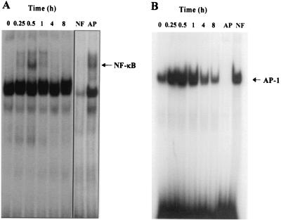
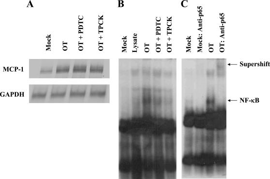
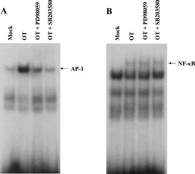
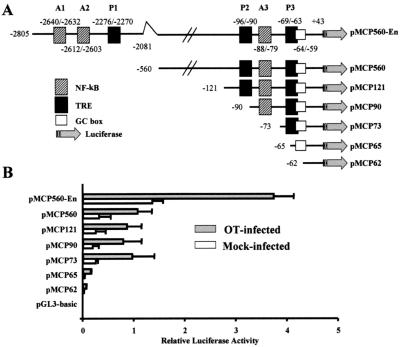
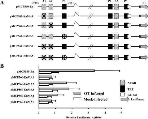
Human macrophage chemoattractant protein 1 (MCP-1) is a potent mediator of macrophage migration and therefore plays an essential role in early events of inflammation. In endothelial cells, at least three independent pathways regulate MCP-1 expression by NF-kappaB and AP-1. Orientia tsutsugamushi causes vasculitis in humans by replicating inside macrophages and endothelial cells. In the present study, we investigated the cis-acting and trans-acting elements involved in O. tsutsugamushi-induced MCP-1 gene expression in human umbilical vein endothelial cells (HUVEC). Although NF-kappaB activation was observed in HUVEC infected with O. tsutsugamushi, inhibition of NF-kappaB activation did not affect the MCP-1 expression. However, treatment of HUVEC with extracellular signal-regulated kinase (ERK) kinase inhibitor or p38 mitogen-activated protein kinase (MAPK) inhibitor suppressed expression of MCP-1 mRNA concomitant with downregulation of activator protein 1 (AP-1) activation. Deletion of triphorbol acetate response elements (TRE) at position -69 to -63 of MCP-1 gene abolished inducible promoter activity. Deletion of TRE at position -69 to -63-96 to -90 or deletion of NF-kappaB-binding site at position -69 to -63-88 to -79 did not affect the inducibility of promoter. Site-directed mutagenesis of the NF-kappaB binding sites at positions -2640 to -2632, -2612 to -2603 in the enhancer region, or the AP-1 biding site at position -2276 to -2270 decreased the inducible activity of the promoter. Taken together, AP-1 activation by both the ERK pathway and the p38 MAPK pathway as well as their binding to TRE at position -69 to -63 in proximal promoter and TRE at position -2276 to -2270 in enhancer region is altogether essential in induction of MCP-1 mRNA in HUVEC infected with O. tsutsugamushi. Although NF-kappaB activation is not essential per se, the kappaB site in the enhancer region is important in MCP-1 induction of HUVEC. This discrepancy in the involvement of the NF-kappaB may be due to the function of chromatin structures and other transcription cofactors in the regulation of MCP-1 gene expression in response to O. tsutsugamushi infectioin.


Figures

FIG. 1.
FIG. 1.
Time course of O. tsutsugamushi-stimulated chemokine induction in HUVEC. (A) The levels of chemokine mRNAs at each time point were assayed by RNase protection. (B) The mRNA levels of MCP-1 gene were analyzed by semiquantitative RT-PCR in 1.2% agarose gel. The levels of β-actin transcripts were also determined to normalize the transcripts of MCP-1. (C) The band densities were determined with TINA software, and the mRNA expression level for MCP-1 was normalized with respect to the intensities of the bands of β-actin. Abbreviations: MIP-1α, macrophage inflammatory protein 1α; GAPDH, glyceraldehyde-3-phosphate dehydrogenase.
FIG. 2.
FIG. 2.
EMSA showing activation of transcription factors, NF-κB (A) and AP-1 (B), in O. tsutsugamushi-infected HUVEC at each time point. A competitive inhibition assay was performed with nuclear extracts that were preincubated with 50-fold molar excesses of either unlabeled NF-κB (NF) or AP-1 (AP) consensus oligonucleotide.
FIG. 3.
FIG. 3.
Effect of PDTC (50 μM) or TPCK (100 μM) on O. tsutsugamushi-induced MCP-1 mRNA level and NF-κB in HUVEC. (A) MCP-1 mRNA level was analyzed using an RNase protection assay with total RNA samples that were prepared from uninfected cells (Mock), cells infected with O. tsutsugamushi for 3 h (OT), and infected cells in the presence of PDTC (OT + PDTC) or TPCK (OT + TPCK). (B) EMSA was performed to analyze the activation of NF-κB using nuclear extracts prepared from HUVEC treated for 2 h with medium (Mock), L-929 cell lysate (Lysate), or O. tsutsugamushi. Nuclear extracts from cells pretreated with PDTC (OT + PDTC) or TPCK (OT + TPCK) for 1 h before infection with O. tsutsugamushi were also analyzed. (C) In the supershift assay, nuclear extracts from HUVEC were preincubated with antibodies against the p65 subunit of NF-κB.
FIG. 4.
FIG. 4.
Effect of PD 098059 and SB 203580 on MCP-1 induction. (A) HUVEC were pretreated for 1 h with the indicated concentrations of PD 098059 or SB 203580 and then infected with O. tsutsugamushi for 3 h. The mRNA levels of MCP-1 gene were analyzed by semiquantitative RT-PCR in 1.2% agarose gel. (B) The levels of β-actin transcripts were also determined to normalize the transcripts of MCP-1. (C) The band densities were determined with TINA software, and the mRNA expression level of MCP-1 was normalized with respect to the intensities of the bands of the β-actin. Abbreviations: M, ΦX174 DNA digested with HaeIII; C, mock-treated HUVEC; N, PCR-negative control (reactions performed without any cDNA).
FIG. 5.
FIG. 5.
Effects of PD 098059 and SB 203580 on the activation of NF-κB or AP-1. (A) HUVEC were pretreated for 1 h with PD 098059 (25 μM), SB 203580 (25 μM), or medium and subsequently exposed to medium (mock) or O. tsutsugamushi for 2 h. Nuclear extracts were analyzed by EMSA using a specific 32P-labeled NF-κB consensus oligonucleotide. (B) The nuclear extracts used in panel A were also analyzed for AP-1 activity by EMSA.
FIG. 6.
FIG. 6.
Deletion analysis of the MCP-1 5′-flanking region. (A) A series of clones was used to characterized the MCP-1 promoter region. pMCP560-En contains a proximal promoter region extending from bp +43 to −560 and an enhancer region extending from bp −2081 to −2805 relative to the transcription start site. Numbers indicate nucleotides relative to transcription start site and the first nucleotide inserted into each plasmid DNA. All of the insert DNA extends to nucleotide +43 of the MCP-1 gene. (B) Luciferase activity of HUVEC transfected transiently with clones in panel A. HUVEC were transiently transfected as described and allowed to recover for 18 h. The cells were then stimulated with O. tsutsugamushi (filled bars) or left untreated (open bars) and allowed to incubate for 24 h. Luciferase activity was normalized according to β-galactosidase activity obtained from the pCMV-βgal plasmid which was cotransfected. Results are given as means + standard deviations (error bars) of three independent transfection experiments.
FIG. 7.
FIG. 7.
Mutational analysis of transcriptional control elements in the MCP-1 enhancer region. (A) A series of clones was used to characterize the MCP-1 enhancer region. pMCP560-En contains insert DNA as described in the legend to Fig. 6. Nucleotides of the NF-κB consensus site (hatched box) and/or AP-1 consensus site (filled box) in the enhancer region were substituted as described in Material and Methods and are indicated (X). (B) Luciferase activity of HUVEC transfected transiently with clones in panel A. The extracts from cells stimulated with O. tsutsugamushi (closed bars) or left untreated (open bars) were assayed for luciferase activity. Luciferase activity was normalized as described in the legend to Fig. 6. Results are given as means + standard deviations (error bars) of three independent transfection experiments.

References

    1. Allen, A. C., and S. Spitz. 1945. A comparative study of the pathology of scrub typhus (tsutsugamushi disease) and other rickettsial disease. Am. J. Pathol. 21:603-681. - PMC - PubMed
    1. Baggiolini, M., B. Dewald, and B. Moser. 1994. Interleukin-8 and related chemotactic cytokines-CXC and CC chemokines. Adv. Immunol. 55:97-179. - PubMed
    1. Baldwin, A. S., Jr. 1996. The NF-kappa B and I kappa B proteins: new discoveries and insights. Annu. Rev. Immunol. 14:649-683. - PubMed
    1. Beilke, M. A. 1989. Vascular endothelium in immunology and infectious disease. Rev. Infect. Dis. 11:273-283. - PubMed
    1. Beyaert, R., A. Cuenda, W. Vanden Berghe, S. Plaisance, J. C. Lee, G. Haegeman, P. Cohen, and W. Fiers. 1996. The p38/RK mitogen-activated protein kinase pathway regulates interleukin-6 synthesis response to tumor necrosis factor. EMBO J. 15:1914-1923. - PMC - PubMed

Publication types

MeSH terms

LinkOut - more resources