. 2002 Sep;70(9):5193-201.
doi: 10.1128/IAI.70.9.5193-5201.2002.

Analysis of pathogen-host cell interactions in purpura fulminans: expression of capsule, type IV pili, and PorA by Neisseria meningitidis in vivo

Affiliations

Analysis of pathogen-host cell interactions in purpura fulminans: expression of capsule, type IV pili, and PorA by Neisseria meningitidis in vivo

O B Harrison et al. Infect Immun. 2002 Sep.

Abstract

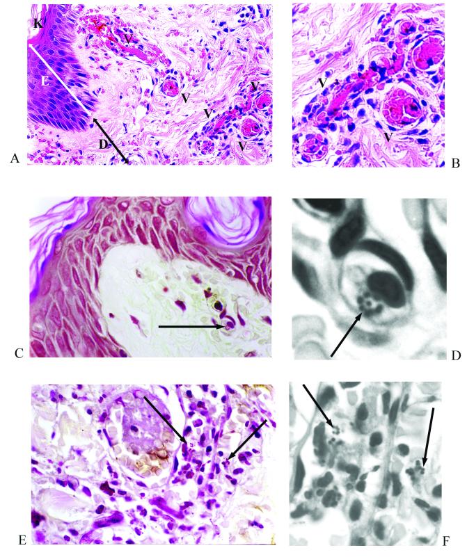
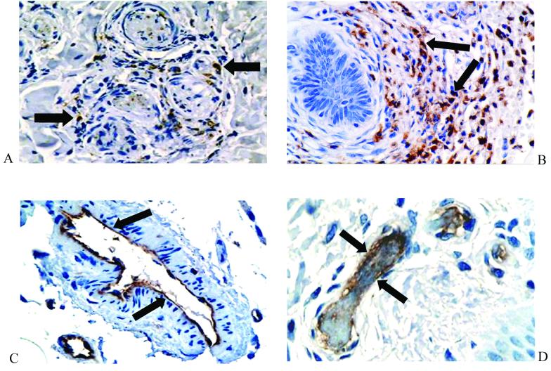
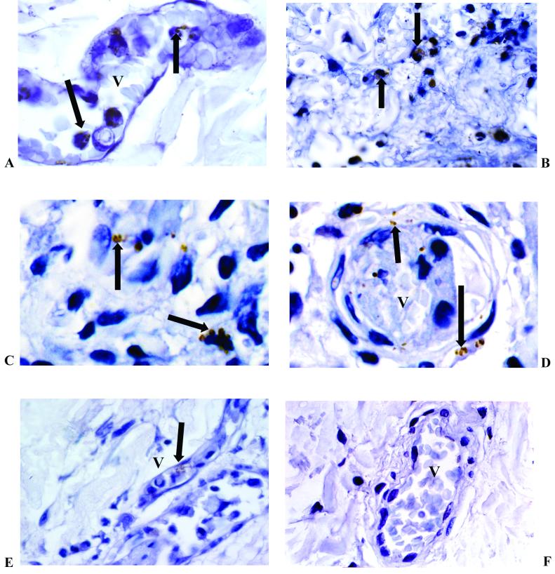
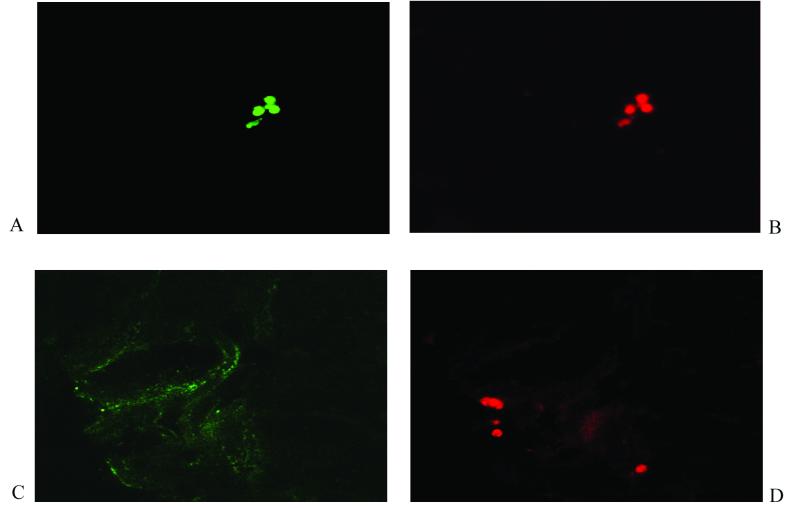
The pattern of meningococcal surface structure expression in different microenvironments following bloodstream invasion in vivo is not known. We used immunohistochemistry to determine the expression of capsule, type IV pili, and PorA by meningococci residing in the skin lesions of children with purpura fulminans. All the skin biopsy samples showed evidence of thrombosis and, frequently, a perivascular inflammatory cell infiltrate consisting of neutrophils (elastase positive) and monocytes/macrophages (CD68 positive). Modified Gram staining revealed 20 to over 100 gram-negative diplococci in each 4-microm-thick section, usually grouped into microcolonies. Immunoperoxidase staining demonstrated that the invading meningococci expressed PorA, capsule, and type IV pilin. Expression of these antigens was not restricted to any particular environment and was found in association with meningococci located in leukocytes, small blood vessels, and the dermal interstitium. Confocal laser scanning microscopy demonstrated coexpression of pilin and capsule by numerous microcolonies. However, there was some discordance in capsule and pilin expression within the microcolonies, suggesting phase variation. The strategy employed in this study will be helpful in investigating invasive bacterial diseases where antigenic and phase variation has a significant impact on virulence and on vaccine design.


Figures

FIG. 1.
FIG. 1.
Skin biopsy specimen from a patient with meningococcal septicemia. (A and B) An area of endothelial swelling, acute inflammation with a perivascular infiltrate, and thrombosis. Panel A shows hemotoxylin and eosin staining at a magnification of ×400; panel B was electronically enlarged. (C through F) Gram-stained sections reveal meningococci associated with a leukocyte (panel C, magnification at ×400; panel D, electronically enlarged) and within blood vessels (panel E, magnification at ×400; panel F, electronically enlarged). Figures were processed with Adobe Photoshop and illustrator software. K, keratin layer; E, epidermis; D, dermis; V, blood vessel. Arrows indicate Gram-stained meningococci.
FIG. 2.
FIG. 2.
Immunohistochemical staining of inflammatory cells and the vascular endothelium in skin biopsy samples from patients with meningococcal disease. The inflammatory infiltrate consisted of a mixture of CD68-positive macrophages (A) (magnification, ×400) and neutrophil elastase-positive polymorphonuclear cells (B) (magnification, ×400). Multiple CD31-positive blood vessels were seen throughout the biopsy samples (C and D) (magnification, ×400 and 600, respectively). Arrows indicate positive immunoperoxidase staining (brown) with the appropriate specific mouse monoclonal antibody (nuclei were counterstained with hematoxylin).
FIG. 3.
FIG. 3.
Immunohistochemical staining of N. meningitidis in skin biopsy samples from patients with meningococcal disease. These examples show capsulated serogroup B visualized in the blood vessel (A) (magnification, ×1,000), inflammatory cells (B) (magnification, ×600), and the interstitium (C) (magnification, ×1,000). PorA stained bacteria in the interstitium and blood vessel (D), and pilin stained meningococci in a small blood vessel (E) (magnification, ×600). A negative control is also shown (F) (magnification, ×400). V, blood vessel. Arrows indicate immunoperoxidase-positive staining of meningococci (brown) with the appropriate specific mouse monoclonal antimeningococcal antibody (nuclei were counterstained in hematoxylin).
FIG. 4.
FIG. 4.
Confocal laser scanning microscopy to detect N. meningitidis in skin biopsy samples from patients with meningococcal disease. Seven-micrometer-thick frozen sections were analyzed as sequential 1-μm-thick sections (×1,000). (A and B) A microcolony of N. meningitidis double stained with anticapsule monoclonal antibody detected with Alexa Fluor 488 (green fluorescence) and antipilin monoclonal antibody detected with Alexa Fluor 594 (red fluorescence), respectively. (C and D) A microcolony staining with antipilin monoclonal antibody (red fluorescence) and some background fluorescence but no specific staining with anticapsule monoclonal antibody (green fluorescence), respectively.

References

    1. Ala'Aldeen, D. A. A., K. R. Neal, K. Ait-Tahar, J. S. Nguyen-Van-Tam, A. English, T. J. Falla, P. M. Hawkey, and R. C. B. Slack. 2000. Dynamics of meningococcal long-term carriage among university students and their implications for mass vaccination. J. Clin. Microbiol. 38:2311-2316. - PMC - PubMed
    1. Birkness, K. A., B. L. Swisher, E. H. White, E. G. Long, E. P. Ewing, Jr., and F. D. Quinn. 1995. A tissue culture bilayer model to study the passage of Neisseria meningitidis. Infect. Immun. 63:402-409. - PMC - PubMed
    1. Bjerknes, R., H. K. Guttormsen, C. O. Solberg, and L. M. Wetzler. 1995. Neisserial porins inhibit human neutrophil actin polymerization, degranulation, opsonin receptor expression, and phagocytosis but prime the neutrophils to increase their oxidative burst. Infect. Immun. 63:160-167. - PMC - PubMed
    1. Bjune, G., E. A. Høiby, J. K. Grønnesby, Ø. Arnesen, J. H. Fredriksen, A. Halstensen, E. Holten, A.-K. Lindbak, H. Nøkleby, E. Rosenqvist, L. K. Solberg, O. Closs, J. Eng, L. O. Frøholm, A. Lystad, L. S. Bakketeig, and B. Hareide. 1991. Effect of outer membrane vesicle vaccine against group B meningococcal disease in Norway. Lancet 338:1093-1096. - PubMed
    1. Brandtzaeg, P. 1995. Pathogenesis of meningococcal disease, p. 71-114. In K. Cartwright (ed.), Meningococcal disease. John Wiley & Sons Ltd., Chichester, United Kingdom.

Publication types

MeSH terms