. 2002 Sep 3;99(18):11670-5.
doi: 10.1073/pnas.172404099. Epub 2002 Aug 16.

Structural insights into peptide bond formation

Affiliations

Structural insights into peptide bond formation

Jeffrey L Hansen et al. Proc Natl Acad Sci U S A. .

Abstract

The large ribosomal subunit catalyzes peptide bond formation and will do so by using small aminoacyl- and peptidyl-RNA fragments of tRNA. We have refined at 3-A resolution the structures of both A and P site substrate and product analogues, as well as an intermediate analogue, bound to the Haloarcula marismortui 50S ribosomal subunit. A P site substrate, CCA-Phe-caproic acid-biotin, binds equally to both sites, but in the presence of sparsomycin binds only to the P site. The CCA portions of these analogues are bound identically by either the A or P loop of the 23S rRNA. Combining the separate P and A site substrate complexes into one model reveals interactions that may occur when both are present simultaneously. The alpha-NH(2) group of an aminoacylated fragment in the A site forms one hydrogen bond with the N3 of A2486 (2451) and may form a second hydrogen bond either with the 2' OH of the A-76 ribose in the P site or with the 2' OH of A2486 (2451). These interactions position the alpha amino group adjacent to the carbonyl carbon of esterified P site substrate in an orientation suitable for a nucleophilic attack.


Figures

Figure 1
Figure 1
Chemical structures of peptidyl transferase substrate analogues. (A) CCA-pcb is active as a P site substrate and binds to only the P site in the presence of the antibiotic, sparsomycin. (B) An aminoacylated RNA minihelix binds to the A site. (C) CCdA-phosphate-puromycin is an intermediate analogue containing A and P site-binding components. (D) CC-puromycin-phenylalanine-caproic acid–biotin and deacylated CCA are products of the peptidyl transferase reaction.
Figure 2
Figure 2
Experimental electron density maps. (A) An FoFo electron density map (blue net) contoured at 4.0 σ shows density corresponding to CCA-pcb (green) in the P site and sparsomycin (yellow). Additional density corresponds to altered conformations of nucleotides such as A2637 (orange). (B) FoFo electron density map of CCA-pcb shows that in the absence of sparsomycin, the P site substrate is bound equally between the P site (green) and the A site (red).
Figure 3
Figure 3
The various peptidyl transferase fragment substrate and product analogues and tRNA bind the 50S and 70S ribosome in the same way. (A) Three P site substrate analogues, CCA-pcb (green), CC-acetylated-puromycin (gray), the CCdA portion of CCdA-phosphate-puromycin (dark blue), which results when ribosomal RNA is superimposed by least squares among the cocrystal structures. Likewise, three A site substrates, stem–loop–CC-puromycin (purple), A site product (20) (light blue and green), and puromycin from the CCdA-phosphate-puromycin (dark blue) structure also superimposed. The P and A site substrates are separated for clearer viewing. (B) The positions of the acceptor ends of tRNA molecules (blue backbone) bound to the A and P sites of the 70S ribosome (28) agree well with the positions of the fragment P site (green) and A site (purple) bound to the 50S subunit. A2486 (2451) is yellow.
Figure 4
Figure 4
A and P site substrates on the peptidyl-transferase center. (A) A model resulting from the superposition of the A and P site substrate complexes places the α amine of the A site substrate (purple) in position for a pro-R attack (black arrow) on the carbonyl carbon of the aminoacyl ester bond of the P site substrate (green). (B) The CCdAp-puromycin intermediate analogue superimposed on an intermediate modeled from the A and P site substrate complexes diverges near the tetrahedral carbon oxyanion of the intermediate.
Figure 5
Figure 5
Model of peptide bond formation pathway. (A theoretical model of how the peptidyl transferase reaction might proceed is illustrated in Movie 1, which is published as supporting information on the PNAS web site, www.pnas.org.) (A) Substrate bound at A site (purple) is in a relative position for a pro-R attack on a P site bound substrate (green), based on superposition of two cocrystal structures. (B) A model of the tetrahedral intermediate with the oxyanion points away from A2486 (2451). (C) The structure of products of the peptidyl transferase reaction bound to the peptidyl transferase center (20).
Figure 6
Figure 6
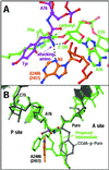
Modeling of A2486 (2451) mutation to C in the A site substrate complex. (A) The structure of the active site shows N3 of A2486 (2451) (orange) forming a hydrogen bond (dotted line) with the attacking amine of an A site substrate (purple). (B) Model of mutant C2486 (2451) (orange) shows that the O2 of a C could substitute as a hydrogen bond acceptor to the α amino group, and that hydrogen bonds between the N4 of a C2486 (2451) to G2101 (2061) and G2482 (2448) might still occur.

Similar articles

Cited by

References

    1. Traut R R, Monro R E. J Mol Biol. 1964;10:63–71. - PubMed
    1. Rychlik I. Biochim Biophys Acta. 1966;114:425–427. - PubMed
    1. Monro R E. J Mol Biol. 1967;26:147–151. - PubMed
    1. Maden B E, Traut R R, Monro R E. J Mol Biol. 1968;35:333–345. - PubMed
    1. Watson J D. Bull Soc Chim Biol. 1964;46:1399–1425. - PubMed

Publication types

Associated data