. 2002 Aug 19;196(4):493-503.
doi: 10.1084/jem.20011836.

Surface cathepsin B protects cytotoxic lymphocytes from self-destruction after degranulation

Affiliations

Surface cathepsin B protects cytotoxic lymphocytes from self-destruction after degranulation

Kithiganahalli N Balaji et al. J Exp Med. .

Abstract

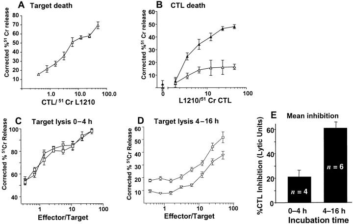
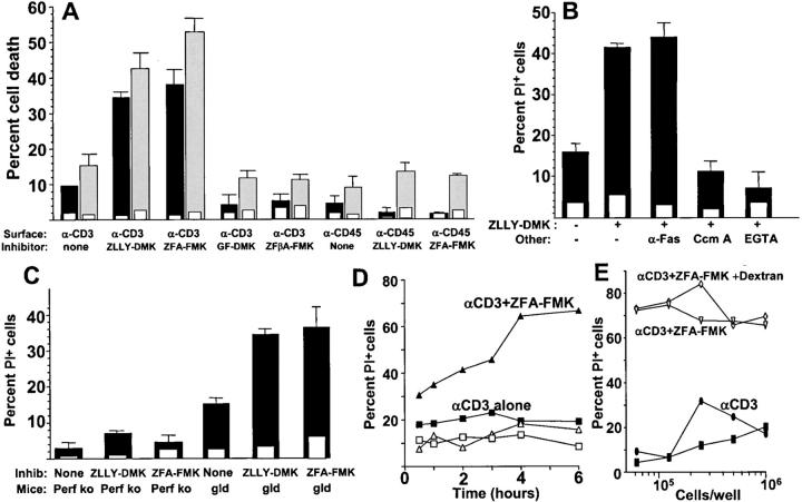
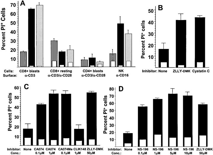
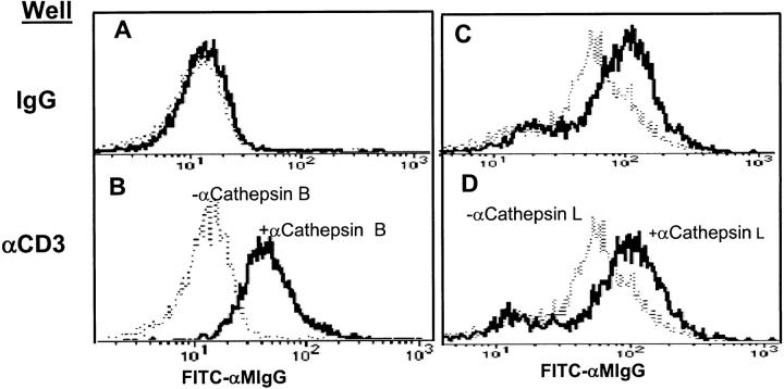
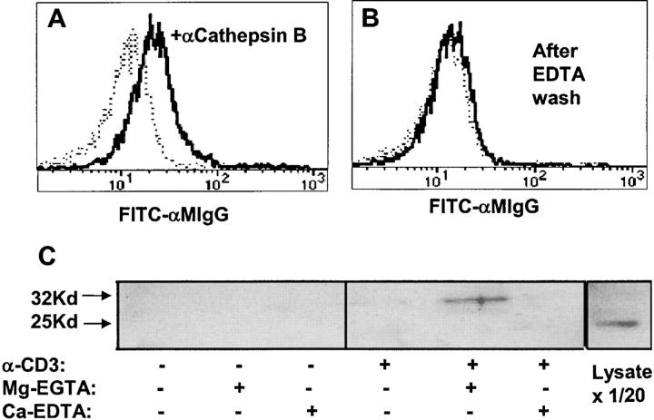
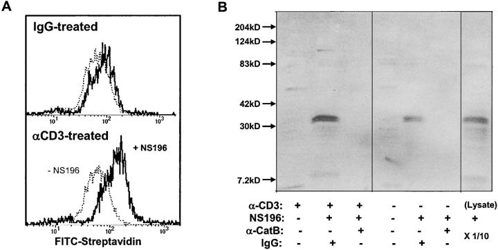
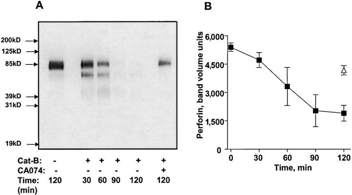
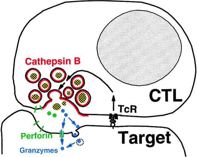
The granule exocytosis cytotoxicity pathway is the major molecular mechanism for cytotoxic T lymphocyte (CTL) and natural killer (NK) cytotoxicity, but the question of how these cytotoxic lymphocytes avoid self-destruction after secreting perforin has remained unresolved. We show that CTL and NK cells die within a few hours if they are triggered to degranulate in the presence of nontoxic thiol cathepsin protease inhibitors. The potent activity of the impermeant, highly cathepsin B-specific membrane inhibitors CA074 and NS-196 strongly implicates extracellular cathepsin B. CTL suicide in the presence of cathepsin inhibitors requires the granule exocytosis cytotoxicity pathway, as it is normal with CTLs from gld mice, but does not occur in CTLs from perforin knockout mice. Flow cytometry shows that CTLs express low to undetectable levels of cathepsin B on their surface before degranulation, with a substantial rapid increase after T cell receptor triggering. Surface cathepsin B eluted from live CTL after degranulation by calcium chelation is the single chain processed form of active cathepsin B. Degranulated CTLs are surface biotinylated by the cathepsin B-specific affinity reagent NS-196, which exclusively labels immunoreactive cathepsin B. These experiments support a model in which granule-derived surface cathepsin B provides self-protection for degranulating cytotoxic lymphocytes.


Figures

Figure 3.
Figure 3.
CTL suicide during target cell lysis. (A) Lysis of H-2d–bearing L1210 tumor cells by H-2d–reactive CTL in a 4-h assay. (B) Lysis of 51Cr-labeled CTL (104/well) used in A when incubated for 4 h with L1210 target cells in the presence of 10 μM CA074. ▴, CA074; ▵, no inhibitor. (C–D) H-2b–reactive CTL were mixed with 51Cr-labeled EL4 cells in the presence or absence of 10 μM CA074. After 4 h, 125 μl supernatant were harvested for counting (C) and replaced with 125 μl of medium for ∼12 h of additional incubation. D shows the second harvest. As a toxicity control for CA074 in D, the CTLs in C were incubated with 10 μM CA074 for 16 h before the cytotoxicity assay with 51Cr-labeled EL-4 cells. □, no inhibitor; ▿, 10 μM CA074. (E) Mean inhibition of CTL lytic activity from multiple experiments like those shown in C and D, calculated by lytic units (horizontal shift between cytotoxicity dose response curves).
Figure 1.
Figure 1.
Alloactivated mouse CD8+ T cell death induced by anti-CD3 in the presence of cathepsin inhibitors. (A) CD8+ T cells from B6 mice activated by a primary MLR culture were incubated for 4 h with and without the indicated cathepsin inhibitors on wells coated with anti-CD3 or anti-CD45 (filled bars) or control hamster or mouse IgG (inset open bars). Cell death was measured microscopically by nuclear propidium iodide staining (measuring membrane integrity, solid bars) or apoptotic nuclear morphology using Hoechst 33342 (gray bars). The cathepsin inhibitors ZLLY-DMK, ZFA-FMK, and controls GF-DMK and ZFβA-FMK, were used at final concentrations of 50 μM. Error bars show SEM of triplicate samples. (B) Murine-alloactivated CD8+ MLR blasts were incubated with 50 μM cathepsin inhibitor ZLLY-DMK in the presence of 10 μg/ml IgG anti-Fas antibody, 1μM concanamycin A (Ccm A), or 2.5 mM EGTA. Cell death was assessed after 4 h by propidium iodide staining in anti-CD3– (solid bars) or control hamster IgG–coated wells (inset open bars). (C) Alloactivated CD8+ MLR blasts from perforin knockout or gld mice were incubated 4 h with or without 50 μM cathepsin inhibitor ZLLY-DMK. Cell death by propidium iodide was measured in wells coated with anti-CD3 (solid bars) or control IgG (open inset bars). (D) Kinetics of death of human CTL clone RS-56 induced by plate-bound anti-CD3 in the presence of 50 μM cathepsin inhibitor ZFA-FMK. □, no anti-CD3 or ZFA-FMK. (E) Density dependence of CTL death as in D, measured at 4 h. ▿, CTL incubated with 50 μM ZFA-FMK on plate-bound anti-CD3; ⋄, same as ▿ with 2 × 10⁶ kD dextran added to a final concentration of 5% to increase medium viscosity to block fratricidal killing; ▪, anti-CD3 with no ZFA-FMK; •, ZFA-FMK with no anti-CD3.
Figure 2.
Figure 2.
Activation-induced suicide of cytotoxic effectors in the presence of membrane-impermeant cathepsin B inhibitors. (A) Human CD8+ T cell blasts, resting blood CD8+ cells, CD4+ T cell blasts, and NK cells were incubated for 4 h with and without 50 μM cathepsin inhibitors ZLLY-DMK or ZFA-FMK. Cell death was assessed by propidium iodide from wells coated with control IgG (open inset bars) or the indicated degranulating stimuli (open bars). Striped bars, no inhibitor; solid bars, ZLLY-DMK; gray bars, ZFA-FMK.(B) Alloactivated mouse CD8+ T cell blasts were incubated for 4 h on anti-CD3–coated wells (solid bars) or control IgG (open inset bars) with or without 10 μM cystatin C or 50 μM ZLLY-DMK, and cell death was assessed by propidium iodide. (C) Alloactivated CD8+ BALB/c T cells were incubated on wells coated with anti-CD3 (solid bars) or control IgG (open inset bars) for 4 h in presence or absence of the indicated cathepsin inhibitors and stained with propidium iodide. (D) Same conditions as in C, comparing NS-196 and ZLLY-DMK.
Figure 4.
Figure 4.
TCR cross-linking rapidly increases CTL surface expression of cathepsin B, but not cathepsin L. (B and D) CD8+ cloned human CTL were incubated on surface-bound anti-CD3, or (A and C) isotype IgG for 2 h at 37°C. (A and B) Cells were then stained with anti-cathepsin B or (C and D) with anti-cathepsin L antibody followed by FITC-anti–mouse IgG (heavy lines). Dashed lines show staining by normal IgG controls.
Figure 5.
Figure 5.
Surface cathepsin B on TCR-activated CTL is released by EGTA and is the single chain form. CD8+ cloned human CTL were stimulated with plate-bound anti-CD3 for 2 h at 37°C, washed with PBS, and (A) incubated with PBS or (B) 0.53 mM EDTA in PBS for 10 min at 37°C. After washing, cells were incubated with anti-cathepsin B (heavy line) followed by anti–mouse Ig-FITC (dashed line shows control with no anticathepsin B). (C) Anti-CD3–treated CTL as in A and B were incubated with 2 mM Mg-EGTA or Ca-EDTA in HBSS for 10 min at 37°C. Supernatants were analyzed by blotting with anti-cathepsin B. The last lane shows whole CTL dissolved in SDS sample buffer and run directly (1/20 of the cell-equivalent input of other lanes).
Figure 6.
Figure 6.
Surface cathepsin B on TCR-activated T cells is active and the target of NS-196. (A) Detection of biotinylated NS-196 on the CTL surface after degranulation. Flow cytometry of CD8+ cloned human CTL RS-56 after culture on wells coated with anti-CD3 or isotope control for 2 h, followed by treatment with or without 1 μM NS-196, which was detected by FITC-streptavidin. (B) Identification of cathepsin B as the molecular target of NS-196. CTL clone RS-56 was incubated for 2 h on anti-CD3– or IgG-coated wells, followed by incubation with or without 0.1 μM NS-196. Cells were lysed with Triton X-100 and immunodepleted with beads containing anticathepsin B antibody or control rabbit IgG. The remaining lysate was run on a 12% nonreduced SDS gel, blotted onto nitrocellulose, probed with Streptavidin-HRP, and developed using ECL. The right lane shows the biotinylation pattern when the whole CTL lysate was labeled with 0.1 μM NS-196 and run directly (1/10 of the cell-equivalent input of other lanes).
Figure 7.
Figure 7.
Perforin cleavage by cathepsin B. (A) Perforin from CTL extracts was treated with 258 ng/ml purified cathepsin B for various times and analyzed by blotting with anti-perforin antibody. (B) Mean densitometric analysis of perforin degradation by cathepsin B. Three experiments are shown.
Figure 8.
Figure 8.
Surface cathepsin B model for cytotoxic lymphocyte self-protection.

References

    1. Henkart, P.A. 1994. Lymphocyte-mediated cytotoxicity: two pathways and multiple effector molecules. Immunity. 1:343–346. - PubMed
    1. Harty, J.T., A.R. Tvinnereim, and D.W. White. 2000. CD8+ T cell effector mechanisms in resistance to infection. Annu. Rev. Immunol. 18:275–308. - PubMed
    1. Smyth, M.J., K.Y. Thia, S.E. Street, D. MacGregor, D.I. Godfrey, and J.A. Trapani. 2000. Perforin-mediated cytotoxicity is critical for surveillance of spontaneous lymphoma. J. Exp. Med. 192:755–760. - PMC - PubMed
    1. Stepp, S.E., R. Dufourcq-Lagelouse, F. Le Deist, S. Bhawan, S. Certain, P.A. Mathew, J.I. Henter, M. Bennett, A. Fischer, G. de Saint Basile, et al. 1999. Perforin gene defects in familial hemophagocytic lymphohistiocytosis. Science. 286:1957–1959. - PubMed
    1. Straus, S.E., M. Sneller, M.J. Lenardo, J.M. Puck, and W. Strober. 1999. An inherited disorder of lymphocyte apoptosis: the autoimmune lymphoproliferative syndrome. Ann. Intern. Med. 130:591–601. - PubMed

Publication types

MeSH terms