. 2002 Aug 19;158(4):639-46.
doi: 10.1083/jcb.200203086. Epub 2002 Aug 19.

A survival pathway for Caenorhabditis elegans with a blocked unfolded protein response

Affiliations

A survival pathway for Caenorhabditis elegans with a blocked unfolded protein response

Fumihiko Urano et al. J Cell Biol. .

Abstract

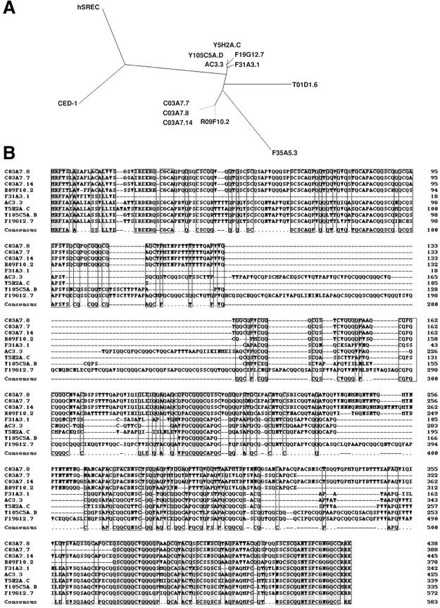
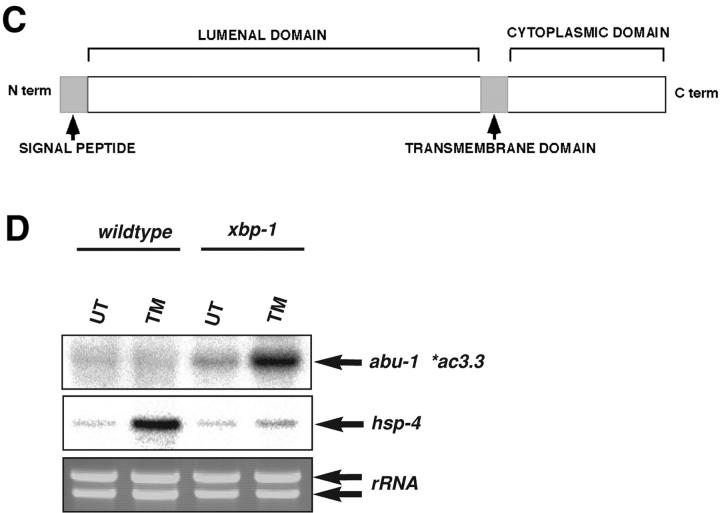
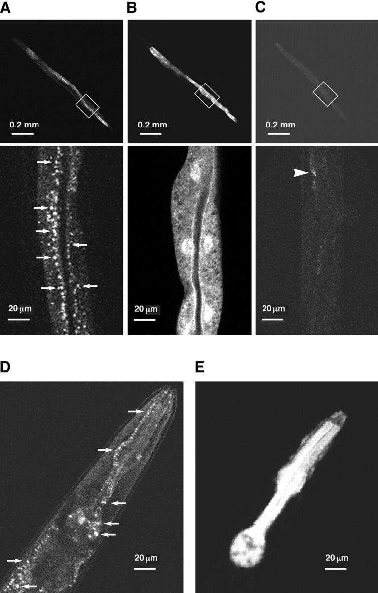
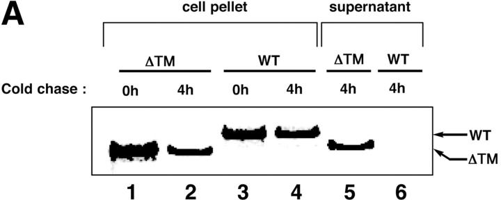
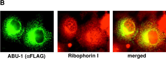
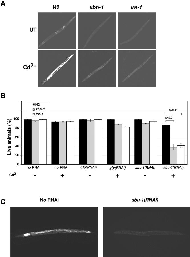
The unfolded protein response (UPR) counteracts stress caused by unprocessed ER client proteins. A genome-wide survey showed impaired induction of many UPR target genes in xbp-1 mutant Caenorhabditis elegans that are unable to signal in the highly conserved IRE1-dependent UPR pathway. However a family of genes, abu (activated in blocked UPR), was induced to higher levels in ER-stressed xbp-1 mutant animals than in ER-stressed wild-type animals. RNA-mediated interference (RNAi) inactivation of a representative abu family member, abu-1 (AC3.3), activated the ER stress marker hsp-4::gfp in otherwise normal animals and killed 50% of ER-stressed ire-1 and xbp-1 mutant animals. Abu-1(RNAi) also enhanced the effect of inactivation of sel-1, an ER-associated protein degradation gene. The nine abu genes encode highly related type I transmembrane proteins whose lumenal domains have sequence similarity to a mammalian cell surface scavenger receptor of endothelial cells that binds chemically modified extracellular proteins and directs their lysosomal degradation. Our findings that ABU-1 is an intracellular protein located within the endomembrane system that is induced by ER stress in xbp-1 mutant animals suggest that ABU proteins may interact with abnormal ER client proteins and this function may be particularly important in animals with an impaired UPR.


Figures

Figure 1.
Figure 1.
The C. elegans activated in blocked UPR (abu) gene family. (A) The amino acid sequence identity relationship of the ABU family members, C. elegans CED-1 (Zhou et al., 2001), and human scavenger receptor of endothelial cells (Adachi et al., 1997) is displayed as a radial tree (Page, 1998). (B) Amino acid sequence alignment of the nine ABU proteins using the Clustal W (1.81) program. (C) Predicted structure of ABU family members. (D) Northern blot analysis of total RNA from untreated and tunicamycin-treated wild-type and xbp-1(zc12)III mutant C. elegans. The blot was hybridized sequentially to labeled abu-1 and hsp-4 cDNA. The ethidium bromide–stained gel (bottom panel) reports on the integrity of the mRNA in all samples.
Figure 1.
Figure 1.
The C. elegans activated in blocked UPR (abu) gene family. (A) The amino acid sequence identity relationship of the ABU family members, C. elegans CED-1 (Zhou et al., 2001), and human scavenger receptor of endothelial cells (Adachi et al., 1997) is displayed as a radial tree (Page, 1998). (B) Amino acid sequence alignment of the nine ABU proteins using the Clustal W (1.81) program. (C) Predicted structure of ABU family members. (D) Northern blot analysis of total RNA from untreated and tunicamycin-treated wild-type and xbp-1(zc12)III mutant C. elegans. The blot was hybridized sequentially to labeled abu-1 and hsp-4 cDNA. The ethidium bromide–stained gel (bottom panel) reports on the integrity of the mRNA in all samples.
Figure 2.
Figure 2.
Confocal fluorescence micrographs of C. elegans expressing fluorescent fusion proteins. (A) ges-1::abu-1::gfp(zcEx7) expressing ABU-1 with GFP fused to its COOH terminus in the intestinal cell. (B) ges-1::gfp(zcEx6) expresses unmodified GFP in the intestinal cell. (C) Animals with no transgene. The arrows point to the vesicular structures positive for ABU-1–GFP and the arrowheads point to the background autofluorescence that is present in worms with no transgene. Shown are representative images obtained using the same settings. (D) The abu-1::gfp(zcEx8) transgene expresses an ABU-1–GFP fusion protein from the endogenous abu-1 promoter. The arrows point to the vesicular structures positive for ABU-1–GFP protein. (E) The abu-1::gfp(zcIs8)X reporter expresses GFP from the abu-1 promoter.
Figure 5.
Figure 5.
Inactivation of abu-1 causes ER stress and enhances the phenotype of C. elegans with defective ER-associated protein degradation. (A) Fluorescence micrographs of untreated (UT) or cadmium-treated (Cd2+) wild-type or xbp-1(zc12)III mutant C. elegans transgenic for an abu-1::gfp(zcIs8)X reporter. (B) Fluorescence micrographs of untreated (UT), abu-1(RNAi), or sel-1(RNAi) animals transgenic for the ER stress reporter gene hsp-4::gfp(zcIs4)V. (C) Lethality at 72 h of wild-type and xbp-1(zc12)III mutant C. elegans untreated (UT), abu-1(RNAi), sel-1(RNAi), or compound abu-1(RNAi); sel-1(RNAi). Shown are mean ± SEM of a typical experiment performed in duplicate on 30 animals of each genotype and reproduced four times. (D) Photomicrographs using Nomarski optics (X400) of the intestine of living young adult C. elegans of the following genotypes: (1) wild type, (2) abu-1(RNAi);sel-1(RNAi), (3) xbp-1(zc12)III, (4) xbp-1(zc12)III; abu-1(RNAi), (5) xbp-1(zc12)III; sel-1(RNAi), and (6) xbp-1(zc12)III;abu-1(RNAi);sel-1(RNAi). The arrow points to the dark large granules that accumulate in the cytoplasm of the compound mutant animals.
Figure 3.
Figure 3.
ABU-1 is retained in the ER of mammalian cells by its transmembrane domain. (A) Autoradiogram of metabolically-labeled wild-type ABU-1 (WT) or ABU-1 lacking the transmembrane domain (ΔTM) expressed in COS1 cells and immunoprecipitated immediately after the labeling period (lanes 1 and 3) or after 4 h of cold chase (lanes 2 and 4–6). The protein in lanes 1–4 was extracted by detergent from disrupted cells, whereas the protein in lanes 5 and 6 was recovered from the culture supernatant. (B) Immunocytochemical staining of FLAG-tagged ABU-1 expressed in COS1 cells using an anti-FLAG monoclonal antibody. Staining of the same cells with ribophorin I outlines the ER and the merged image reveals the colocalization of ABU-1 (anti-FLAG) and ribophorin 1.
Figure 3.
Figure 3.
ABU-1 is retained in the ER of mammalian cells by its transmembrane domain. (A) Autoradiogram of metabolically-labeled wild-type ABU-1 (WT) or ABU-1 lacking the transmembrane domain (ΔTM) expressed in COS1 cells and immunoprecipitated immediately after the labeling period (lanes 1 and 3) or after 4 h of cold chase (lanes 2 and 4–6). The protein in lanes 1–4 was extracted by detergent from disrupted cells, whereas the protein in lanes 5 and 6 was recovered from the culture supernatant. (B) Immunocytochemical staining of FLAG-tagged ABU-1 expressed in COS1 cells using an anti-FLAG monoclonal antibody. Staining of the same cells with ribophorin I outlines the ER and the merged image reveals the colocalization of ABU-1 (anti-FLAG) and ribophorin 1.
Figure 4.
Figure 4.
Inactivation of abu-1 by RNAi reduces the viability of C. elegans with a blocked UPR. (A) Fluorescence micrographs of untreated (UT) or cadmium-treated (Cd2+) wild-type, xbp-1(zc12)III mutant, or ire-1(zc14)II mutant C. elegans transgenic for the ER stress reporter gene hsp-4::gfp(zcIs4)V. (B) Survival at 48 h of untreated (UT) or cadmium-treated (Cd2+) wild-type, xbp-1(zc12)III mutant, or ire-1(zc14)II mutant C. elegans at the L4 stage that had been subjected to abu-1(RNAi), gfp(RNAi), or no RNAi. Shown are mean ± SEM of a typical experiment performed in duplicate on 30 animals in each group and reproduced four times. (C) Fluorescence micrograph of untreated (UT) and abu-1(RNAi)–treated ges-1::abu-1:: gfp(zcEx7) animals.

References

    1. Adachi, H., M. Tsujimoto, H. Arai, and K. Inoue. 1997. Expression cloning of a novel scavenger receptor from human endothelial cells. J. Biol. Chem. 272:31217–31220. - PubMed
    1. Aridor, M., and W.E. Balch. 1999. Integration of endoplasmic reticulum signaling in health and disease. Nat. Med. 5:745–751. - PubMed
    1. Bertolotti, A., X. Wang, I. Novoa, R. Jungreis, K. Schlessinger, J.H. Cho, A.B. West, and D. Ron. 2001. Increased sensitivity to dextran sodium sulfate colitis in IRE1β-deficient mice. J. Clin. Invest. 107:585–593. - PMC - PubMed
    1. Calfon, M., H. Zeng, F. Urano, J.H. Till, S.R. Hubbard, H.P. Harding, S.G. Clark, and D. Ron. 2002. IRE1 couples endoplasmic reticulum load to secretory capacity by processing the XBP-1 mRNA. Nature. 415:92–96. - PubMed
    1. Carrell, R.W., and D.A. Lomas. 1997. Conformational disease. Lancet. 350:134–138. - PubMed

Publication types

MeSH terms