. 2002 Aug 19;158(4):709-18.
doi: 10.1083/jcb.200202002. Epub 2002 Aug 19.

Notch1 control of oligodendrocyte differentiation in the spinal cord

Affiliations

Notch1 control of oligodendrocyte differentiation in the spinal cord

Stephane Genoud et al. J Cell Biol. .

Abstract

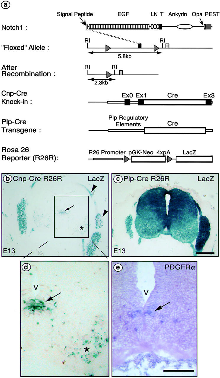
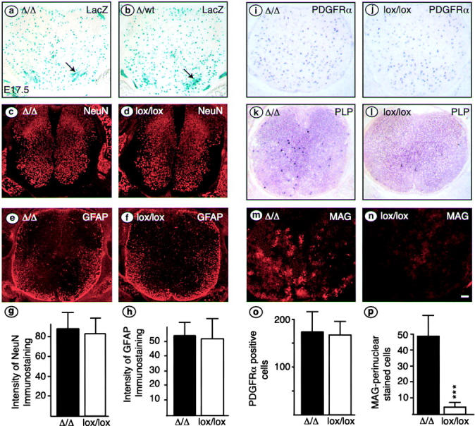
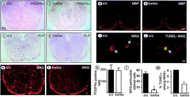
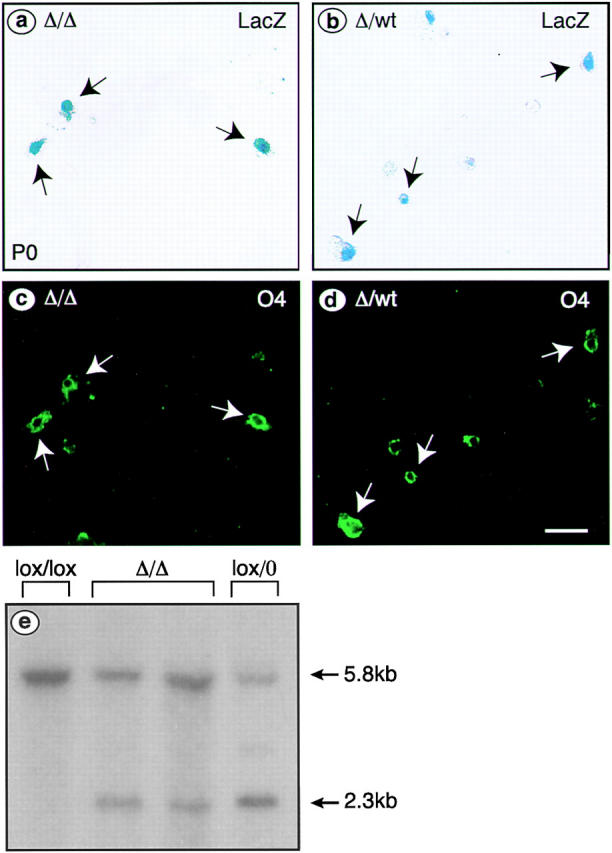
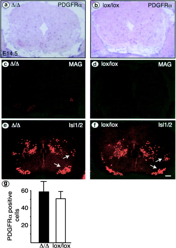
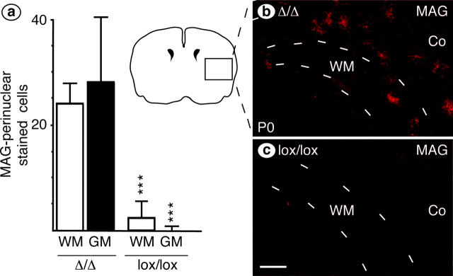
We have selectively inhibited Notch1 signaling in oligodendrocyte precursors (OPCs) using the Cre/loxP system in transgenic mice to investigate the role of Notch1 in oligodendrocyte (OL) development and differentiation. Early development of OPCs appeared normal in the spinal cord. However, at embryonic day 17.5, premature OL differentiation was observed and ectopic immature OLs were present in the gray matter. At birth, OL apoptosis was strongly increased in Notch1 mutant animals. Premature OL differentiation was also observed in the cerebrum, indicating that Notch1 is required for the correct spatial and temporal regulation of OL differentiation in various regions of the central nervous system. These findings establish a widespread function of Notch1 in the late steps of mammalian OPC development in vivo.


Figures

Figure 1.
Figure 1.
Experimental strategy and Cre-mediated recombination in OPCs at E13. (a) Schematic representation of the murine Notch1 protein and LoxP/Cre-mediated deletion strategy. The Notch protein contains 2,531 amino acid residues that encompass a signal peptide, 36 EGF repeats (EGF), 3 Lin/Notch domains (LN), a transmembrane domain (T), cytoplasmic ankyrin repeats, a polyglutamine stretch (Opa), and a PEST sequence. In the floxed Notch1 allele (Radtke et al., 1999), the first coding exon is flanked by LoxP sequences (gray triangles). After Cre-mediated recombination, a null allele is generated. Arrows indicate EcoRI fragments that differ in size between the floxed locus compared with the locus after deletion of the 3.5-kb segment flanked by loxP sites. The Cre recombinase gene was inserted into the Cnp locus (Cnp-Cre) or is driven from Plp/DM20 regulatory elements (Spassky et al., 1998) (Plp-Cre). The ROSA26 reporter mouse (Soriano, 1999) (R26R) was used to follow recombination events: Cre-mediated recombination deletes the neomycin phosphotransferase gene (PGK-neo) plus four polyadenylation sites (4xpA), activating the production of β-gal encoded by LacZ. RI indicates EcoRI restriction sites. (b–d) Cre-induced β-gal expression patterns. Cnp-Cre or Plp-cre mice were crossed with R26R reporter mice. Cells in which Cre had been expressed were detected by X-gal staining for β-galactosidase activity in sections of E13 embryos. The Cnp-Cre line shows expression in the ventral ventricular zone (b, arrow) and motoneurons (b, asterisk); these regions are enlarged in panel d. The area indicated by an arrow (b and d) is the site of origin of the OPCs consistent with the expected endogenous CNP expression pattern and confirmed by PDGFR-α in situ hybridization (e). (b) Arrowheads indicate the nerve roots and dorsal root ganglia that are part of the PNS. Expression in the Plp-Cre line used is much broader, affecting most cells of the spinal cord (c). V, ventricle. Bars: 10 μm.
Figure 2.
Figure 2.
Precocious differentiation of immature OLs in E17.5 spinal cord of Cnp-CreΔ/Δ mice. (a and b) Sections were cut from triple transgenic animals carrying Cnp-Cre, R26R, and one (b) or two copies (a) of the floxed Notch1 allele. X-gal stainings indicate recombination with a similar pattern in homozygous mutant (a, Δ/Δ) or heterozygous mutant (b, Δ/wt) spinal cords; motoneurons are indicated by arrows. Neurons, astrocytes, OPCs, and immature OLs were compared in Cnp-Cre Δ/Δ and control (lox/lox) spinal cords by immunohistochemistry (c–f, m, and n) or in situ hybridization (i–l). Neurons, astrocytes, and OPCs, assessed by the markers NeuN (c and d), GFAP (e and f), and PDGFR-α (i and j), respectively, appear normal in conditional mutant animals (Δ/Δ) compared with control (lox/lox) littermates. Total image intensity was determined for 10 sections of each genotype. For NeuN (g), we integrated only in the region of the gray matter, and for GFAP (h) intensity was averaged over the whole spinal cord section. Intensities are in arbitrary units. The number of cells positive for PDGFR-α mRNA was similar in mutant and control animals (o). By contrast, the number of immature OLs, assessed by counting cells positive for perinuclear MAG immunoreactivity (m and n) is significantly increased in mutant compared with control spinal cord (p, mean ± SD; ***, P < 0.001, t test). The precocious differentiation was confirmed by in situ hybridization for Plp/DM20 (k and l). Bar: (a–f and i–n) 10 μm.
Figure 3.
Figure 3.
OPCs and immature OLs in P0 Cnp-CreΔ/Δ spinal cord. Sections of thoracic spinal cord of Cnp-Cre Δ/Δ or control (lox/lox) mice were labeled with PDGFR-α cRNA or PLP cRNA (a–d), with anti-MAG (e, f, and i) or anti-MBP (g and h) antibodies, or with MAG antibodies combined with TUNEL staining (j). The number and location of PDGFR-α–positive OPCs was similar in mutant and control animals (a, b, and k). Also, the number and location of mature MBP-positive OLs in the developing white matter of the cervical spinal cord was not altered (g and h). In contrast, in the gray matter, there was a significant increase in ectopic immature OLs with extensive perinuclear MAG staining (i, quantified in panel l, mean ± SD; ***, P < 0.001). A subpopulation of these cells were TUNEL-positive (j, arrow). The percentage of apoptotic cells among perinuclear MAG-positive cells was significantly higher in mutant than in control spinal cord (m, 1,077 cells counted in mutant mice and 303 cells in control mice; [χ2 distribution] χ2= 86.29; **, P < 0.005). Bars: (a–h) 100 μm; (i and j) 10 μm.
Figure 4.
Figure 4.
Recombination in cultured OL and efficiency of ablation of Notch1 in P0 Cnp-CreΔ/Δ spinal cord. Acutely dissociated cells isolated from individual P0 Cnp-Cre R26R mice homozygous for the floxed Notch1 allele (a and c, Δ/Δ) or from Cnp-Cre R26R heterozygous littermates (b and d, Δ/wt) were stained for X-gal (a and b) and O4 (c and d). Arrows mark double-labeled cells. (e) Southern blot analysis of DNA isolated from P0 spinal cord using a probe derived from the 5′ upstream region of the Notch1 locus revealed a 5.8-kb fragment from the wild-type allele after digestion with EcoRI. After recombination, a 2.3-kb fragment can be detected. Genotypes of the animals used are indicated above the lanes.
Figure 5.
Figure 5.
E14.5 spinal cord appears normal in Cnp-CreΔ/Δ mice. Transverse sections of E14.5 Cnp-Cre Δ/Δ and control (lox/lox) spinal cords following in situ hybridization (a and b) or immunohistochemistry (c–f). OPCs, OLs, and motoneurons were analyzed by the expression of PDGFR-α mRNA (a and b), MAG (c and d), and Isl1/2, respectively. Comparable numbers of OPCs (g) and no MAG- positive cells were observed. Motoneurons (e and f, arrows) appear normal; some interneurons and dorsal root ganglia are also marked by Isl1/2. Bar: 10 μm.
Figure 6.
Figure 6.
Precocious development of immature OLs in the cerebrum of P0 Cnp-CreΔ/Δ mice. Transverse sections of P0 cerebrum were immunostained for MAG. (b) At P0, there were many immature (perinuclear MAG-positive) OLs present in the mutant brain (Δ/Δ), both in white matter (WM) and in gray matter (GM, here cortex, Co), whereas these were rare in the control (lox/lox) brain (c). These results are quantitated in panel a, which shows the mean number of cells (±SD) with perinuclear MAG staining. Bar: 20 μm.

References

    1. Apelqvist, A., H. Li, L. Sommer, P. Beatus, D.J. Anderson, T. Honjo, M. Hrabe de Angelis, U. Lendahl, and H. Edlund. 1999. Notch signalling controls pancreatic cell differentiation. Nature. 400:877–881. - PubMed
    1. Armstrong, R.C., H.H. Dorn, C.V. Kufta, E. Friedman, and M.E. Dubois-Dalcq. 1992. Pre-oligodendrocytes from adult human CNS. J. Neurosci. 12:1538–1547. - PMC - PubMed
    1. Artavanis-Tsakonas, S., K. Matsuno, and M.E. Fortini. 1995. Notch signaling. Science. 268:225–232. - PubMed
    1. Artavanis-Tsakonas, S., M.D. Rand, and R.J. Lake. 1999. Notch signaling: cell fate control and signal integration in development. Science. 284:770–776. - PubMed
    1. Barres, B.A., and M.C. Raff. 1993. Proliferation of oligodendrocyte precursor cells depends on electrical activity in axons. Nature. 361:258–260. - PubMed

Publication types