. 2002 Sep;76(18):9207-17.
doi: 10.1128/jvi.76.18.9207-9217.2002.

Subversion of cell signaling pathways by hepatitis C virus nonstructural 5A protein via interaction with Grb2 and P85 phosphatidylinositol 3-kinase

Affiliations

Subversion of cell signaling pathways by hepatitis C virus nonstructural 5A protein via interaction with Grb2 and P85 phosphatidylinositol 3-kinase

Yupeng He et al. J Virol. 2002 Sep.

Abstract

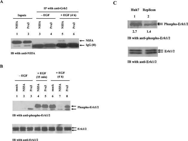
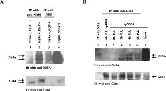
Hepatitis C virus (HCV) sets up a persistent infection in patients that likely involves a complex virus-host interaction. We previously found that the HCV nonstructural 5A (NS5A) protein interacts with growth factor receptor-binding protein 2 (Grb2) adaptor protein and inhibits the activation of extracellular signal-regulated kinases 1 and 2 (ERK1/2) by epidermal growth factor (EGF). In the present study, we extended this analysis and investigated the specificity of the Grb2-NS5A interaction and whether the subversion of mitogenic signaling involves additional pathways. NS5A containing mutations within the C-terminal proline-rich motif neither bound Grb2 nor inhibited ERK1/2 activation by EGF, demonstrating that NS5A-Grb2 binding and downstream effects were due to direct interactions. Interestingly, NS5A could also form a complex with the Grb2-associated binder 1 (Gab1) protein in an EGF treatment-dependent manner. However, the NS5A-Gab1 association, which appeared indirect, was not mediated by direct NS5A-Grb2 interaction but was likely dependent on direct NS5A interaction with the p85 subunit of phosphatidylinositol 3-kinase (PI3K). The in vivo association of NS5A with p85 PI3K required the N-terminal, but not the C-terminal, region of NS5A. The downstream effects of the NS5A-p85 PI3K interaction included increased tyrosine phosphorylation of p85 PI3K in response to EGF. Consistent with this observation and the antiapoptotic properties of NS5A, we also detected enhanced tyrosine phosphorylation of the downstream AKT protein kinase and increased serine phosphorylation of BAD, a proapoptotic factor and an AKT substrate, in the presence of NS5A. These results collectively suggest a model in which NS5A interacts with Grb2 to inhibit mitogenic signaling while simultaneously promoting the PI3K-AKT cell survival pathway by interaction with p85 PI3K, which may represent a crucial step in HCV persistence and pathogenesis.


Figures

FIG. 1.
FIG. 1.
The C-terminal proline-rich, SH3 domain-binding motif of NS5A is required for NS5A-Grb2 interaction and inhibition of ERK1/2 MAPK activation. (A) Tet-Off HeLa cells were transiently transfected with either pTRE-NS5A-wild type (NS5A) or pTRE-NS5A-Pro3 (Pro3). At 24 h posttransfection, the cells were either treated with EGF (lanes 5 and 6) or left untreated (lanes 3 and 4). At 4 h post-EGF treatment, cell lysates were collected and coimmunoprecipitation assays were performed with anti-Grb2 antibody. The immunocomplexes were resolved by SDS-12% PAGE and transferred to a nitrocellulose membrane. IgG (H) denotes the heavy chain of mouse immunoglobulin G. Lanes 1 and 2 show total lysates from cells transfected with either pTRE-NS5A-wild type (NS5A) or pTRE-NS5A-Pro3 (Pro3), respectively, probed with anti-NS5A antibody to show NS5A expression levels. (B) Cell lysates from the experiment described above were resolved by SDS-PAGE and subjected to immunoblot (IB) analysis with antibody specific to the dually phosphorylated, activated forms of Erk1/2 (top). The same blot was stripped and probed with anti-Erk1/2 antibody to show total Erk1/2 protein levels (bottom). (C) Equal amounts (30 μg) of protein from actively proliferating (∼70% confluent) Huh7 and HCV replicon cells were resolved by SDS-PAGE, transferred to nitrocellulose membrane, and probed with antibody specific to phosphorylated Erk1/2 MAPKs (top). The same membrane was stripped and reprobed with antibody specific to Erk1/2 MAPKs (bottom). The values under the antiphospho-Erk1/2 blot are the relative levels of Erk1/2 phosphorylation. IP, immunoprecipitation.
FIG. 2.
FIG. 2.
NS5A associates with the Gab1 signaling complex. (A) Lysates from wild-type NS5A-expressing Tet-Off HeLa cells treated with EGF (lanes 2 and 3) or left untreated (lane 1) were immunoprecipitated with either anti-Gab1 antibody (lanes 1 and 2) or a normal rabbit serum (NRS) control (lane 3). The immunocomplexes were resolved by SDS-12% PAGE, transferred to a nitrocellulose membrane, and subjected to immunoblot (IB) analysis with anti-NS5A antibody (top). Lane 4 shows NS5A expression in total cell lysates. The same membrane was stripped and probed with anti-Gab1 antibody (bottom) to show Gab1 protein levels. (B) HeLa S3 cells were infected with either a recombinant VV carrying the NS5A gene (vpNS5A) or the control VV (vp1080). Cell lysates were collected at 2, 4, and 6 h postinfection (P.I.) and used for coimmunoprecipitation assays with either anti-Gab1 antibody (lanes 2 to 6) or NRS (lane 1); this was followed by immunoblot analysis as described above. Lane 7 shows expression of NS5A (top) and Gab1 (bottom) in total lysates from vpNS5A-infected cells at 6 h postinfection. IP, immunoprecipitation.
FIG. 3.
FIG. 3.
The NS5A-Gab1 association is indirect and independent of NS5A-Grb2 interaction. (A) Tet-Off HeLa cells stably expressing wild-type NS5A (lanes 1, 2, 4, and 5) or not expressing NS5A (lane 3) were either treated with EGF (lanes 1, 3, and 5) or left untreated (lanes 2 and 4), and cell lysates were collected at 4 h post-EGF treatment. GST pulldown assays were performed with either GST-Gab1 (lanes 1, 2, and 3) or a GST control (lanes 4 and 5). The pulldown products were resolved by SDS-12% PAGE and transferred to nitrocellulose membrane; this was followed by immunoblot (IB) analysis with NS5A-specific antibody. Lane 6 shows NS5A expression in total lysates from NS5A-expressing cells. (B) Recombinant NS5A protein purified from insect cells was used for GST pulldown assays with either GST-Gab1 (lanes 3 to 6) or GST-Grb2 (lane 2) as described above. Lysates from actively proliferating HeLa S3 cells were added in increasing amounts (10, 30, and 50 μg) to reconstitute the NS5A-Gab1 association (lanes 4 to 6). Lane 1 shows purified NS5A protein immunoblotted with NS5A-specific antibody. (C) Recombinant NS5A protein purified from insect cells was used for GST pulldown assays with either GST-Gab1 (lanes 3 and 4) or GST-Grb2 (lane 2) as described above. Recombinant Grb2 protein purified from Escherichia coli was added to the reaction mixture in lane 4. Lane 1 shows purified NS5A protein immunoblotted with NS5A-specific antibody. (D) Tet-Off HeLa cells were transiently transfected with the pTRE vector (vector lanes), pTRE-NS5A-wild type (NS5A lanes), or pTRE-NS5A-Pro3 (Pro3 lanes). At 24 h after transfection, the cells were either treated with EGF for an additional 4 h or left untreated. Cell lysates were then used for coimmunoprecipitation analysis with anti-Gab1 antibody or a normal rabbit serum (NRS) control. The immunocomplexes were then resolved by SDS-12% PAGE; this was followed by Western transfer and immunoblot analysis with anti-NS5A antibody. Lanes 1 to 3 show total lysates of cells transfected with pTRE, pTRE-NS5A-wild type, or pTRE-NS5A-Pro3 immunoblotted with anti-NS5A antibody. IP, immunoprecipitation.
FIG. 4.
FIG. 4.
NS5A interacts with the p85 subunit of PI3K. (A) Organization of the functional domains of p85 PI3K and PLC-γ. (B) Recombinant NS5A protein purified from insect cells was used for GST pulldown assays with GST-Grb2 (lane 2), GST-Gab1 (lane 3), GST-p85 PI3K (lane 4), GST-C-terminal SH2 domain of p85 (lane 5), GST-N-terminal SH2 domain of p85 (lane 6), or GST-PLC-γ1 (lane 7). The precipitates were resolved by SDS-12% PAGE; this was followed by Western transfer and immunoblot (IB) analysis with anti-NS5A antibody. Lane 1 shows purified NS5A protein immunoblotted with NS5A-specific antibody. The same blot was stripped and probed with anti-GST antibody to show GST fusion protein levels (bottom). The values to the left are molecular masses in kilodaltons. (C) Tet-Off HeLa cells either expressing (lanes 3 to 6) or not expressing wild-type NS5A (lanes 1 and 2) were either treated with EGF (lanes 1 to 3, 5, and 6) or left untreated (lane 4), and cell lysates were collected at different time points post-EGF treatment as indicated. Coimmunoprecipitation assays were performed with p85 PI3K-specific antibody (lanes 1, 2, and 4 to 6) or a normal rabbit serum (NRS) control (lane 3); this was followed by SDS-12% PAGE and immunoblot analysis with NS5A-specific antibody (top). The same blot was stripped and probed with anti-p85 antibody to show p85 protein levels (bottom). (D) HeLa S3 cells were infected with either vpNS5A (lanes 3 to 7) or vp1080 (lanes 1 and 2). Cell lysates were collected at different time points postinfection (P.I.), as indicated, and used for coimmunoprecipitation assays with p85-specific antibody (lanes 1 to 6) or an NRS control (lane 7); this was followed by SDS-12% PAGE and immunoblot analysis with anti-NS5A antibody (top). The same blot was stripped and probed with anti-p85 antibody (bottom). Lane 8 shows NS5A (top) and p85 PI3K (bottom) expression in total lysates from vpNS5A-infected cells. (E and F) HCV replicon cells were treated with EGF for 1 h (lanes 1, 2, and 4) or left untreated (lane 3), and cell lysates were subjected to coimmunoprecipitation analysis with p85 PI3K-specific antibody (lanes 3 and 4) or an NRS control (lane 2). The immunocomplexes were resolved by SDS-12% PAGE; this was followed by immunoblot analysis with anti-NS5A (E) or anti-NS4 (F) antibody. Lane 1 shows NS5A expression in total lysates of HCV replicon cells. IP, immunoprecipitation.
FIG. 5.
FIG. 5.
The NS5A-Gab1 association requires NS5A-p85 PI3K interaction. Tet-Off HeLa cells were transiently transfected with pTRE-NS5A-NR (full length) (lane 1), pTRE-NS5A-ΔN110 (lane 2), or pTRE-NS5A-ΔC117 (lane 3), and at 24 h posttransfection, the cells were treated with EGF for 1 h. Cell lysates were then collected and used for coimmunoprecipitation analysis with either anti-p85 PI3K antibody (A) or anti-Gab1 antibody (B); this was followed by immunoblot (IB) analysis with human serum from HCV patients to detect the presence of NS5A proteins. IgG(H) denotes the heavy chain of human immunoglobulin G. wt, wild type; IP, immunoprecipitation.
FIG. 6.
FIG. 6.
NS5A enhances activation of the PI3K-AKT pathway and modulates the downstream apoptosis pathway. (A) Tet-Off HeLa cells stably expressing wild-type NS5A (lanes 1 to 4) or not expressing NS5A (lanes 5 to 8) were either treated with EGF (lanes 2 to 4 and 6 to 8) or left untreated (lanes 1 and 5). Cell lysates were collected at different time points post-EGF treatment, as indicated. Coimmunoprecipitation assays were performed with antibody specific to p85 PI3K. The immunocomplexes were resolved by SDS-12%c PAGE; this was followed by immunoblot (IB) analysis with phosphotyrosine-specific antibody (top). The same membrane was stripped and probed with p85-specific antibody to show total p85 protein levels (bottom). The values under the antiphosphotyrosine blot are the relative levels of p85 PI3K phosphorylation. (B) Tet-Off HeLa cells stably expressing wild-type NS5A (lanes 1 and 2) or not expressing NS5A (lanes 3 and 4) were either treated with EGF (lanes 2 to 4) or left untreated (lanes 1 and 3), and cell lysates were collected at 30 min after EGF treatment. The cell lysates were resolved by SDS-12% PAGE; this was followed by immunoblot analysis with antibody specific to the phosphorylated, activated form of AKT (top). The same blot was stripped and probed with anti-AKT antibody to show AKT levels (bottom). The values under the antiphospho-AKT blot are the relative levels of AKT phosphorylation. (C) HeLa S3 cells were infected with either a recombinant VV carrying the NS5A-encoding gene (vpNS5A) (lanes 4 to 6) or the control VV (vp1080) (lanes 1 to 3). Cell lysates were collected at 2, 4, and 6 h postinfection (p.i.) and resolved by SDS-12% PAGE (12%); this was followed by immunoblot analysis with antibody specific to the phosphorylated form of AKT (top). The same blot was stripped and probed with anti-AKT antibody to show total AKT protein levels (bottom). The values under the anti-phospho-AKT blot are the relative levels of AKT phosphorylation. (D) Stable Tet-Off HeLa cell lines carrying either full-length NS5A-NR (lanes 1 to 4) or N-terminal deletion mutant NS5A (lanes 5 to 8) were either induced to express NS5A (lanes 3, 4, 7, and 8) or not induced (lanes 1, 2, 5, and 6). At 24 h after induction, the cells were either treated with EGF for 30 min (lanes 2, 4, 6, and 8) or left untreated (lanes 1, 3, 5, and 7). Cell lysates were collected and subjected to immunoblot analysis with anti-phospho-AKT antibody (top). The same blot was stripped and probed with anti-AKT antibody to show AKT levels (bottom). The values under the antiphospho-AKT blot are the relative levels of AKT phosphorylation. (E) Tet-Off HeLa cells stably expressing wild-type NS5A (lanes 3 and 4) or not expressing NS5A (lanes 1 and 2) were either treated with EGF (lanes 2 to 4) or left untreated (lanes 1 and 3). Cell lysates were collected 30 min after EGF treatment and resolved by SDS-12% PAGE; this was followed by immunoblot analysis with antibody specific to the phosphorylated form of BAD (top). The same blot was stripped and reprobed with anti-BAD antibody to show BAD levels (bottom). The values under the antiphospho-BAD blot are the relative levels of BAD phosphorylation. (F) HeLa S3 cells were infected with either a recombinant VV carrying the NS5A-encoding gene (vpNS5A) (lanes 4 to 6) or the control VV (vp1080) (lanes 1 to 3). Cell lysates were collected at 2, 4, and 6 h postinfection and resolved by SDS-12% PAGE (12%); this was followed by immunoblot analysis with antibody specific to the phosphorylated form of BAD (top). The same blot was stripped and probed with anti-BAD antibody to show total BAD protein levels (bottom). The values under the antiphospho-BAD blot are the relative levels of BAD phosphorylation. IP, immunoprecipitation; ND, not detectable.
FIG. 7.
FIG. 7.
Model of the modulation of cellular signaling pathways by NS5A. NS5A, through its C-terminal proline-rich SH3-binding motif, directly interacts with Grb2 to perturb the downstream MAPK pathway, which may contribute to regulation of translation (25) and perturbation of IFN signaling (24). On the other hand, the N-terminal region of NS5A directly interacts with the p85 subunit of PI3K, which enhances the PI3K-AKT pathway and regulates the downstream apoptosis machinery. The modulation of the PI3K-AKT pathway by NS5A may contribute to cell survival and inhibition of apoptosis in virus-infected cells, resulting in viral persistence and contributing to pathogenesis.

References

    1. Aizaki, H., S. Saito, T. Ogino, N. Miyajima, T. Harada, Y. Matsuura, T. Miyamura, and M. Kohase. 2000. Suppression of interferon-induced antiviral activity in cells expressing hepatitis C virus proteins. J. Interferon Cytokine Res. 20:1111-1120. - PubMed
    1. Blight, K. J., A. A. Kolykhalov, and C. M. Rice. 2000. Efficient initiation of HCV RNA replication in cell culture. Science 290:1972-1974. - PubMed
    1. Borgatti, P., G. Zauli, M. L. Colamussi, D. Gibellini, M. Previati, L. L. Cantley, and S. Capitani. 1997. Extracellular HIV-1 Tat protein activates phosphatidylinositol 3- and Akt/PKB kinases in CD4+ T lymphoblastoid Jurkat cells. Eur. J. Immunol. 27:2805-2811. - PubMed
    1. Brown, J. P., D. R. Twardzik, H. Marquardt, and G. J. Todaro. 1985. Vaccinia virus encodes a polypeptide homologous to epidermal growth factor and transforming growth factor. Nature 313:491-492. - PubMed
    1. Cantrell, D. A. 2001. Phosphoinositide 3-kinase signalling pathways. J. Cell Sci. 114:1439-1445. - PubMed

Publication types

MeSH terms