. 2002 Sep;76(18):9284-97.
doi: 10.1128/jvi.76.18.9284-9297.2002.

Roles for the cytoplasmic tails of the fusion and hemagglutinin-neuraminidase proteins in budding of the paramyxovirus simian virus 5

Affiliations

Roles for the cytoplasmic tails of the fusion and hemagglutinin-neuraminidase proteins in budding of the paramyxovirus simian virus 5

David L Waning et al. J Virol. 2002 Sep.

Abstract

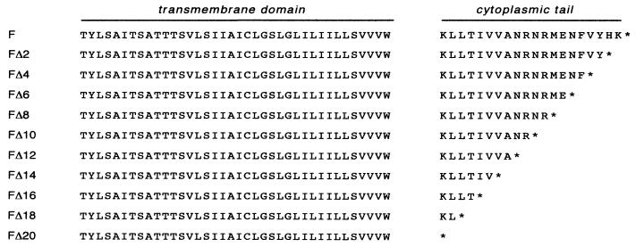
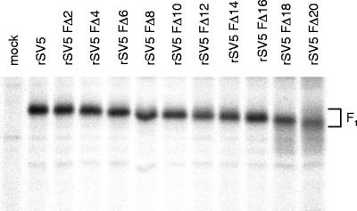
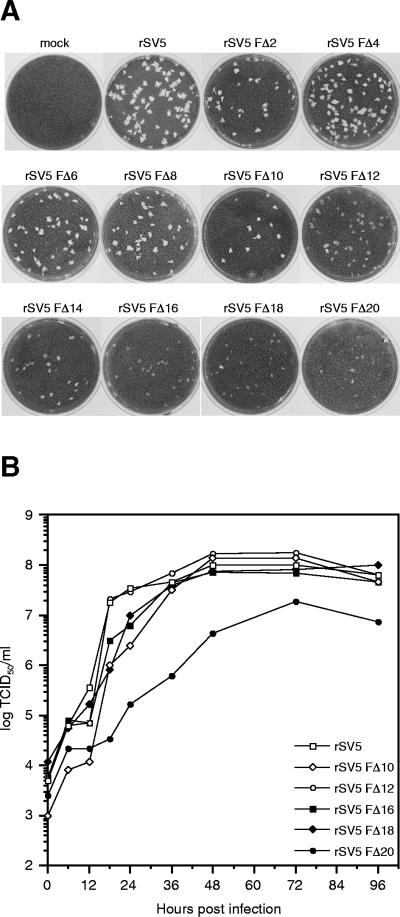
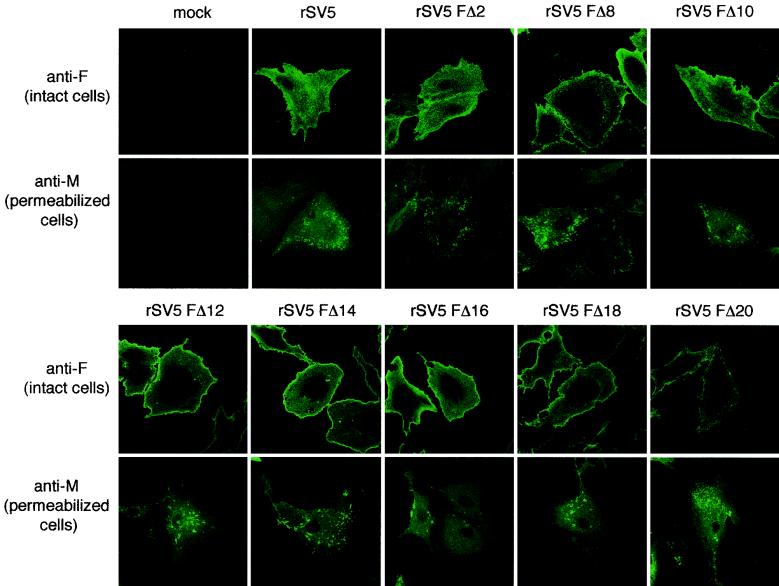
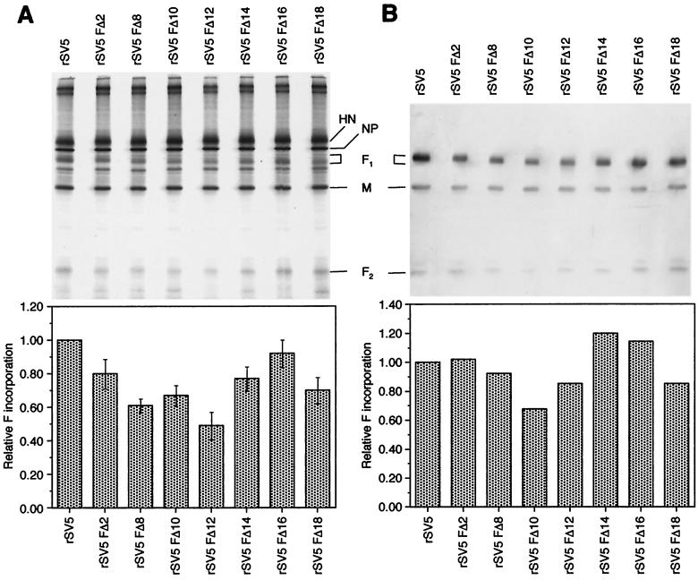
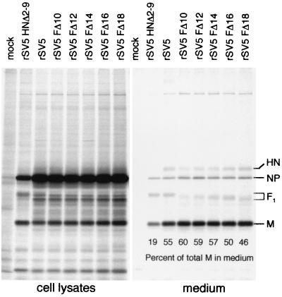
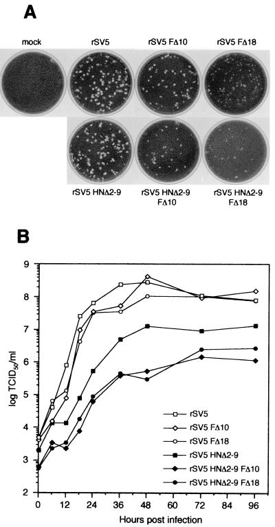
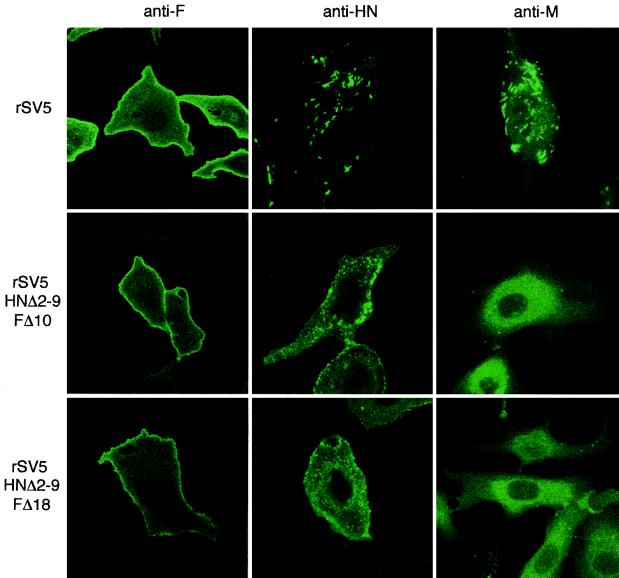
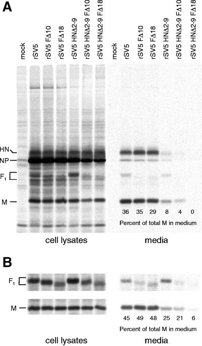
The efficient release of many enveloped viruses from cells involves the coalescence of viral components at sites of budding on the plasma membrane of infected cells. This coalescence is believed to require interactions between the cytoplasmic tails of surface glycoproteins and the matrix (M) protein. For the paramyxovirus simian virus 5 (SV5), the cytoplasmic tail of the hemagglutinin-neuraminidase (HN) protein has been shown previously to be important for normal virus budding. To investigate a role for the cytoplasmic tail of the fusion (F) protein in virus assembly and budding, we generated a series of F cytoplasmic tail-truncated recombinant viruses. Analysis of these viruses in tissue culture indicated that the cytoplasmic tail of the F protein was dispensable for normal virus replication and budding. To investigate further the requirements for assembly and budding of SV5, we generated two double-mutant recombinant viruses that lack 8 amino acids of the predicted 17-amino-acid HN protein cytoplasmic tail in combination with truncation of either 10 or 18 amino acids from the predicted 20-amino-acid F protein cytoplasmic tail. Both of the double mutant recombinant viruses displayed a replication defect in tissue culture and a budding defect, the extent of which was dependent on the length of the remaining F cytoplasmic tail. Taken together, this work and our earlier data on virus-like particle formation (A. P. Schmitt, G. P. Leser, D. L. Waning, and R. A. Lamb, J. Virol. 76:3953-3964, 2002) suggest a redundant role for the cytoplasmic tails of the HN and F proteins in virus assembly and budding.


Figures

FIG. 1.
FIG. 1.
Schematic diagram of the amino acid sequences of SV5 F proteins with truncated cytoplasmic tails. The cytoplasmic tail of the F protein is predicted to comprise the C-terminal 20 amino acids of the protein. The indicated nested set of F proteins containing progressive C-terminal deletions FΔ2 to FΔ20 was generated by site-directed mutagenesis of the F cDNA by using a four-primer PCR strategy.
FIG. 2.
FIG. 2.
Expression of SV5 F protein from cells infected with rSV5 harboring F protein cytoplasmic tail-truncations. CV-1 cells were infected with wt rSV5 and rSV5 FΔ2 to rSV5 FΔ20 and, at 18 h p.i., cells were radiolabeled with [35S]-Promix for 2 h. Cells were lysed in RIPA buffer, and F proteins were immunoprecipitated and analyzed by SDS-PAGE on a 10% gel as described in Materials and Methods. The F1 polypeptide chain of the F cytoplasmic tail-truncated proteins exhibits a progressively faster electrophoretic mobility consistent with successive removal of residues from the F protein cytoplasmic tail.
FIG. 3.
FIG. 3.
Plaque formation and growth kinetics of rSV5 harboring F protein cytoplasmic tail truncations. (A) Plaques formed by rSV5 harboring F protein cytoplasmic tail truncations after 5 days in BHK-21F cells. (B) Growth curves of rSV5 F cytoplasmic tail-truncated viruses. MDBK cells were infected with the indicated viruses at an MOI of 5.0 TCID50/cell, and the culture media was harvested at the indicted times p.i. Due to the fuzzy morphology of plaques formed by rSV5 FΔ12 to rSV5 FΔ20, all virus titers were determined by endpoint dilution (i.e., the TCID50) and calculated as described in Materials and Methods. Titers at each time point are the average of duplicate experiments.
FIG. 4.
FIG. 4.
Subcellular localization of F and M proteins in cells infected with rSV5 harboring F protein cytoplasmic tail truncations. CV-1 cells on glass coverslips were infected with the indicated viruses. At 16 h p.i., cells were fixed with formaldehyde (and for M protein staining, the cells were permeabilized with 0.1% saponin) and bound with MAbs specific for SV5 F (Fla) or M (M-h) and then with FITC-conjugated goat anti-mouse secondary antibody. Fluorescence was examined with a Zeiss LSM 410 confocal microscope.
FIG. 5.
FIG. 5.
Polypeptide composition of rSV5 harboring F protein cytoplasmic tail truncations. rSV5 and representative rSV5 F cytoplasmic tail-truncated viruses were grown in MDBK cells. For total viral polypeptide composition, cells were metabolically labeled with [35S]-Promix. Virions were purified through a 35% sucrose cushion. (A) Radiolabeled virions were fractionated by SDS-PAGE, and polypeptide radioactivity was quantified on a Fuji BioImager 1000. Incorporation of F protein into virions was calculated by quantification of the F1 band. The histogram indicates the average data from three independent experiments. (B) Nonradiolabeled virions were fractionated by SDS-PAGE, transferred to a polyvinylidene difluoride membrane, and probed with antibodies specific for F1, F2, and M, followed by the addition of alkaline phosphatase-conjugated secondary antibodies. Chemiluminescence was imaged on a STORM 860 system.
FIG. 6.
FIG. 6.
Analysis of budding of rSV5 harboring F cytoplasmic tail truncations. 293T cells were infected with wt rSV5, rSV5 HNΔ2-9 (which contains an HN cytoplasmic tail truncation), and selected examples (rSV5 FΔ10 to rSV5 FΔ20) of the F protein cytoplasmic tail-truncated viruses. At 24 h p.i., cells were radiolabeled with [35S]-Promix as described in Materials and Methods. Culture media and the cell lysates were harvested separately, and virions in the media were pelleted through a 35% sucrose cushion. Viral polypeptides (HN, F, NP, and M) were immunoprecipitated from both cell lysates and media as described in Materials and Methods, and polypeptides were analyzed by SDS-PAGE. The amount of M protein released into the medium was quantified and was used as a measure of viral budding competence. The percentage of total M protein in the medium was calculated from the total amount of M protein present in the cell lysate plus the medium.
FIG. 7.
FIG. 7.
Plaque formation and growth kinetics of rSV5 harboring HN and F protein double cytoplasmic tail truncations. (A) Plaques formed by the double mutants rSV5 HNΔ2-9 FΔ10 and rSV5 HNΔ2-9 FΔ18 and related single-mutant recombinant viruses after 5 days in BHK-21F cells. (B) Growth curves of rSV5 harboring HN and F double cytoplasmic tail truncations. MDBK cells were infected with the indicated viruses at an MOI of 5.0 TCID50/cell, and the culture media was harvested at the indicated times p.i. Virus titers were determined by endpoint dilution (i.e., the TCID50) and calculated as described in Materials and Methods. The titers at each time point are the average of duplicate experiments.
FIG. 8.
FIG. 8.
Localization of HN, F, and M in cells infected with rSV5 harboring HN and F double cytoplasmic tail truncations. CV-1 cells on glass coverslips were infected with rSV5, rSV5 HNΔ2-9 FΔ10, and rSV5 HNΔ2-9 FΔ18. At 16 h p.i., the surface localization of HN and F proteins was detected by binding HN and F specific MAbs (HN1b and F1a) to intact cells. Intracellular localization of M protein was detected by binding of M-h MAb to saponin-permeabilized cells. The cells were then stained with a FITC-conjugated goat anti-mouse secondary antibody. Fluorescence was detected by using a Zeiss LSM 410 confocal microscope.
FIG. 9.
FIG. 9.
Budding efficiency of rSV5 harboring cytoplasmic tail truncations in both the HN and F proteins. (A) 293T cells were infected with wt rSV5 or rSV5 harboring single or double cytoplasmic tail truncations to the HN and F proteins as indicated. At 24 h p.i., cells were radiolabeled for 16 h with [35S]-Promix, followed by collection of both cell lysates and culture media. Culture media was clarified by low-speed centrifugation, centrifuged through 35% sucrose cushions, and separated on sucrose flotation gradients. Proteins in the top 2.1 ml of the flotation gradient were analyzed by immunoprecipitation. Cells were disrupted by Dounce homogenization and centrifuged at a low speed to remove nuclei and debris. SV5 proteins were immunoprecipitated from the samples, and polypeptides were analyzed by SDS-PAGE on 10% gels as described in Materials and Methods. (B) Analysis of polypeptides in cell lysates and media in an experiment similar to that described in panel A except that the flotation gradient was not performed. Proteins in the media were analyzed directly after pelleting through a 35% sucrose cushion. In panels A and B, the budding efficiency was quantified as the percentage of M protein detected in the medium compared to the total M protein in the cell lysate plus the medium. In panel A, the time of phosphorimaging was longer for the medium samples than for the cell lysates, but quantitation of M protein was done on similarly exposed gels.
FIG. 10.
FIG. 10.
Morphology of rSV5 harboring cytoplasmic tail truncations in both the HN and F proteins. The virions wt rSV5 (A), rSV5 HNΔ2-9 (B), rSV5 FΔ18 (C), and rSV5 HNΔ2-9 FΔ18 (D) were grown in MDBK cells and purified on sucrose density gradients. Virions were adsorbed onto copper grids and negatively stained with phosphotungstic acid and examined by electron microscopy as described in Materials and Methods by using a JEOL JEM-100CX II electron microscope. Bar, 50 nm.

Similar articles

Cited by

References

    1. Ali, A., and D. P. Nayak. 2000. Assembly of Sendai virus: M protein interacts with F and HN proteins and with the cytoplasmic tail and transmembrane domain of F protein. Virology 276:289-303. - PubMed
    1. Bagai, S., and R. A. Lamb. 1996. Truncation of the COOH-terminal region of the paramyxovirus SV5 fusion protein leads to hemifusion but not complete fusion. J. Cell Biol. 135:73-84. - PMC - PubMed
    1. Bavari, S., C. M. Bosio, E. Wiegand, G. Ruthel, A. B. Will, T. W. Geisbert, M. Hevey, C. Schmaljohn, A. Schmaljohn, and M. J. Aman. 2002. Lipid raft microdomains: a gateway for compartmentalized trafficking of Ebola and Marburg viruses. J. Exp. Med. 195:593-602. - PMC - PubMed
    1. Buchholz, U. J., S. Finke, and K.-K. Conzelmann. 1999. Generation of bovine respiratory syncytial virus (BRSV) from cDNA: BRSV NS2 is not essential for virus replication in tissue culture, and the human RSV leader region acts as a functional BRSV genome promoter. J. Virol. 73:251-259. - PMC - PubMed
    1. Calain, P., and L. Roux. 1993. The rule of six, a basic feature for efficient replication of Sendai virus defective interfering RNA. J. Virol. 67:4822-4830. - PMC - PubMed

Publication types

LinkOut - more resources