. 2002 Aug 21;3(1):25.
doi: 10.1186/1471-2164-3-25.

In silico and in situ characterization of the zebrafish (Danio rerio) gnrh3 (sGnRH) gene

Affiliations

In silico and in situ characterization of the zebrafish (Danio rerio) gnrh3 (sGnRH) gene

Jacob Torgersen et al. BMC Genomics. .

Abstract

Background: Gonadotropin releasing hormone (GnRH) is responsible for stimulation of gonadotropic hormone (GtH) in the hypothalamus-pituitary-gonadal axis (HPG). The regulatory mechanisms responsible for brain specificity make the promoter attractive for in silico analysis and reporter gene studies in zebrafish (Danio rerio).

Results: We have characterized a zebrafish [Trp7, Leu8] or salmon (s) GnRH variant, gnrh3. The gene includes a 1.6 Kb upstream regulatory region and displays the conserved structure of 4 exons and 3 introns, as seen in other species. An in silico defined enhancer at -976 in the zebrafish promoter, containing adjacent binding sites for Oct-1, CREB and Sp1, was predicted in 2 mammalian and 5 teleost GnRH promoters. Reporter gene studies confirmed the importance of this enhancer for cell specific expression in zebrafish. Interestingly the promoter of human GnRH-I, known as mammalian GnRH (mGnRH), was shown capable of driving cell specific reporter gene expression in transgenic zebrafish.

Conclusions: The characterized zebrafish Gnrh3 decapeptide exhibits complete homology to the Atlantic salmon (Salmo salar) GnRH-III variant. In silico analysis of mammalian and teleost GnRH promoters revealed a conserved enhancer possessing binding sites for Oct-1, CREB and Sp1. Transgenic and transient reporter gene expression in zebrafish larvae, confirmed the importance of the in silico defined zebrafish enhancer at -976. The capability of the human GnRH-I promoter of directing cell specific reporter gene expression in zebrafish supports orthology between GnRH-I and GnRH-III.


Figures

Figure 1
Figure 1
GnRH decapeptide conservation Sequence comparison of mature GnRH peptides [13-15], show complete similarity between Atlantic salmon GnRH-III and zebrafish Gnrh3. The common name, species of origin nomenclature, scientific name, GnRH-I, -II or -III nomenclature and corresponding mature GnRH peptides are shown.
Figure 2
Figure 2
A) Schematic intron-exon diagram of the zebrafish gnrh3 gene The exons and introns are marked as boxes and lines, respectively. The intron lengths are shown underneath the lines. B) Exon lengths are shown above the mRNA sequence. The functional domains of the prepro polypeptide and untranslated regions (UTR) are marked. C) Post-translational cleavage of the signal peptide and GAP at the proteolytic cleavage site (GKR), generate the mature Gnrh3 decapeptide from the prepro polypeptide.
Figure 3
Figure 3
Overview of the PCR promoter capture method A) Sheared genomic DNA of 4–10 Kb harboring the gene of interest (thick line) is used as template in B) asymmetrical PCR reactions. The reactions are conducted with a biotinylated high Tm anti-sense primer and C) the resulting single stranded fragments are purified with streptavidin coated magnetic particles. D) A secondary PCR reaction is conducted with an oligo dT20 and a nested anti-sense primer.
Figure 4
Figure 4
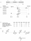
GnRH tissue specific enhancer and promoter constructs A) The in silico predicted zebrafish gnrh3 enhancer region located between -976 and -929, featuring binding sites for Oct-1, CREB and Sp1. Coordinates are negatively numbered with the experimental TSS as +1. B) Schematic overview of all zebrafish constructs tested, with nomenclature by sense primer used. The human RFP construct is not shown. The 91 bp deletion (-960 to -869) in the pΔ(A25)-GFP is denoted by an angle in the thin line. The gray and the white boxes correspond to the enhancer and exon 1, respectively. Arrows describe PCR primers, showing name and distance from the upstream end relative to TSS. Restriction sites at the bottom correspond to the tail of the PCR primers used for amplification of alternative promoter fragments. Transient expression data are indicated with +, - or (+), the latter denoting broader cell specificity. PCR primer sequences: A23 (5'aagcttggagtttgcatgttctccct3'), A24 (5'aagcttcctttcttaaaatattgaattatgat3'), A25 (5'ggatcccttcagggatgccaggtctt3'), A26 (5'ctcgaggctgtgtttgttcaagatgagttct3'), A33 (5'aagcttcagggagaacatgcaaact3') and A42 (5'aagcttggaatcagagaccttcttgct3'). C) The putative enhancer featuring binding sites for Oct-1, CREB and Sp1 is 100 % conserved with respect to presence of Oct-1 and CREB for the promoters tested [22,29,36-38]. The distance to TSS (+1), the transcription factor binding site and number of nucleotides between binding sites (space), are shown for each promoter. The location of the predicted enhancer in rat GnRH-I is according to data published [54]. D) Schematic overview of primer binding sites and the orientation of the respective primers. The upper line denotes genomic DNA and the lower mRNA, where both are displayed in 5'-3' direction. Exons are shown as gray boxes and primers as arrows.
Figure 5
Figure 5
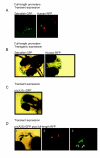
Reporter gene expression A) Transient GFP and RFP expression driven by the full-length zebrafish and human promoters. B) Zebrafish and human full-length -GFP and -RFP transgenics (F1) show reporter gene activity in the same cells. C) Retained cell specificity of pΔ(A33)-GFP. D) Expression patterns of pΔ(A25)-GFP. (1) Co-localized GFP and RFP activity (yellow) of pΔ(A25)-GFP and full length RFP. (2) Some of the co-injected larvae displayed broader cell specificity and lack of co-localization. (3) GFP activity in midbrain/hindbrain located neuronal cells (arrows).

References

    1. Sherwood N. Evolution of a neuropeptide family, Gonadotropin-releasing hormone. Amer Zool. 1986. pp. 1041–1054.
    1. Seeburg PH, Mason AJ, Stewart TA, Nikolics K. The mammalian GnRH gene and its pivotal role in reproduction. Recent Prog Horm Res. 1987;43:69–98. - PubMed
    1. King JA, Millar RP. Gonadotropin-Releasing hormones. In: Pang PKT, Schreibman MP, editor. Vertebrate Endocrinology: Fundamental and Biomedical Implications. Orlando, Academic Press; 1991. pp. 1–33.
    1. Amano M, Oka Y, Aida K, Okumoto N, Kawashima S, Hasegawa Y. Immunocytochemical demonstration of salmon GnRH and chicken GnRH-II in the brain of masu salmon, Oncorhynchus masou. J Comp Neurol. 1991;314:587–597. - PubMed
    1. Bailhache T, Arazam A, Klungland H, Alestrom P, Breton B, Jego P. Localization of salmon gonadotropin-releasing hormone mRNA and peptide in the brain of Atlantic salmon and rainbow trout. J Comp Neurol. 1994;347:444–454. - PubMed

LinkOut - more resources