. 2002 Sep;3(9):862-8.
doi: 10.1093/embo-reports/kvf174. Epub 2002 Aug 16.

Functional human CFTR produced by a stable minichromosome

Affiliations

Functional human CFTR produced by a stable minichromosome

Cristina Auriche et al. EMBO Rep. 2002 Sep.

Abstract

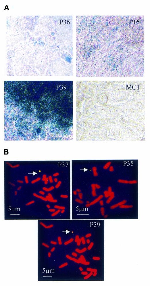
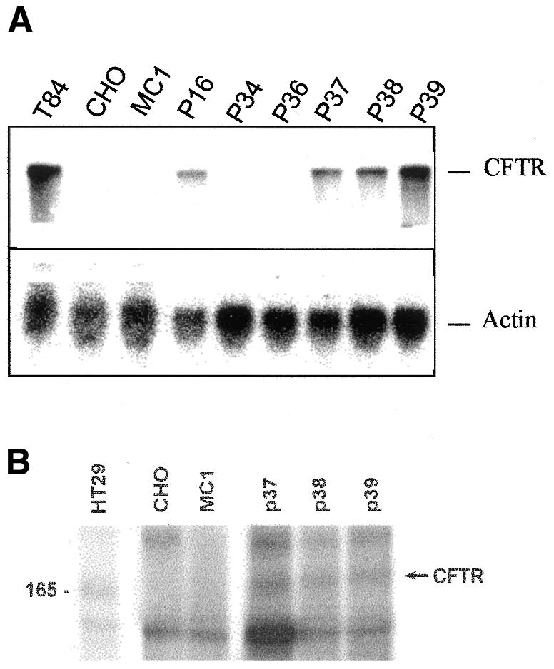
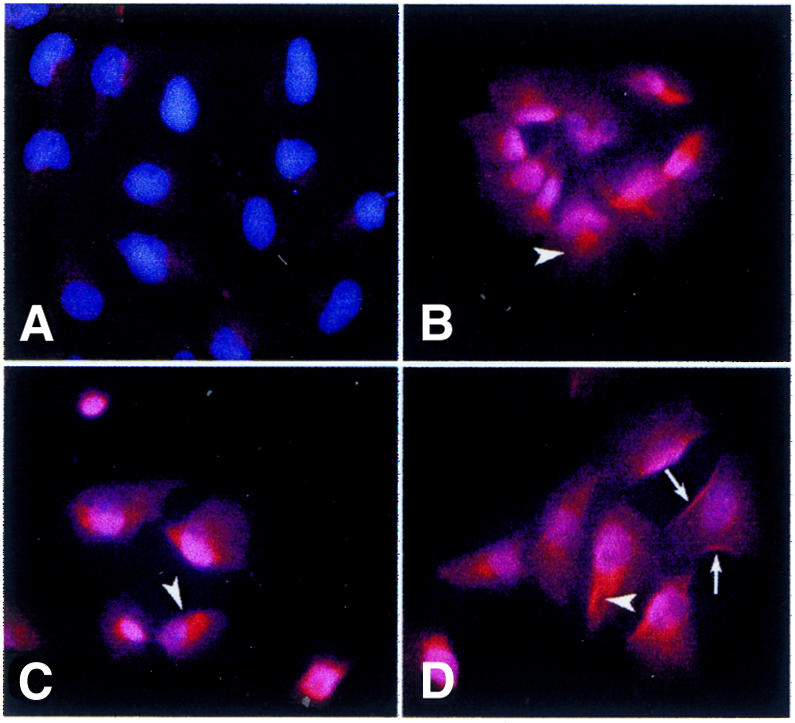
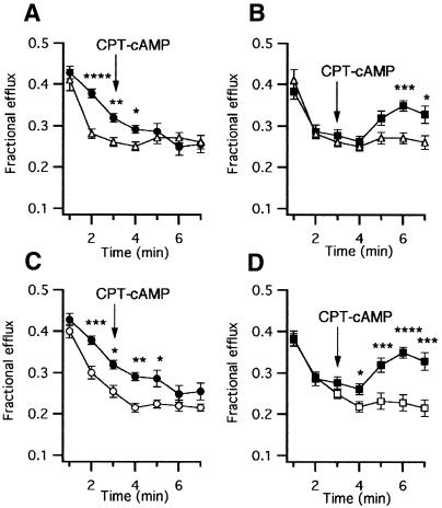
Artificial chromosomes have been claimed to be the ideal vector for gene therapy, but their use has been hampered by an inability to produce stable and well designed molecules. We have used a structurally defined minichromosome to clone the human cystic fybrosis transmembrane conductance regulator (CFTR) locus. To guarantee the presence of the proper regulatory elements, we used the 320 kb yeast artificial chromosome (YAC) 37AB12 with the intact CFTR gene and upstream sequences. The resulting minichromosome was analyzed for the presence of the entire CFTR gene and for its functional activity by molecular and functional methods. We have identified clones showing the presence of both the transcript and the CFTR protein. Moreover, in the same clones, a chloride secretory response to cAMP was detected. Mitotic and molecular stability after prolonged growth without selection demonstrated that the constructs were stable. This is the first example of a structurally known minichromosome made to contain an active therapeutic gene.


Figures

None
Fig.1. Analysis of MC1-CFTR clones. (A) β-galactosidase staining with X-gal for the indicated clones. MC1 (CHO-MC1 cells) represents the negative control. (B) FISH analysis of the clones indicated in each panel with a CFTR probe. Chromosomes were counterstained with propidium iodide and the probe was revealed with avidin-FITC. The arrow points to the minichromosome.
None
Fig. 2. Structural analysis of MC1-CFTR. (A) PCR products from the indicated CFTR exons (1, 2, 5, 7, 10, 13, 16, 20 and 23; see Methods for primers). For each exon MC1 (negative control), T84 (positive control) and P39 DNA samples are reported from left to right. (B) Logarithmic plot of the ratio T/S (CFTR band intensity/competitor band intensity) versus competitor DNA. The data refer to 15 µl aliquots of each PCR. Dashed lines represent P39 (squares) and T84 (triangles), respectively; the corresponding continuous lines represent the regression. (C) Mitotic stability of MC1-CFTR after growth with (+) and without (–) selection for the indicated number of generations. The majority of the cells contained one minichromosome per cell. (D) Alu-PCR products from MC1, P37, P38 and P39 after 60 generations of growth in the presence (+G418) and absence (–G418) of selection. MC1 represents CHO-MC1 cells.
None
Fig. 3. CFTR activity in MC1-CFTR clones. (A) Northern blot analysis of the indicated RNA samples using a CFTR probe (upper panel). RNA loading control using an actin probe (lower panel). (B) SDS–PAGE of cell lysates immunoprecipitated with MATG1105 and phosphorylated with [γ-32P]ATP. The arrow points to mature CFTR, and the molecular marker on the left side of the figure is indicated in kilodaltons. Samples: T84 (A) and HT29 (B) cells, positive controls; CHO and MC1 (CHO-MC1 cells), negative controls.
None
Fig. 4. CFTR subcellular localization in MC1-CFTR clones. (A) CHO-MC1; (BD) P37–P39. Cells were immunostained with MATG1031, revealed with a second antibody (Texas Red) and counterstained with Hoechst 33258. Arrowheads point to specific staining around nuclei in a position reminiscent of Golgi; arrows point to plasma membrane staining.
None
Fig. 5 36Cl efflux from cells stimulated with 500 µM CPT-cAMP. (A) P38 (n = 8) and MC1 (n = 20); (B) P39 (n = 10) and CHO-MC1 (n = 20); (C) P38 in the absence (n = 8) and presence (n = 4) of glibenclamide; (D) P39 in the absence (n = 10) and presence (n = 6) of glibenclamide. P38 in the absence (filled circles) and presence (open circles) of glibenclamide; P39 in the absence (filled squares) and presence (open squares) of glibenclamide; CHO-MC1 (triangles). Vertical bars are SEM. Asterisks indicate statistically significant differences (*P < 0.05, **P < 0.01, ***P < 0.005 and ****P < 0.001).

References

    1. Alton E. and Geddes, D. (1998) Cystic fibrosis clinical trials. Adv. Drug Deliv. Rev., 30, 205–217. - PubMed
    1. Auriche C., Donini, P. and Ascenzioni, F. (2001) Molecular and cytological analysis of a 5.5 Mb minichromosome. EMBO rep., 2, 102–107. - PMC - PubMed
    1. Barriere H., Poujeol, C., Tauc, M., Blasi, J.M., Counillon, L. and Poujeol, P. (2001) CFTR modulates programmed cell death by decreasing intracellular pH in Chinese hamster lung fibroblasts. Am. J. Physiol. Cell Physiol., 281, C810–C824. - PubMed
    1. Carine K., Solus, J., Waltzer, E., Manch-Citron, J., Hamkalo, B.A. and Scheffler, I.E. (1986) Chinese hamster cells with a minichromosome containing the centromeric region of human chromosome 1. Somat. Cell Mol. Genet., 12, 479–491. - PubMed
    1. Dechecchi M.C., Tamanini, A., Berton, G. and Cabrini, G. (1993) Protein kinase C activates chloride conductance in c127 cells stably expressing the cystic fibrosis gene. J. Biol. Chem., 268, 11321–11325. - PubMed

Publication types

Grants and funding