Comparative Study
. 2002 Sep 3;99(18):11700-5.
doi: 10.1073/pnas.182372299. Epub 2002 Aug 20.

The histone deacetylase inhibitor SAHA arrests cancer cell growth, up-regulates thioredoxin-binding protein-2, and down-regulates thioredoxin

Affiliations
Comparative Study

The histone deacetylase inhibitor SAHA arrests cancer cell growth, up-regulates thioredoxin-binding protein-2, and down-regulates thioredoxin

Lisa M Butler et al. Proc Natl Acad Sci U S A. .

Abstract

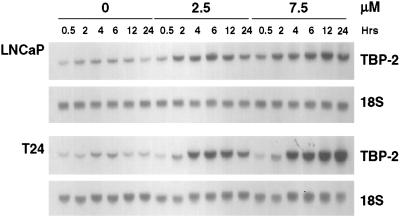
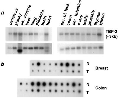
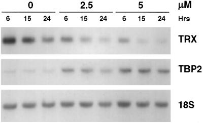
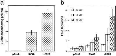
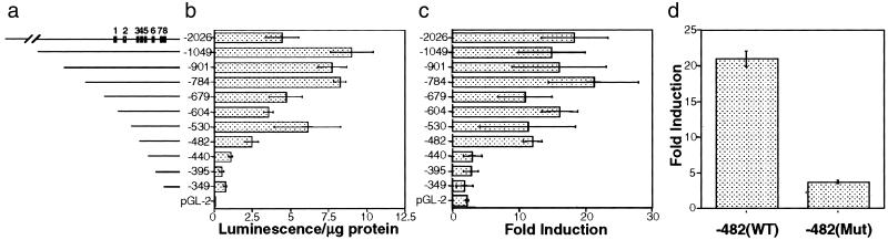
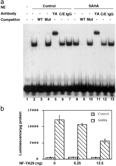
Suberoylanilide hydroxamic acid (SAHA) is a potent inhibitor of histone deacetylases (HDACs) that causes growth arrest, differentiation, and/or apoptosis of many tumor types in vitro and in vivo. SAHA is in clinical trials for the treatment of cancer. HDAC inhibitors induce the expression of less than 2% of genes in cultured cells. In this study we show that SAHA induces the expression of vitamin D-up-regulated protein 1/thioredoxin-binding protein-2 (TBP-2) in transformed cells. As the expression of TBP-2 mRNA is increased, the expression of a second gene, thioredoxin, is decreased. In transient transfection assays, HDAC inhibitors induce TBP-2 promoter constructs, and this induction requires an NF-Y binding site. We report here that TBP-2 expression is reduced in human primary breast and colon tumors compared with adjacent tissue. These results support a model in which the expression of a subset of genes (i.e., including TBP-2) is repressed in transformed cells, leading to a block in differentiation, and culture of transformed cells with SAHA causes re-expression of these genes, leading to induction of growth arrest, differentiation, and/or apoptosis.


Figures

Figure 1
Figure 1
Induction of TBP-2 mRNA levels in transformed cells by SAHA. LNCaP prostate carcinoma and T24 bladder carcinoma cells were cultured with vehicle alone (0) or SAHA (2.5 or 7.5 μM) for the indicated times. Total RNA was extracted from the cells, and the levels of TBP-2 were determined by Northern blotting using a 1.1-kb 32P-labeled TBP-2 cDNA probe (Upper for each cell line). Blots were rehybridized with a γ-32P-labeled 18S oligonucleotide probe to indicate RNA loading and are shown (Lower for each cell line). Similar results were obtained for a total of six transformed cell lines.
Figure 2
Figure 2
Expression of TBP-2 in normal and tumor tissues. (a) Multiple tissue Northern blots, containing poly(A)+ RNA from the indicated tissues (CLONTECH), were hybridized with a 1.1-kb 32P-labeled TBP-2 cDNA probe (Upper). The bolts were rehybridized with a 2.0-kb probe for β-actin, as a control for loading (Lower). sk. muscle, skeletal muscle; per. bl. leuk., peripheral blood leukocytes. (b) A dot blot containing matched samples of cDNA extracted from normal human tissues and tumors (CLONTECH) was hybridized with a 1.1-kb 32P-labeled TBP-2 cDNA probe. Samples of colon and breast tumors (T) are shown, with the cDNA from the normal tissue (N) shown directly above each corresponding tumor sample.
Figure 3
Figure 3
Expression of TRX in transformed cells cultured with SAHA. T24 bladder carcinoma cells were cultured with vehicle alone (0) or 2.5 or 5 μM SAHA for 6, 15, or 24 h. RNA was extracted and analyzed by Northern blotting for levels of TRX, using a 500-bp 32P-labeled cDNA probe (Top). The blots were subsequently rehybridized with the 1.1-kb 32P-labeled TBP-2 cDNA probe to confirm induction of TBP-2 (Middle) and a γ-32P-labeled 18S oligonucleotide probe to indicate RNA loading (Bottom).
Figure 4
Figure 4
The cloned TBP-2 promoter is functional and induced by SAHA. 293T cells were transfected with 100 ng of either an empty pGL2 vector, a pGL2-SV40 positive control vector, or the TBP-2 construct (−2026). (a) Firefly luminescence was measured 24 h after transfection. (b) The transfection medium was removed 12 h after transfection and replaced with medium containing DMSO alone or SAHA (0.5, 1, or 2 μM). Luminescence was measured 24 h later and normalized for total protein concentration of each sample. Fold induction is obtained by normalizing the luciferase value in the presence of SAHA against luciferase value in the absence of SAHA.
Figure 5
Figure 5
Deletion and mutation analyses of TBP-2 promoter. (a) Schematic representation of the putative TBP-2 promoter region and the deletion mutants. The positions of putative transcription factors binding sites in the promoter are shown: 1, NF-κB binding site; 2, vitamin D receptor/retinoid X receptor responsive element; 3, E2F binding site; 4, E box; 5, inverted CCAAT box; 6, CCAAT box; 7, E box; 8, TATA box. (b) Activity of deletion mutants. Different lengths of the 5′ flanking region of human TBP-2 gene were amplified by PCR and cloned upstream of the luciferase gene in the pGL-2 vector. The constructs were transfected into 293T cells and luciferase activity was measured. (c) SAHA induction of deletion mutants. Parallel experiments were carried out as in b except SAHA was added 12 h after transfection. Luciferase activity was normalized against total protein. (d) The inverted CCAAT box is critical for SAHA inducibility. PCR-based mutagenesis was performed to mutate the inverted CCAAT box in the isolated TBP-2 promoter. The mutant promoter was then cloned into pGL-2 and transfected into 293T cells with or without SAHA (2 μM) treatment. The TBP-2 promoter construct with the mutations in the inverted CCAAT box has about 8% of wild-type promoter activity (not shown). Fold induction was calculated as in b and results shown are the average of three independent transfections ± SD.
Figure 6
Figure 6
Role of NF-Y in induction of TBP-2. (a) Binding of NF-Y to the inverted CCAAT box in TBP-2 promoter. Electrophoretic mobility-shift assay (lanes 1–4 and 8–10) detects specific complex formation at the inverted CCAAT box. 32P-labeled wild-type probe (20,000 cpm, ≈0.5 ng; lane 1) was incubated with 10 μg nuclear extracts prepared from untreated (lanes 2–7) or 7.5 μM SAHA-treated (12 h) (lanes 8–13) T24 cells, in the absence (lanes 2 and 8) or presence of 25 ng (×50) wild-type (lanes 3 and 9) or mutant (lanes 4 and 10) oligonucleotide competitors. For supershift assays, nuclear extracts were incubated with 2 μg rabbit anti-NF-YA (lanes 5 and 11), 2 μg goat anti-C/EBP (lanes 6 and 12), or 2 μg normal rabbit IgG (lanes 7 and 13). WT, wild-type probe competitor; Mut, mutant probe competitor; YA, anti-NF-YA; C/E, anti-C/EBP. (b) Dominant negative NF-Y mutant (NF-YA29) decreases the promoter induction by SAHA. The pGL2-TBP-2 −2026 promoter construct (100 ng) was cotransfected with NF-YA29 expression vector as indicated, and then treated with or without SAHA (2 μM) for 24 h.

References

    1. Marks P A, Rifkind R A, Richon V M, Breslow R, Miller T, Kelly W K. Nat Rev Cancer. 2001;1:194–202. - PubMed
    1. Grunstein M. Nature (London) 1997;389:349–352. - PubMed
    1. Richon V M, Emiliani S, Verdin E, Webb Y, Breslow R, Rifkind R A, Marks P A. Proc Natl Acad Sci USA. 1998;95:3003–3007. - PMC - PubMed
    1. Richon V M, Webb Y, Merger R, Sheppard T, Jursic B, Ngo L, Civoli F, Breslow R, Rifkind R A, Marks P A. Proc Natl Acad Sci USA. 1996;93:5705–5708. - PMC - PubMed
    1. Coffey D C, Kutko M C, Glick R D, Swendeman S L, Butler L, Rifkind R, Marks P A, Richon V M, LaQuaglia M P. Med Pediatr Oncol. 2000;35:577–581. - PubMed

Publication types

Associated data

LinkOut - more resources