. 2002 Sep 3;99(18):11830-5.
doi: 10.1073/pnas.192203399. Epub 2002 Aug 20.

The Alzheimer's A beta -peptide is deposited at sites of complement activation in pathologic deposits associated with aging and age-related macular degeneration

Affiliations

The Alzheimer's A beta -peptide is deposited at sites of complement activation in pathologic deposits associated with aging and age-related macular degeneration

Lincoln V Johnson et al. Proc Natl Acad Sci U S A. .

Abstract

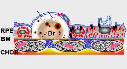
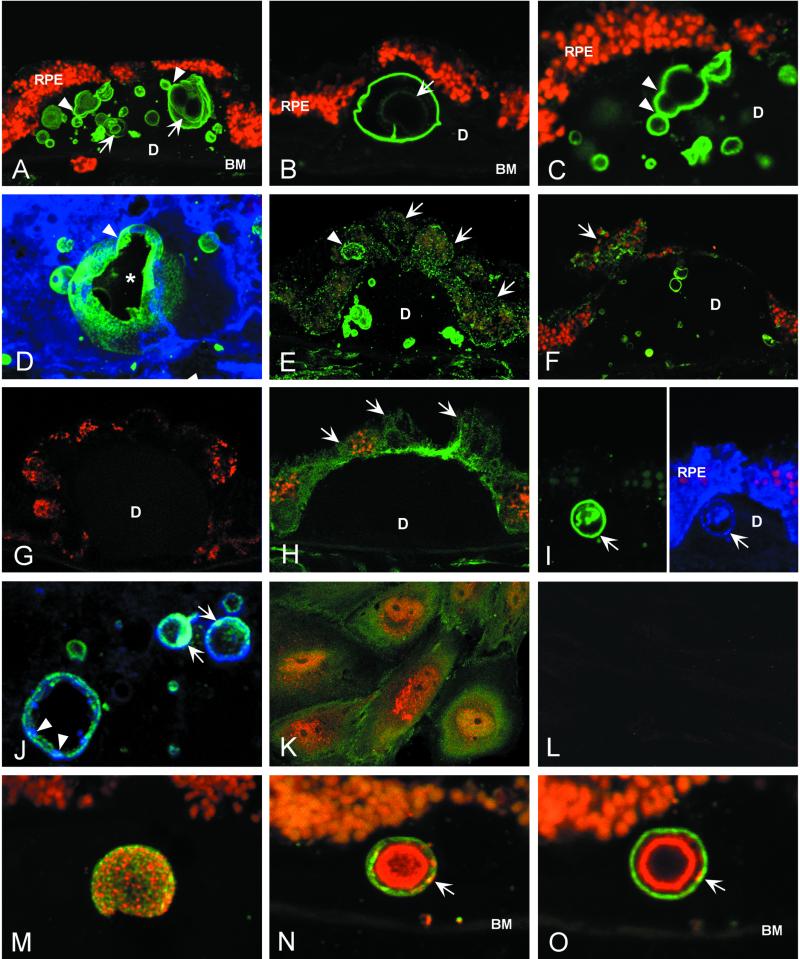
Age-related macular degeneration (AMD) is a leading cause of irreversible vision loss in older individuals worldwide. The disease is characterized by abnormal extracellular deposits, known as drusen, that accumulate along the basal surface of the retinal pigmented epithelium. Although drusen deposition is common in older individuals, large numbers of drusen and/or extensive areas of confluent drusen represent a significant risk factor for AMD. Widespread drusen deposition is associated with retinal pigmented epithelial cell dysfunction and degeneration of the photoreceptor cells of the neural retina. Recent studies have shown that drusen contain a variety of immunomodulatory molecules, suggesting that the process of drusen formation involves local inflammatory events, including activation of the complement cascade. Similar observations in Alzheimer's disease (AD) have lead to the hypothesis that chronic localized inflammation is an important element of AD pathogenesis, with significant neurodegenerative consequences. Accordingly, the amyloid beta (A beta) peptide, a major constituent of neuritic plaques in AD, has been implicated as a primary activator of complement in AD. Here we show that A beta is associated with a substructural vesicular component within drusen. A beta colocalizes with activated complement components in these "amyloid vesicles," thereby identifying them as potential primary sites of complement activation. Thus, A beta deposition could be an important component of the local inflammatory events that contribute to atrophy of the retinal pigmented epithelium, drusen biogenesis, and the pathogenesis of AMD.


Figures

Figure 1
Figure 1
Diagram depicting the anatomical relationships among the retinal pigmented epithelium (RPE), Bruch's membrane (BM), and drusen (Dr). Vesicles containing amyloid β that likely represent primary sites of complement activation are identified within drusen (arrows). CHOR, choroidal vasculature; CAP, capilllary.
Figure 2
Figure 2
Immunolocalization of APP and Aβ in drusen by laser scanning confocal immunofluorescence microscopy. (AC) Anti-Aβ (6E10) binds vesicular elements within drusen (green; Cy 2 channel) of an 84-year-old male with clinical diagnosis of atrophic AMD. These “amyloid vesicles” frequently exhibit a multilamellar structure (arrows in A and B). Aβ immunoreactivity is concentrated on the outer shell of the vesicle; varying levels of Aβ are associated with the inner lamellae. Images suggesting the fusion and/or budding of vesicles are often observed (arrowheads in A and C). Autofluorescent lipofuscin particles are concentrated in the RPE cytoplasm (red; Cy3 channel). (D) A serial reconstruction of 34 0.3-μm optical sections showing a large amyloid vesicle and several smaller vesicles labeled with anti-Aβ (6E10); sectioning has partially exposed the central cavity (asterisk). Evidence of fusion or budding is apparent near the top of the vesicle (arrowhead) and on the far interior wall. (E and F) Anti-Aβ labeling of RPE cells (E, 6E10; F, 4G8). Fine, granular Aβ immunoreactivity is observed in the cytoplasm of RPE cells overlying drusen (arrows). One RPE cell contains an Aβ-positive structure that is similar in size and shape to the amyloid vesicles within drusen (arrowhead in E). (G) Secondary antibody control illustrating the absence of binding to drusen or RPE cells by the Cy2-conjugated donkey anti-mouse Ig used for the detection of anti-Aβ and anti-APP monoclonal antibodies. Autofluorescent lipofuscin particles (red; Cy3 channel) mark the RPE cells. (H) Anti-APP (22C11) localizes APP to the RPE cell cytoplasm (arrows); however, drusen are not labeled. (I and J) Double-label images illustrating labeling patterns for APP (blue; Cy5 channel) and Aβ (green; Cy2 channel). (I Left) Anti-Aβ (6E10) strongly labels a single large amyloid vesicle (arrow); (Right) In the same section, anti-APP (N-terminal peptide 44–63 polyclonal antibody) binds the same vesicle (arrow), as well as the RPE cytoplasm. (J) In merged images of different amyloid vesicles, punctate regions of both APP (blue, arrowheads) and Aβ immunoreactivity (green) are apparent. Areas of light blue fluorescence are indicative of APP and Aβ colocalization (arrows). (K) Double-label image showing the distributions of Aβ (red; Cy3) and APP (green; Cy2) in cultured human RPE cells. APP shows diffuse cytoplasmic staining, whereas punctate Aβ immunoreactivity is concentrated in the perinuclear region. (L) Anti-Aβ labeling of cultured human RPE cell cytoplasm is abrogated by preadsorption of anti-Aβ antibody (4G8) with Aβ1→42 peptide. (MO) Double-label confocal immunofluorescence images showing the distributions of Aβ (green; Cy2) and iC3b, an activation-specific fragment of complement C3 (red; Cy3), in amyloid vesicles. The vesicular surface is punctated by discrete, as well as overlapping, areas of Aβ and/or iC3b immunoreactivity (M). A merged projection series (N) shows that most iC3b immunoreactivity is concentrated in the interior of the vesicles, whereas Aβ predominates in the outer shell. This is illustrated most dramatically in a single optical section (O) through the same vesicle shown in N. Note that discrete areas of colocalization (yellow) are also present on the outer shell (arrows in M and N). BM, Bruch's membrane; D, drusen.
Figure 3
Figure 3
PCR analyses of APP and β-secretase expression by human RPE cells relative to human brain and primary human fibroblasts. (A) Endpoint PCR analysis of APP isoform expression. Primary RPE cell cultures (lane B), as well as an SV40 transformed RPE cell line (lane C), express transcripts for the three major APP isoforms (APP770, APP751, and APP695). A similar pattern of expression is observed in primary fibroblast cultures (lane F). The APP695 isoform predominates in both the adult (lane D) and fetal (lane E) brain, as has been reported (25). A fourth unidentified amplicon (asterisk) was detected in all samples examined. Lane A is a 100-bp size reference ladder. (B) Quantitative PCR analyses of the expression of APP751, APP695, and β-secretase. APP751 transcripts are expressed at similar levels in primary RPE cell cultures (HFRPE), SV40 transformed RPE cells (SVRPE), fibroblasts, and in both adult and fetal brain. In contrast, the expression level of APP695 in primary RPE cells is 20-fold lower than in adult brain, and more than 40-fold lower than in fetal brain. Similar expression levels are exhibited by SVRPE cells and normal human fibroblasts. The β-secretase expression level in primary RPE cells is approximately one-half of the level in the brain samples. In comparison to the primary RPE cells, SVRPE cells and fibroblasts have somewhat lower and higher β-secretase expression levels respectively. (see also Table 1). (C) Gel analysis of PCR products from the quantitative PCR analyses demonstrating the amplification of appropriately sized products for β-secretase (121 bp), APP751 (378 bp), APP695 (125 bp), and 18s rRNA (67 bp). (Lane 1, adult brain; Lane 2, fetal brain; lane 3, primary RPE; lane 4, SV40 RPE; lane 5, fibroblasts).

Similar articles

Cited by

References

    1. Akiyama H, Barger S, Barnum S, Bradt B, Bauer J, Cole G M, Cooper N R, Eikelenboom P, Emmerling M, Fiebich B L, et al. Neurobiol Aging. 2000;21:383–421. - PMC - PubMed
    1. Bradt B M, Kolb W P, Cooper N R. J Exp Med. 1998;188:431–438. - PMC - PubMed
    1. Webster S, Lue L F, Brachova L, Tenner A J, McGeer P L, Terai K, Walker D G, Bradt B, Cooper N R, Rogers J. Neurobiol Aging. 1997;18:415–421. - PubMed
    1. Rogers J, Cooper N R, Webster S, Schultz J, McGeer P L, Styren S D, Civin W H, Brachova L, Bradt B, Ward P, et al. Proc Natl Acad Sci USA. 1992;89:10016–20. - PMC - PubMed
    1. McGeer P L, Akiyama H, Itagaki S, McGeer E G. Can J Neurol Sci. 1989;16:516–527. - PubMed

Publication types