. 2002 Aug;110(4):515-26.
doi: 10.1172/JCI13847.

Identification of ARTS-1 as a novel TNFR1-binding protein that promotes TNFR1 ectodomain shedding

Affiliations

Identification of ARTS-1 as a novel TNFR1-binding protein that promotes TNFR1 ectodomain shedding

Xinle Cui et al. J Clin Invest. 2002 Aug.

Abstract

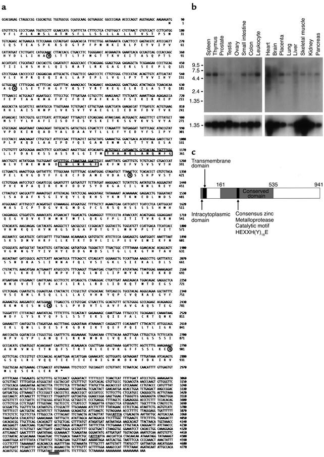
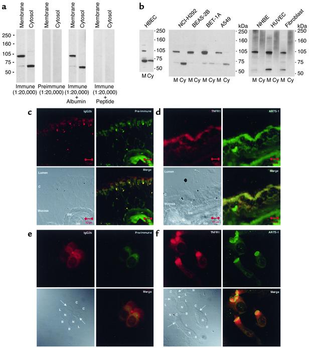
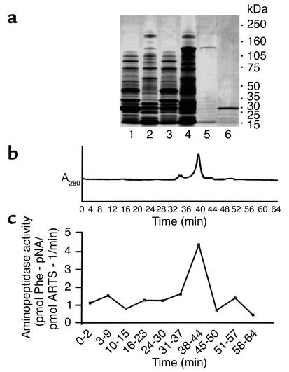
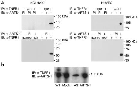
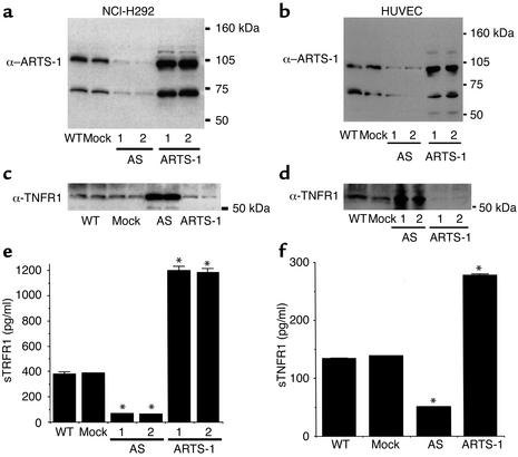
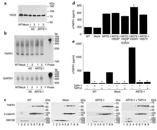
Proteolytic cleavage of TNF receptor 1 (TNFR1) generates soluble receptors that regulate TNF bioactivity. We hypothesized that the mechanism of TNFR1 shedding might involve interactions with regulatory ectoproteins. Using a yeast two-hybrid approach, we identified ARTS-1 (aminopeptidase regulator of TNFR1 shedding) as a type II integral membrane protein that binds to the TNFR1 extracellular domain. In vivo binding of membrane-associated ARTS-1 to TNFR1 was confirmed by coimmunoprecipitation experiments using human pulmonary epithelial and umbilical vein endothelial cells. A direct relationship exists between membrane-associated ARTS-1 protein levels and concordant changes in TNFR1 shedding. Cells overexpressing ARTS-1 demonstrated increased TNFR1 shedding and decreased membrane-associated TNFR1, while cells expressing antisense ARTS-1 mRNA demonstrated decreased membrane-associated ARTS-1, decreased TNFR1 shedding, and increased membrane-associated TNFR1. ARTS-1 neither bound to TNFR2 nor altered its shedding, suggesting specificity for TNFR1. Although a recombinant ARTS-1 protein demonstrated selective aminopeptidase activity toward nonpolar amino acids, multiple lines of negative evidence suggest that ARTS-1 does not possess TNFR1 sheddase activity. These data indicate that ARTS-1 is a multifunctional ectoprotein capable of binding to and promoting TNFR1 shedding. We propose that formation of a TNFR1-ARTS-1 molecular complex represents a novel mechanism by which TNFR1 shedding is regulated.


Figures

Figure 1
Figure 1
Characterization of ARTS-1 mRNA and protein. (a) ARTS-1 nucleotide and amino acid sequences. The full-length (4,845 bp) ARTS-1 cDNA includes a 2,823-bp open reading frame encoding a 941-amino-acid protein. The putative transmembrane domain (amino acids 5 and 28) is underlined, the consensus zinc metalloprotease catalytic motif HEXXH(Y)18E is boxed, five potential N-glycosylation sites are circled, two mRNA destabilization motifs in the 3′ UTR are in bold and underlined, and the putative polyadenylation signal is double underlined. The ARTS-1 sequence data were submitted to GenBank under accession number AF222340. (b) Tissue distribution of ARTS-1 mRNA expression. Northern blot analysis of mRNA from multiple human tissues hybridized with 32P-labeled ARTS-1 cDNA is shown in the top panel, and GAPDH mRNA is shown below. (c) ARTS-1 protein structure. The ARTS-1 protein is predicted to be a type II integral membrane protein with a very short, amino-terminal intracytoplasmic domain, followed by a hydrophobic transmembrane α-helical domain. Located within the large 913-amino-acid extracellular domain is a 375-amino-acid region containing the consensus zinc metalloprotease catalytic motif HEXXH(Y)18E, which is highly conserved among aminopeptidase family members.
Figure 2
Figure 2
Characterization of ARTS-1 as a type II integral membrane protein. (a) Specificity of anti–ARTS-1 serum. Immunoblots were performed on membrane and cytosolic fractions from NCI-H292 cells using anti–ARTS-1 immune or preimmune serum. Competitive inhibition experiments were conducted by preincubation of anti–ARTS-1 immune serum with either BSA or the peptide epitope against which the anti–ARTS-1 immune serum was raised. (b) ARTS-1 is a membrane-associated protein. Membrane (M) and cytosolic (CY) protein fractions of human bronchial epithelial cells (HBECs) obtained via bronchial brushings (left panel), human bronchial epithelial cell lines (NCI-H292, BEAS-2B, BET-1A, and A549) (center panel), and primary cultures of normal human bronchial epithelial cells (NHBEs), HUVECs, and fibroblasts (right panel) were separated by SDS-PAGE, transferred to nitrocellulose membranes, and reacted with anti–ARTS-1 immune serum. (cf) Colocalization of membrane-associated ARTS-1 and TNFR1 in human bronchial epithelial cells. Confocal immunofluorescence laser microscopy was performed on nonfixed, nonpermeabilized frozen sections of normal human bronchi (c and d) and on nonfixed, nonpermeabilized cytospin preparations of normal human bronchial epithelial cells obtained via bronchial brushings (e and f) using a murine IgG2b isotype control and preimmune serum (c and e) and anti-TNFR1 and anti–ARTS-1 antibodies (d and f). An annotated differential interference contrast image is shown in the bottom left panels. Arrows denote the apical cell membrane. C, cilia; BM, basement membrane; SM, submucosa; N, nucleus; L, lateral cell membrane; B, basal cell membrane.
Figure 3
Figure 3
Characterization of GST–ARTS-1 aminopeptidase activity. (a) Generation of GST–ARTS-1. Soluble and insoluble protein fractions were isolated from BL21 E. coli transformed with empty pGEX-6P-1 (Lane 1, soluble fraction; Lane 2, insoluble fraction) or ARTS-1 pGEX-6P-1 (Lane 3, soluble fraction; Lane 4, insoluble fraction). Proteins were subjected to SDS-PAGE and stained with Coomassie brilliant blue. Purified GST–ARTS-1 fusion protein from the insoluble fraction is shown as a predominant 130-kDa band in Lane 5, and the 26-kDa purified control GST tag is shown in Lane 6. (b) FPLC analysis of purified recombinant GST–ARTS-1 fusion protein revealed a major peak that eluted at approximately 40 minutes. (c) Assay of GST–ARTS-1 aminopeptidase activity. FPLC fractions were assessed for aminopeptidase activity using a phenylalanine–p-nitroanilide (Phe-pNA) substrate. Phenylalanine aminopeptidase activity was present in pooled fractions eluting from 38 to 44 minutes, which correlated with the major FPLC peak.
Figure 4
Figure 4
In vivo binding of ARTS-1 to TNFR1 in human epithelial and endothelial cells. (a) ARTS-1 binds to TNFR1 in vivo. Coimmunoprecipitation experiments were performed on membrane proteins from NCI-H292 cells (left) and HUVECs (right). As shown in the top panels, immunoprecipitations were performed with either an anti-TNFR1 monoclonal antibody (+) or a murine IgG1 isotype control (IgG1) and immunoblotted with either anti–ARTS-1 preimmune (PI) or immune (+) serum. As shown in the bottom panels, reciprocal coimmunoprecipitations were performed with either anti-ARTS-1 preimmune (PI) or immune (+) serum and immunoblotted with either an anti-TNFR1 monoclonal antibody (+) or a murine IgG1 isotype control (IgG1). IP indicates the antibody used for immunoprecipitation and IB indicates the antibody used for immunoblotting. (b) Effect of ARTS-1 protein expression on in vivo binding of ARTS-1 to TNFR1 in NCI-H292 cell lines. Membrane proteins of wild-type NCI-H292 cells, mock-transfected cells, and ARTS-1 cell lines were immunoprecipitated with an anti-TNFR1 monoclonal antibody and immunoblotted with anti–ARTS-1 serum.
Figure 5
Figure 5
ARTS-1 promotes TNFR1 shedding from human epithelial and endothelial cells. (a) ARTS-1 protein expression by NCI-H292 cell lines. Immunoblots were performed on membrane fractions from wild-type NCI-H292 cells (WT) or cells stably transfected with either empty pTarget (Mock), or pTarget encoding either sense (ARTS-1) or antisense (AS) ARTS-1 coding sequence. Samples 1 and 2 are from representative clonal lines. (b) ARTS-1 protein expression by transiently transfected HUVECs. Proteins were prepared the same way as for a. Samples 1 and 2 are from two representative transient transfections. (c) Effect of ARTS-1 protein expression on membrane-associated TNFR1 levels in ARTS-1 cell lines. Immunoblots of membrane fractions were performed in duplicate with an anti-TNFR1 antibody. (d) Effect of ARTS-1 protein expression on membrane-associated TNFR1 levels in HUVECs. Immunoblots of membrane fractions of transiently transfected HUVECs were performed with an anti-TNFR1 antibody. Samples 1 and 2 are from two representative transient transfections. (e) Effect of ARTS-1 on TNFR1 shedding from ARTS-1 cell lines. The amounts of sTNFR1 present in cell culture supernatants from two antisense (AS) and two sense ARTS-1 cell lines over a 24-hour period were determined by ELISA (n = 5). *P < 0.05 as compared to mock transfected cells. (f) Effect of ARTS-1 on TNFR1 shedding from HUVECs. The amount of sTNFR1 present in cell culture supernatants from transiently transfected HUVECs over a 24-hour period was determined by ELISA (n = 5). *P < 0.05 as compared with mock-transfected cells.
Figure 6
Figure 6
Characterization of TNFR1 shedding from ARTS-1 cell lines. (a) ARTS-1 expression does not alter TACE protein levels. Immunoblots were performed on membrane fractions of ARTS-1 cell lines, as described in the legend to Figure 5a, and reacted with an anti-TACE antibody. Samples 1 and 2 are from representative clone cell lines. (b) ARTS-1 expression does not alter TNFR1 mRNA levels. Ribonuclease protection assays were performed on total RNA isolated from ARTS-1 cell lines. Probe, undigested riboprobe; Y, yeast tRNA negative control. (c) ARTS-1 expression does not alter TNFR1 subcellular localization. Crude membrane fractions from ARTS-1 cell lines, with or without TAPI-2 (25 μm) treatment, were centrifuged through a discontinuous sucrose gradient. TCA-precipitated proteins were immunoblotted with antibodies against TNFR1, β-catenin, and GM130. Discontinuous sucrose gradient fractions are as follows: Lane 1, 0.25 M; lane 2, 0.25/0.5 M interface; lane 3, 0.5 M; lane 4, 0.5/0.86 M interface; lane 5, 0.86 M; lane 6, 0.86/1.15 M interface; lane 7, 1.15 M; lane 8, 1.15/1.4 M interface; lane 9, pellet. (d) Increased TNFR1 shedding is preserved in ARTS-1 catalytic site mutants. Cell culture supernatants were collected after 24 hours and the amount of sTNFR1 present was determined by ELISA (n = 5). *P < 0.02 compared with ARTS-1. (e) TAPI inhibits ARTS-1–mediated increases in TNFR1 shedding. ARTS-1 cell lines were treated for 24 hours with TAPI-1 or TAPI-2 (25 μM). The amount of sTNFR1 present in cell culture supernatants was determined by ELISA and compared with untreated cells (n = 5). * P < 0.05.

References

    1. Vilcek J, Lee TH. Tumor necrosis factor. New insights into the molecular mechanisms of its multiple actions. J Biol Chem. 1991;266:7313–7316. - PubMed
    1. Smith CA, Farrah T, Goodwin RG. The TNF receptor superfamily of cellular and viral proteins: activation, costimulation, and death. Cell. 1994;76:959–962. - PubMed
    1. Hsu H, Xiong J, Goeddel DV. The TNF receptor 1-associated protein TRADD signals cell death and NF-kappa B activation. Cell. 1995;81:495–504. - PubMed
    1. Nophar Y, et al. Soluble forms of tumor necrosis factor receptors (TNF-Rs). The cDNA for the type I TNF-R, cloned using amino acid sequence data of its soluble form, encodes both the cell surface and a soluble form of the receptor. EMBO J. 1990;9:3269–3278. - PMC - PubMed
    1. Aderka D, Engelmann H, Maor Y, Brakebusch C, Wallach D. Stabilization of the bioactivity of tumor necrosis factor by its soluble receptors. J Exp Med. 1992;175:323–329. - PMC - PubMed

MeSH terms

Associated data