

Review
doi: 10.1128/MCB.22.18.6321-6335.2002.
Classification of human B-ZIP proteins based on dimerization properties
Affiliations
- PMID: 12192032
- PMCID: PMC135624
- DOI: 10.1128/MCB.22.18.6321-6335.2002
Item in Clipboard
Review
Classification of human B-ZIP proteins based on dimerization properties
Mol Cell Biol.
2002 Sep.
No abstract available
Figures
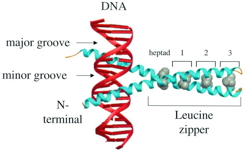
X-ray structure of the yeast B-ZIP homodimer, GCN4 (blue α-helices) bound to DNA (red helices). The N-terminal DNA recognition helix lies in the major groove of the DNA. An almost invariant leucine present every two turns of the C-terminal α-helix (at the d position) is shown in gray.
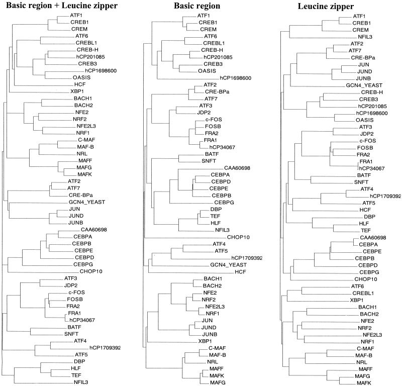
Dendrogram of 53 human B-ZIP proteins. Phylogenetic trees were generated by Align X module of Vector NTI, 7.0 with default parameters. (A) Alignment based on the entire B-ZIP domain. (B) Alignment based on the basic region. (C) Alignment based on the leucine zipper defined from the first leucine shown in the consensus on Fig. 3.
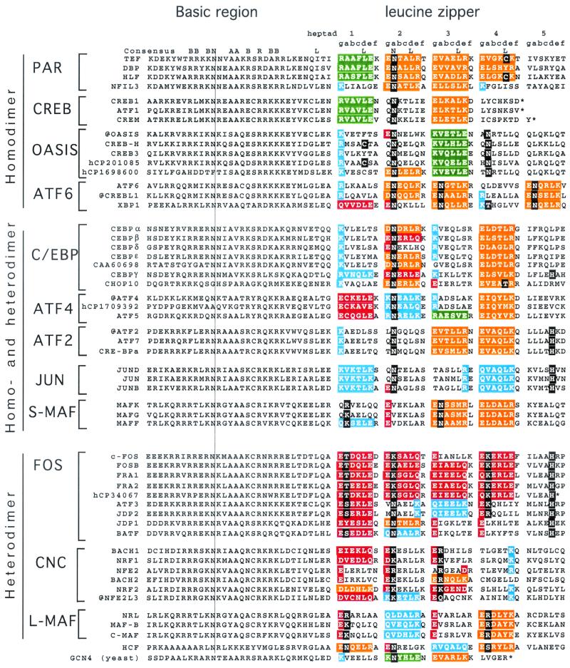
Amino acid sequence of all 53 human B-ZIP domains. Proteins are placed in to 12 groups based on predicted dimerization properties. We have placed the well-studied yeast B-ZIP protein GCN4 at the bottom of the figure for reference. The natural C terminus is denoted with an asterisk. Leucine zipper heptads are grouped (gabcdef) to help visualize potential g↔e′ pairs. If both the g and e positions contain charged amino acids, we have colored the gabcde amino acids. Our description of “attractive” and “repulsive” refers to pairs that would be present in the homodimer. We use the four colors to describe the g↔e′ pairs. Green is for the attractive basic-acidic pairs (R↔E and K↔E), orange is for the attractive acidic-basic pairs (E↔R, E↔K, D↔R, and D↔K), red is for the repulsive acidic pairs (E↔E, E↔D, E↔Q, and Q↔E), and blue is for the repulsive basic pairs (K↔K, R↔K, Q↔K, R↔Q, and K↔Q (Table 3). If only one of the two amino acids in the g↔e′ pair is charged, we color only that amino acid: red if it is acidic and blue if it is basic. If the a or d positions contain polar or charged amino acids, they are colored black.
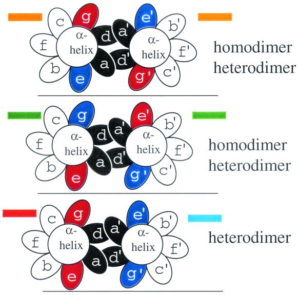
Schematic of leucine zipper dimerization according to the color code used in Fig. 3. End view of a coiled coil with the seven unique positions of the heptad presented as ellipses, looking from the N terminus to the C terminus. The a and d positions are colored black. Three possible combinations of acidic and basic amino acids in the g and e positions are presented and color-coded as in Fig. 3. (Top panel) A coiled coil with a g↔e′ pair containing an acidic amino acid in the g position and a basic amino acid in the following e position (orange in Fig. 3) can form a homo- or heterodimer with a similarly charged α-helix. (Middle panel) An α-helix with a g↔e′ pair containing a basic amino acid in the g position and an acidic amino acid in the following e position (green in Fig. 3) can form a homo- or heterodimer with a similarly charged α-helix. (Bottom panel) A heterodimer between an acidic g↔e′ pair (red in Fig. 3) and a basic g↔e′ pair (blue in Fig. 3).
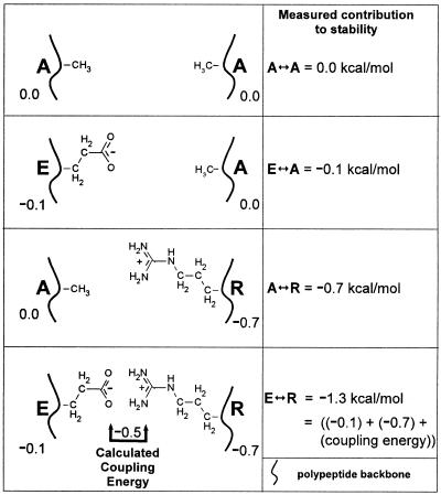
Schematic describing the four proteins used for a double-mutant thermodynamic cycle. The top panel depicts two alanines in the g and e′ positions of a g↔e′. The second panel shows that an E↔A pair is 0.1 kcal/mol more stable than an A↔A pair. The third panel shows that a A↔R pair is 0.7 kcal/mol more stable than an A↔A pair. The fourth panel shows a E↔R pair. Instead of being 0.8 kcal/mol more stable than A↔A, as would be expected if the two amino acids did not interact, E↔R is 1.3 kcal/mol more stable. The additional 0.5 kcal/mol is described as the coupling energy indicative of an physical interaction between the E and R side chains.
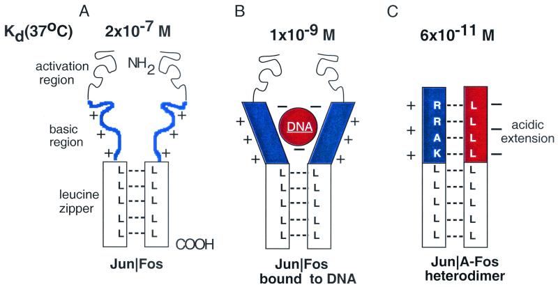
Schematic of the A-ZIP dominant negative. (A) A model of a B-ZIP dimer with the basic region (blue) unstructured. (B) A B-ZIP dimer bound to DNA with the basic region now α-helical. (C) A B-ZIP|A-ZIP heterodimer with the protein-protein interface extending N terminally into the basic region. The basic region (blue) and the designed acidic amphipathic region (red) interact as α-helices to extend the coiled-coil domain.
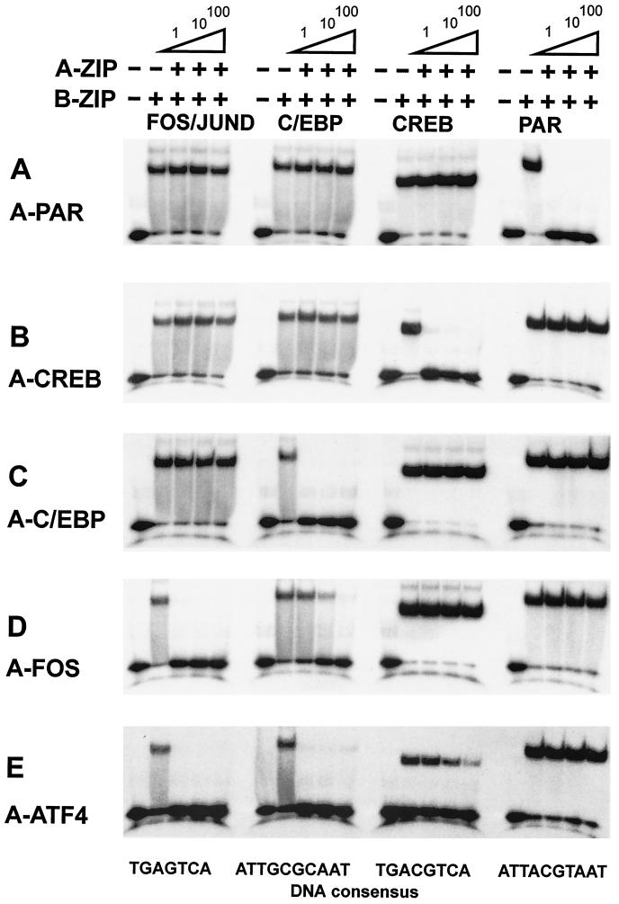
B-ZIP DNA binding is inhibited by specific A-ZIP dominant negatives. Four B-ZIP dimers, the PAR, CREB, and C/EBPα homodimers, and the FOS|JUND heterodimer are bound to a 28-bp DNA containing the optimal binding site for each B-ZIP dimer. One, 10, or 100 molar equivalents of the A-ZIP is added to the B-ZIP before addition of DNA, and the solution is electrophoresed with an electrophoretic mobility shift assay. The sequence of the 28-mer DNA probes is shown below with the consensus binding sites underlined: GTCAGTCAGAATGACTCA TATCGGTCAG (AP-1), GTCAGTCAGATGACGTCA TATCGGTCAG (CREB), GTCAGTCAGATTACGTAAT ATCGGTCAG (VBP), and GTCAGTCAGATTGCGCAAT ATCGGTCAG (C/EBP).
References
-
- Alber, T. 1992. Structure of the leucine zipper. Curr. Opin. Genet. Dev. 2:205-210. - PubMed
-
- Baxevanis, A. D., and C. R. Vinson. 1993. Interactions of coiled coils in transcription factors: where is the specificity? Curr. Opin. Genet. Dev. 3:278-285. - PubMed
-
- Bohmann, D., T. J. Bos, A. Admon, T. Nishimura, P. K. Vogt, and R. Tjian. 1987. Human proto-oncogene c-jun encodes a DNA binding protein with structural and functional properties of transcription factor AP-1. Science 238:1386-1392. - PubMed
-
- Cao, Z., R. M. Umek, and S. L. McKnight. 1991. Regulated expression of three C/EBP isoforms during adipose conversion of 3T3-L1 cells. Genes Dev. 5:1538-1552. - PubMed
Publication types
MeSH terms
Substances
LinkOut - more resources
Full Text Sources
Other Literature Sources