. 2002 Sep;22(18):6354-62.
doi: 10.1128/MCB.22.18.6354-6362.2002.

Mechanism of rapid transcriptional induction of tumor necrosis factor alpha-responsive genes by NF-kappaB

Affiliations

Mechanism of rapid transcriptional induction of tumor necrosis factor alpha-responsive genes by NF-kappaB

Elena Ainbinder et al. Mol Cell Biol. 2002 Sep.

Abstract

NF-kappaB induces the expression of genes involved in immune response, apoptosis, inflammation, and the cell cycle. Certain NF-kappaB-responsive genes are activated rapidly after the cell is stimulated by cytokines and other extracellular signals. However, the mechanism by which these genes are activated is not entirely understood. Here we report that even though NF-kappaB interacts directly with TAF(II)s, induction of NF-kappaB by tumor necrosis factor alpha (TNF-alpha) does not enhance TFIID recruitment and preinitiation complex formation on some NF-kappaB-responsive promoters. These promoters are bound by the transcription apparatus prior to TNF-alpha stimulus. Using the immediate-early TNF-alpha-responsive gene A20 as a prototype promoter, we found that the constitutive association of the general transcription apparatus is mediated by Sp1 and that this is crucial for rapid transcriptional induction by NF-kappaB. In vitro transcription assays confirmed that NF-kappaB plays a postinitiation role since it enhances the transcription reinitiation rate whereas Sp1 is required for the initiation step. Thus, the consecutive effects of Sp1 and NF-kappaB on the transcription process underlie the mechanism of their synergy and allow rapid transcriptional induction in response to cytokines.


Figures

FIG. 1.
FIG. 1.
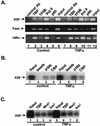
Induction of the NF-κB-A20 promoter interaction does not enhance TFIID binding. (A) TNF-α induces transport of NF-κB into the nucleus. Jurkat cells were stimulated for 1 h with TNF-α (10 ng/ml), and nuclear extracts from untreated or treated cells were used in an electrophoretic mobility shift assay with a labeled NF-κB-binding site. The arrow indicates the NF-κB complex. (B) The promoter of the A20 gene is preoccupied by TFIID in vivo. Jurkat cells were stimulated for 1 h with TNF-α or not stimulated and cross-linked in vivo with 1% formaldehyde. Soluble chromatin was prepared from these cells and used for immunoprecipitation with the indicated antibodies (CHIP assay). The immunoprecipitated DNAs were analyzed for the enrichment of the A20 of the promoter by PCR. In the middle panel, the input extract (0.1%) was subjected to increasing numbers of PCR cycles with the same primer set to check for the relative amount of the control or TNF-α-induced extracts used for immunoprecipitations. In the lower panel, samples from the same immunoprecipitated DNAs were subjected to PCR analysis using primers corresponding to the coding region of the A20 gene. (C) Analysis of a subset of NF-κB-responsive promoters by the CHIP assay as in panel B. The BLR1 gene is an NF-κB regulated gene that is specifically expressed in B cells and is used as a control for specificity.
FIG. 2.
FIG. 2.
Effect of NF-κB induction on promoter occupancy by the transcription apparatus. (A) CHIP assay using soluble chromatin extract from control or TNF-α-treated Jurkat cells and antibodies directed against TBP, TFIIB, Pol II, and the p65 component of NF-κB. The precipitated DNAs were used for PCR amplifications with primers spanning the core promoters of the indicated NF-κB target genes. (B) CHIP assays with antibodies directed against the coactivators CBP and p300 and analysis of A20 promoter by PCR. (C) CHIP assay with antibodies against the Mediator components Med7, Srb7, and TBP, which serve as a control. To confirm the amplification of the A20 promoter, the PCR products were analyzed by Southern blotting.
FIG. 3.
FIG. 3.
Sp1 enhances recruitment of TFIID to the A20 promoter. (A) Schematic representation of A20 promoter organization and the mutated promoters used for the subsequent experiments. (B) NF-κB does not change core promoter occupancy by TFIID in vitro. DNA fragments of wild-type (WT) and NF-κB-mutated promoter were immobilized on magnetic beads and used for a binding reaction with Jurkat nuclear extract supplemented with purified recombinant p65/Re1A. The bound proteins were eluted from the beads and analyzed by immunoblotting with antibodies against p65/Re1A, TBP, and TAFII250. (C) DNA binding by Sp1 is correlated with increased TFIID association with the A20 promoter. Immobilized template-binding reactions with wild-type and Sp1-mutated A20 promoter and Jurkat cell nuclear extract prepared from nonstimulated cells are shown.
FIG. 4.
FIG. 4.
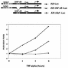
Sp1 is required for rapid transcriptional induction of the A20 promoter. Luciferase reporter genes under the control of wild-type A20 promoter (A20-Luc) or a promoter that is mutated in either NF-κB or Sp1 site (A20-LucΔNF-κB and A20-LucΔSp1, respectively) were transfected into human 293 cells. A schematic representation of the promoter constructs is shown in the upper panel. At 16 h after transfection, TNF-α was added to the cells for the indicated time points and luciferase activity was measured. For each of the reporter constructs, the noninduced activity was set to 1. This is a representative experiment of three independent transfection experiments (done in duplicate), which gave similar results.
FIG. 5.
FIG. 5.
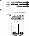
NF-κB increases the rate of transcription cycles. (A) Scheme of the A20 promoters used in the in vitro transcription assays. (B) In vitro transcription reactions were performed with nuclear extract prepared from either control (lanes 1 to 4) or 1-h-TNF-α-treated (lanes 5 to 8) Jurkat cells. The template used in each reaction is shown at the top of each lane. α-Amanitin (5 μM) was added to the reaction mixtures in lanes 2 and 6 to verify that the synthesized transcript is Pol II specific. In the lower panel, the transcription products were quantified by Phosphoimager and normalized to the noninducible cyclin A promoter activity. The noninduced wild-type (WT) promoter activity was set to 1. (C) In vitro transcription reactions with the respective A20 promoter plasmids (indicated at the bottom) and nuclear extract from untreated or TNF-α-treated Jurkat cells. In columns 3, 4, 7, 8, 11, and 12, Sarkosyl (0.2%) was added exactly 1.5 mins after the addition of nucleotides (see Materials and Methods). The results are presented as relative Phosphoimager measurements of the Pol II-specific transcript for each reaction. The noninduced wild-type promoter activity was set to 1. The amount of transcription extract used was normalized according to cyclin A promoter activity. (D) In the upper panel, transcription reaction mixtures were assembled with either wild-type or NF-κB site-mutated A20 promoter plasmids, as indicated at the top, nuclear extract from nonstimulated cells, and the indicated amounts of purified recombinant p65 homodimer. In the lower panel, purified Sp1 was added to the transcription reaction mixtures. To achieve the Sp1 effect, it was necessary to use smaller amounts of nuclear extract, and so the basal activity is not detected. These experiments are representative transcription assays, which were reproduced two to five times with similar results.
FIG. 5.
FIG. 5.
NF-κB increases the rate of transcription cycles. (A) Scheme of the A20 promoters used in the in vitro transcription assays. (B) In vitro transcription reactions were performed with nuclear extract prepared from either control (lanes 1 to 4) or 1-h-TNF-α-treated (lanes 5 to 8) Jurkat cells. The template used in each reaction is shown at the top of each lane. α-Amanitin (5 μM) was added to the reaction mixtures in lanes 2 and 6 to verify that the synthesized transcript is Pol II specific. In the lower panel, the transcription products were quantified by Phosphoimager and normalized to the noninducible cyclin A promoter activity. The noninduced wild-type (WT) promoter activity was set to 1. (C) In vitro transcription reactions with the respective A20 promoter plasmids (indicated at the bottom) and nuclear extract from untreated or TNF-α-treated Jurkat cells. In columns 3, 4, 7, 8, 11, and 12, Sarkosyl (0.2%) was added exactly 1.5 mins after the addition of nucleotides (see Materials and Methods). The results are presented as relative Phosphoimager measurements of the Pol II-specific transcript for each reaction. The noninduced wild-type promoter activity was set to 1. The amount of transcription extract used was normalized according to cyclin A promoter activity. (D) In the upper panel, transcription reaction mixtures were assembled with either wild-type or NF-κB site-mutated A20 promoter plasmids, as indicated at the top, nuclear extract from nonstimulated cells, and the indicated amounts of purified recombinant p65 homodimer. In the lower panel, purified Sp1 was added to the transcription reaction mixtures. To achieve the Sp1 effect, it was necessary to use smaller amounts of nuclear extract, and so the basal activity is not detected. These experiments are representative transcription assays, which were reproduced two to five times with similar results.
FIG. 6.
FIG. 6.
Model for transcriptional induction of NF-κB-responsive genes. In unstimulated cells, the promoters of NF-κB target genes are pre-occupied by the general transcription apparatus due to the function of a constitutively active transcription factor such as Sp1 that acts to recruit TFIID to the promoter. This complex is inadequate for multiple transcription rounds and thus can maintain only a low level of basal transcription. In stimulated cells, the association of NF-κB with the promoter is followed by multiple contacts with components of the transcription apparatus such as the TAF subunits, CBP, p300, and Mediator. These interactions promote multiple transcription cycles, resulting in high level of transcription.

Similar articles

Cited by

References

    1. Agalioti, T., S. Lomvardas, B. Parekh, J. Yie, T. Maniatis, and D. Thanos. 2000. Ordered recruitment of chromatin modifying and general transcription factors to the IFN-beta promoter. Cell 103:667-678. - PubMed
    1. Albright, S. R., and R. Tjian. 2000. TAFs revisited: more data reveal new twists and confirm old ideas. Gene 242:1-13. - PubMed
    1. Berk, A. J. 1999. Activation of RNA polymerase II transcription. Curr. Opin. Cell Biol. 11:330-335. - PubMed
    1. Carey, M., Y. S. Lin, M. R. Green, and M. Ptashne. 1990. A mechanism for synergistic activation of a mammalian gene by GAL4 derivatives. Nature 345:361-364. - PubMed
    1. Dikstein, R., S. Zhou, and R. Tjian. 1996. Human TAFII105 is a cell type-specific TFIID subunit related to hTAFII130. Cell 87:137-146. - PubMed

Publication types