. 2002 Sep;22(18):6406-16.
doi: 10.1128/MCB.22.18.6406-6416.2002.

Targeted histone acetylation at the yeast CUP1 promoter requires the transcriptional activator, the TATA boxes, and the putative histone acetylase encoded by SPT10

Affiliations

Targeted histone acetylation at the yeast CUP1 promoter requires the transcriptional activator, the TATA boxes, and the putative histone acetylase encoded by SPT10

Chang-Hui Shen et al. Mol Cell Biol. 2002 Sep.

Abstract

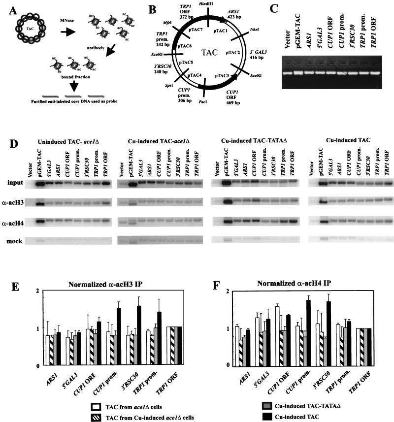
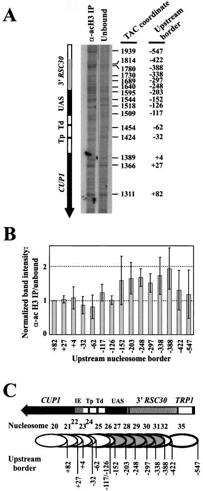
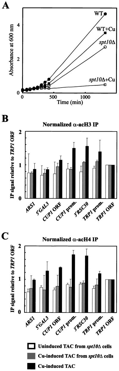
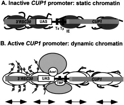
The relationship between chromatin remodeling and histone acetylation at the yeast CUP1 gene was addressed. CUP1 encodes a metallothionein required for cell growth at high copper concentrations. Induction of CUP1 with copper resulted in targeted acetylation of both H3 and H4 at the CUP1 promoter. Nucleosomes containing upstream activating sequences and sequences farther upstream were the targets for H3 acetylation. Targeted acetylation of H3 and H4 required the transcriptional activator (Ace1p) and the TATA boxes, suggesting that targeted acetylation occurs when TATA-binding protein binds to the TATA box or at a later stage in initiation. We have shown previously that induction results in nucleosome repositioning over the entire CUP1 gene, which requires Ace1p but not the TATA boxes. Therefore, the movement of nucleosomes occurring on CUP1 induction is independent of targeted acetylation. Targeted acetylation of both H3 and H4 also required the product of the SPT10 gene, which encodes a putative histone acetylase implicated in regulation at core promoters. Disruption of SPT10 was lethal at high copper concentrations and correlated with slower induction and reduced maximum levels of CUP1 mRNA. These observations constitute evidence for a novel mechanism of chromatin activation at CUP1, with a major role for the TATA box.


Figures

FIG. 1.
FIG. 1.
Chromatin structure of CUP1. A map of the TAC minichromosome. TAC is based on the yeast plasmid TRP1ARS1; TRP1 is a selection marker, and ARS1 is the origin of replication. TRP1ARS1 also contains the upstream region of neighboring GAL3, including a binding site for Gal4p (UASGAL). CUP1 was inserted at the EcoRI site, together with the 3′ flanking region of RSC30, the gene neighboring CUP1 in the genome. The SpeI site marks the limit of the CUP1 promoter, which contains two TATA boxes, five Ace1p binding sites (UAS), and an initiation element (IE). (B) Positioned nucleosomes on CUP1 in TAC minichromosomes purified from copper-induced WT cells and ace1Δ cells (42). The CUP1 and TRP1 promoters are shown as dark boxes. Grey ovals indicate positioned nucleosomes drawn to scale, numbered relative to the HindIII site in TRP1 (not all of TAC is shown; see panel A). TAC from ace1Δ cells (inactive CUP1) is organized into clusters of overlapping positions separated by linkers of various lengths. In copper-induced TAC (active CUP1), nucleosomes occupy additional positions in the linkers, implying that induction results in the movement of nucleosomes over the entire CUP1 gene, including flanking regions. This movement does not require the TATA boxes. It was proposed that nucleosome movement reflects the creation of a dynamic chromatin structure via the Ace1p-dependent recruitment of a nucleosome repositioning activity. Note: nucleosome positions on CUP1 are determined primarily by DNA sequence (41).
FIG. 2.
FIG. 2.
Mutation of both TATA boxes inactivates the CUP1 gene. (A) Sequences of the proximal and distal TATA boxes in the CUP1 promoter, with mutations (TATAΔ) shown in lowercase. Nucleotide positions are reported with respect to the major upstream start site (18). (B) Effect of copper on growth of yeast cells in synthetic complete medium lacking tryptophan. Filled symbols, no copper; open symbols, +1 mM CuSO4; circles, WT TAC; squares, TAC (proximal TATAΔ);diamonds, TAC (distal TATAΔ); triangles, TAC-TATAΔ (double mutant). (C) Mung bean nuclease mapping of CUP1 transcripts. RNA was prepared from cells before and after induction with 1 mM CuSO4 for 15 min. A phosphorimager scan is shown (all lanes are from the same gel). Multiple closely spaced start sites were observed. CUP1 transcripts were quantified relative to PGK1. The level of expression relative to uninduced WT cells (set at 1.0) is shown at bottom. The band marked with an asterisk is derived from PGK1.
FIG. 3.
FIG. 3.
Targeted acetylation of H3 and H4 at the CUP1 promoter requires Ace1p and the TATA boxes. (A) Schematic of the TAC IP experiment. (B) Map of the TAC episome indicating the seven subcloned regions, defined by the restriction sites indicated: (i) ARS1 (also containing the 3′ end of TRP1); (ii) 5′GAL3, the upstream region of GAL3; (iii) CUP1 ORF; (iv) CUP1 promoter; (v) 3′ RSC30; (vi) TRP1 promoter; and (vii) TRP1 ORF. The size of each fragment is indicated. (C) Example of a gel used for Southern blotting, stained with ethidium bromide. Vector, plasmid with no insert. pGEM-TAC, a HindIII digest of this plasmid (bands corresponding to the TAC insert and the vector). Equal amounts of linearized plasmid were loaded. (D) Hybridization of labeled core particle DNA from purified TAC minichromosomes to Southern blots of gels like that shown in panel C. TAC minichromosomes were purified from ace1Δ cells (with or without copper induction) and copper-induced WT cells; TAC-TATAΔ minichromosomes were purified from copper-induced cells. TAC chromatin was digested to core particles for use in IP experiments. Labeled core DNAs derived from input core particles, core particles immunoprecipitated with the anti-acetylated H3 antibody or the anti-hyperacetylated H4 antibody, and mock immunoprecipitates (no antibody) were used as probes. Phosphorimages are shown. For TAC-TATAΔ blots, pTAC4dubR was used instead of pTAC4 (pTAC4dubR is the same as pTAC4, except that it carries the TATA box mutations). (E and F) Quantitative analysis of IP data in panel D. Panel E shows acetylated H3; panel F shows hyperacetylated H4. TAC from uninduced (white bars) and copper-induced (striped bars) ace1Δ cells, TAC-TATAΔ from copper-induced cells (grey bars), and TAC from copper-induced WT cells (black bars) are analyzed. For each data set, the bands were quantified by a phosphorimager and normalized to the TRP1 ORF, which was set equal to 1. These numbers were then divided by the input values normalized identically. Error bars represent the standard error for at least three independent experiments for H3 and two independent experiments for H4.
FIG. 4.
FIG. 4.
Nucleosomes containing CUP1 UASs and sequences farther upstream are the targets for H3 acetylation. (A) Identification of positioned nucleosomes enriched in the acetylated H3 IP obtained with copper-induced TAC from WT cells: monomer extension of nucleosomes in the IP with those in the unbound fraction. A phosphorimage is shown (the exposures of the two lanes are slightly different tofacilitate comparison). The size of each band equals the distance of the farthest border of a positioned nucleosome from the unique SspI site. The coordinates of each border with respect to the HindIII site in TRP1 are given at right (42), and the coordinates with respect to the major upstream start site of CUP1 (+1) are given at far right. Tp, proximal TATA box; Td, distal TATA box. (B) Quantitative analysis of nucleosomes in the acetylated H3 IP (data in panel A). The intensities of the bands in each monomer extension track were normalized to the band at +82 (in the CUP1 ORF), and the ratio of the normalized band in the IP to the normalized band in the unbound fraction was calculated for each nucleosome. (A ratio of 1.0 indicates that this nucleosome is not enriched in the IP.) Error bars represent the standard error for data from four independent experiments. Coordinates are given with respect to the major upstream start site (+1). (C) Schematic showing nucleosomes targeted for H3 acetylation. Nucleosomes are numbered as in Fig. 1B; upstream borders are indicated. Nucleosomes enriched in the IP are shown shaded. The bands at −117 and −126 both derive from nucleosome 25. Nucleosomes 33 and 34 were quantitatively minor and are not included in the analysis.
FIG. 5.
FIG. 5.
SPT10 is required for maximal induction of CUP1 and survival at high copper concentrations. (A) Effect of copper on growth of WT (filled symbols) and spt10Δ (open symbols) BY4741 cells in synthetic complete medium with (squares) or without (circles) 1 mM CuSO4. Growth was measured by absorbance at 600 nm. (B) Time course of copper induction: effect of copper on growth of WT (filled squares) and spt10Δ (open squares) BY4741 cells in synthetic complete medium inoculated with 1.25 mM CuSO4 at time zero. (C) Induction of CUP1 mRNA during growth shown in panel B. Phosphorimages of Northern blots of RNA purified from WT and spt10Δ cells probed for CUP1 or ACT1 mRNA. Total RNA loadings were compared by staining of an agarose gel with ethidium bromide. (D) Quantification of Northern blots in panel C: filled squares indicate CUP1 mRNA in WT cells and open squares indicate CUP1 mRNA in spt10Δ cells after normalization to total RNA.
FIG. 6.
FIG. 6.
SPT10 is required for targeted acetylation of both H3 and H4 at the CUP1 promoter. (A) Effect of copper on growth of WT (filled symbols) and spt10Δ (open symbols) cells containing TAC in synthetic complete medium lacking tryptophan, with (squares) or without (circles) 1 mM CuSO4. Growth was measured by absorbance at 600 nm. Quantitative analysis of IP data is shown for acetylated H3 (B) and hyperacetylated H4 (C). Data for TAC from uninduced (open bars) and copper-induced (grey bars) spt10Δ cells are compared with datafrom Fig. 3 for TAC from copper-induced cells (black bars). Error bars for spt10Δ data represent the standard error for two (uninduced) or three (induced) independent experiments.
FIG. 7.
FIG. 7.
A model for chromatin remodeling events occurring during induction of CUP1. Only the CUP1 promoter region of TAC is shown, with nucleosomes drawn approximately to scale. Nucleosomes can occupy any of several possible positions in vivo (Fig. 1B); four are shown here. (A) Inactive CUP1: the nucleosomes are immobile and the chromatin structure is static. The histone tail domains are mostly bound to nucleosomal DNA or linker DNA and are acetylated at background (global) levels. (B) Active CUP1: copper-activated Ace1p locates a binding site in the UAS region. A remodeling activity is recruited, and nucleosomes begin to slide back and forth, creating a dynamic chromatin structure over the entire gene. TBP with its associated factors binds to one of the two TATA boxes at a moment when it is nucleosome free. A HAT activity (probably Spt10p) is activated and acetylates H3 and H4 in nucleosomes upstream of the TATA box.

Similar articles

Cited by

References

    1. Angelov, D., J. M. Vitolo, V. Mutskov, S. Dimitrov, and J. J. Hayes. 2001. Preferential interaction of the core histone tail domains with linker DNA. Proc. Natl. Acad. Sci. USA 98:6599-6604. - PMC - PubMed
    1. Angus-Hill, M. L., A. Schlichter, D. Roberts, H. Erdjument-Bromage, P. Tempst, and B. R. Cairns. 2001. A Rsc3/Rsc30 zinc cluster dimer reveals novel roles for the chromatin remodeler RSC in gene expression and cell cycle control. Mol. Cell 7:741-751. - PubMed
    1. Berk, A. J., and P. A. Sharp. 1977. Sizing and mapping of early adenovirus mRNAs by gel electrophoresis of S1 endonuclease digest hybrids. Cell 12:721-732. - PubMed
    1. Brachmann, C. B., A. Davies, G. J. Cost, E. Caputo, J. Li, P. Hieter, and J. D. Boeke. 1998. Designer deletion strains derived from Saccharomyces cerevisiae S288C: a useful set of strains and plasmids for PCR-mediated gene disruption and other applications. Yeast 14:115-132. - PubMed
    1. Brownell, J. E., J. Zhou, T. Ranalli, R. Kobayashi, D. G. Edmondson, S. Y. Roth, and C. D. Allis. 1996. Tetrahymena histone acetyltransferase A: a homolog to yeast Gcn5p linking histone acetylation to gene activation. Cell 84:843-851. - PubMed

MeSH terms