. 2002 Sep;22(18):6521-32.
doi: 10.1128/MCB.22.18.6521-6532.2002.

Chk2 is a tumor suppressor that regulates apoptosis in both an ataxia telangiectasia mutated (ATM)-dependent and an ATM-independent manner

Affiliations

Chk2 is a tumor suppressor that regulates apoptosis in both an ataxia telangiectasia mutated (ATM)-dependent and an ATM-independent manner

Atsushi Hirao et al. Mol Cell Biol. 2002 Sep.

Abstract

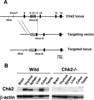
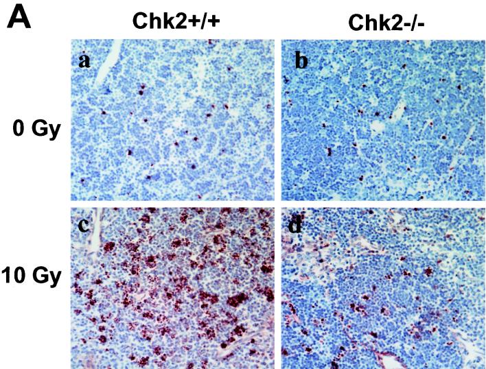
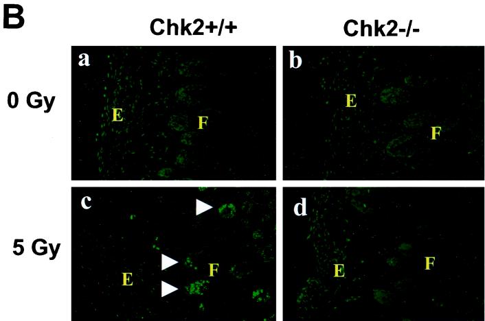
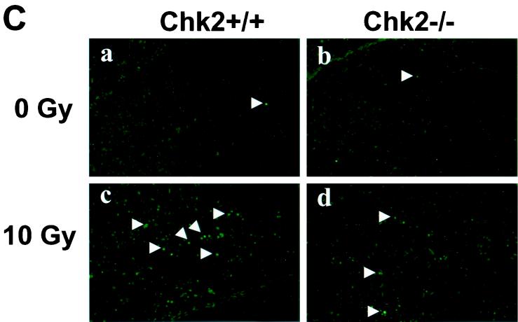
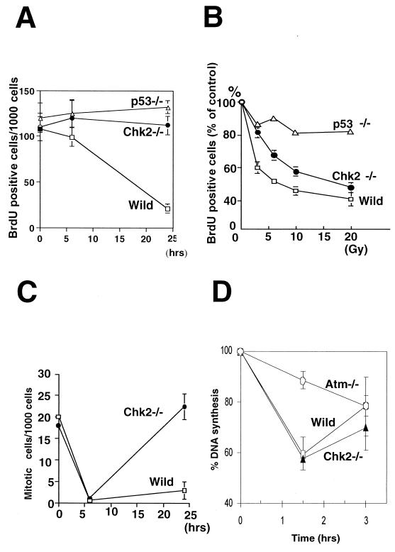
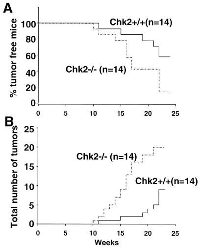
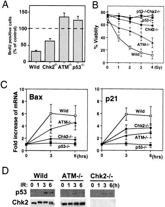
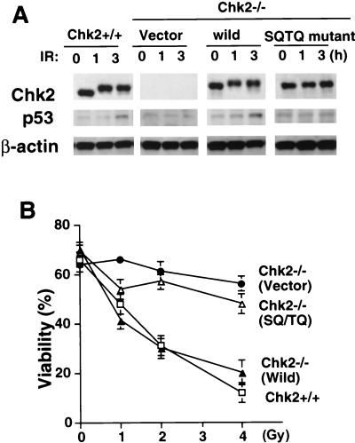
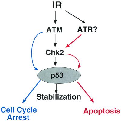
In response to ionizing radiation (IR), the tumor suppressor p53 is stabilized and promotes either cell cycle arrest or apoptosis. Chk2 activated by IR contributes to this stabilization, possibly by direct phosphorylation. Like p53, Chk2 is mutated in patients with Li-Fraumeni syndrome. Since the ataxia telangiectasia mutated (ATM) gene is required for IR-induced activation of Chk2, it has been assumed that ATM and Chk2 act in a linear pathway leading to p53 activation. To clarify the role of Chk2 in tumorigenesis, we generated gene-targeted Chk2-deficient mice. Unlike ATM(-/-) and p53(-/-) mice, Chk2(-/-) mice do not spontaneously develop tumors, although Chk2 does suppress 7,12-dimethylbenzanthracene-induced skin tumors. Tissues from Chk2(-/-) mice, including those from the thymus, central nervous system, fibroblasts, epidermis, and hair follicles, show significant defects in IR-induced apoptosis or impaired G(1)/S arrest. Quantitative comparison of the G(1)/S checkpoint, apoptosis, and expression of p53 proteins in Chk2(-/-) versus ATM(-/-) thymocytes suggested that Chk2 can regulate p53-dependent apoptosis in an ATM-independent manner. IR-induced apoptosis was restored in Chk2(-/-) thymocytes by reintroduction of the wild-type Chk2 gene but not by a Chk2 gene in which the sites phosphorylated by ATM and ataxia telangiectasia and rad3(+) related (ATR) were mutated to alanine. ATR may thus selectively contribute to p53-mediated apoptosis. These data indicate that distinct pathways regulate the activation of p53 leading to cell cycle arrest or apoptosis.


Figures

FIG. 1.
FIG. 1.
Targeted disruption of the Chk2 gene in mice. (A) Targeting strategy. The genomic configuration of the germ line Chk2 locus is shown at the top. The targeting vector is shown in the center; exons 8 to 11 were replaced with a neomycin cassette (Neo). The mutated Chk2 locus is shown at the bottom. (B) Western blot showing the expression of Chk2 and β-actin proteins in various tissues of wild-type (Wild) and Chk2−/− mice.
FIG. 2.
FIG. 2.
Induction of apoptosis in response to γ-irradiation. (A) Mice (8 weeks old) were mock irradiated (a and b) or irradiated with 10 Gy of irradiation (c and d), and apoptosis in thymi was evaluated at 10 h post-IR. (B) Mice (P7) were mock irradiated (a and b) or irradiated with 5 Gy of irradiation (c and d), and apoptosis in skin was evaluated at 6 h post-IR by in situ TUNEL staining. E and F indicate epidermis and hair follicle, respectively. (C) Mice (P5) were mock irradiated (a and b) or irradiated with 10 Gy of irradiation (c and d), and apoptosis in the hippocampal dentate gyrus was evaluated at 24 h post-IR by in situ TUNEL staining. Arrows indicate apoptotic TUNEL-positive cells.
FIG. 2.
FIG. 2.
Induction of apoptosis in response to γ-irradiation. (A) Mice (8 weeks old) were mock irradiated (a and b) or irradiated with 10 Gy of irradiation (c and d), and apoptosis in thymi was evaluated at 10 h post-IR. (B) Mice (P7) were mock irradiated (a and b) or irradiated with 5 Gy of irradiation (c and d), and apoptosis in skin was evaluated at 6 h post-IR by in situ TUNEL staining. E and F indicate epidermis and hair follicle, respectively. (C) Mice (P5) were mock irradiated (a and b) or irradiated with 10 Gy of irradiation (c and d), and apoptosis in the hippocampal dentate gyrus was evaluated at 24 h post-IR by in situ TUNEL staining. Arrows indicate apoptotic TUNEL-positive cells.
FIG. 3.
FIG. 3.
Since the loss of Chk2 leads to suppressed apoptosis and cell cycle arrest in response to IR, one would expect to find a higher incidence of tumor formation in Chk2−/− mice, perhaps comparable to that in p53−/− mice. However, by age 1 year, Chk2−/− mice had not developed tumors of any kind. We speculate that tumors caused by the loss of Chk2 are either too rare to be detected or require a prolonged period to develop. We therefore challenged Chk2−/− mice with the chemical carcinogen DMBA, an agent that damages DNA and efficiently induces skin tumors (15, 40). More Chk2−/− mice than wild-type animals developed skin tumors in response to DMBA treatment (wild type versus Chk2−/−: 6 of 14 versus 12 of 14 at 25 weeks) (Fig. 4A
FIG. 4.
FIG. 4.
Skin tumor formation in mice treated with DMBA. The back skin of 6- to 8-week-old mice was shaved and painted with 10 μg of DMBA once a week for 25 weeks. Tumors in skin were scored once a week. (A) Kaplan-Meier plot of tumor incidence in wild-type (n = 14) and Chk2−/− (n = 14) mice. (B) Total numbers of tumors in mice for which data are shown in panel A.
FIG. 5.
FIG. 5.
IR-induced p53 activation and p53 stabilization in Chk2−/− and ATM−/− thymocytes. (A) G1/S checkpoint induced by γ-irradiation. The indicated strains of mice were irradiated with 10 Gy of irradiation and injected 1 h later with BrdU. At 2 h post-IR, thymocytes were isolated, stained with anti-BrdU Ab, and subjected to flow cytometry. Each value represents the mean percentage ± standard deviation (SD) of BrdU-positive (S phase) cells present after IR in 4 samples per group relative to the unirradiated controls. (B) Apoptosis induced by γ-irradiation. Isolated thymocytes were treated with the indicated doses of γ-irradiation, and apoptotic cells were evaluated by flow cytometry after Annexin V and PI staining. Each value represents the mean percentage ± SD of viable cells (Annexin V negative and PI negative) for 4 samples per group. (C) Induction of p53 downstream molecules. Total RNA was isolated from thymocytes before and after treatment with 5 Gy of irradiation. The expression of mRNA for p21, Bax, and β-actin was evaluated by Northern blotting. The amount of p21 or Bax mRNA was quantified by using a PhosphorImager analyzer and normalized to β-actin expression. Each value represents the mean increase (n-fold) ± SD of the expression of Bax or p21 mRNA after IR in 3 to 5 samples per group relative to the unirradiated controls. (D) p53 protein stabilization and Chk2 phosphorylation in thymocytes after irradiation. Isolated thymocytes were irradiated with 5 Gy of irradiation and lysed at the indicated times. p53 and Chk2 proteins were detected by Western blotting with anti-p53 or anti-Chk2 Ab. Wild, wild type.
FIG. 6.
FIG. 6.
Effect of reintroduced mutant Chk2 on IR-induced apoptosis. Chk2−/− ES cells transfected with either empty vector (vector), vector containing wild-type Chk2 (wild), or vector containing the Chk2 SQ/TQ mutant gene (SQ/TQ mutant) were injected into blastocysts from Rag1−/− mice to generate chimeric animals expressing the corresponding proteins in thymocytes. (A) Expression of reintroduced Chk2 protein and stabilization of p53 following 5 Gy of γ-irradiation for the indicated times. (B) Thymocytes isolated from the chimeric mice for which data are shown in panel A were irradiated at the indicated doses. Apoptosis was analyzed by Annexin V-PI staining at 24 h post-IR. Each value represents the mean percentage ± standard deviation of viable cells in 3 cultures per group.
FIG. 7.
FIG. 7.
Model of the regulation of p53 activation by Chk2 in response to IR. Chk2-mediated stabilization of p53 induced by IR and leading to apoptosis is controlled independently of ATM, possibly by ATR. ATM appears to stabilize p53, leading to cell cycle arrest without involving Chk2.

Comment in

  • CHK2: a tumor suppressor or not?
    McGowan CH. McGowan CH. Cell Cycle. 2002 Nov-Dec;1(6):401-3. doi: 10.4161/cc.1.6.264. Cell Cycle. 2002. PMID: 12548013 Review. No abstract available.

References

    1. Ahn, J. Y., J. K. Schwarz, H. Piwnica-Worms, and C. E. Canman. 2000. Threonine 68 phosphorylation by ataxia telangiectasia mutated is required for efficient activation of Chk2 in response to ionizing radiation. Cancer Res. 60:5934-5936. - PubMed
    1. Banin, S., L. Moyal, S. Shieh, Y. Taya, C. W. Anderson, L. Chessa, N. I. Smorodinsky, C. Prives, Y. Reiss, Y. Shiloh, and Y. Ziv. 1998. Enhanced phosphorylation of p53 by ATM in response to DNA damage. Science 281:1674-1677. - PubMed
    1. Barlow, C., K. D. Brown, C. X. Deng, D. A. Tagle, and A. Wynshaw-Boris. 1997. Atm selectively regulates distinct p53-dependent cell-cycle checkpoint and apoptotic pathways. Nat. Genet. 17:453-456. - PubMed
    1. Barlow, C., S. Hirotsune, R. Paylor, M. Liyanage, M. Eckhaus, F. Collins, Y. Shiloh, J. N. Crawley, T. Ried, D. Tagle, and A. Wynshaw-Boris. 1996. Atm-deficient mice: a paradigm of ataxia telangiectasia. Cell 86:159-171. - PubMed
    1. Bell, D. W., J. M. Varley, T. E. Szydlo, D. H. Kang, D. C. Wahrer, K. E. Shannon, M. Lubratovich, S. J. Verselis, K. J. Isselbacher, J. F. Fraumeni, J. M. Birch, F. P. Li, J. E. Garber, and D. A. Haber. 1999. Heterozygous germ line hCHK2 mutations in Li-Fraumeni syndrome. Science 286:2528-2531. - PubMed

Publication types

MeSH terms