. 2002 Sep;22(18):6611-26.
doi: 10.1128/MCB.22.18.6611-6626.2002.

Microtubule-dependent subcellular redistribution of the transcriptional coactivator p/CIP

Affiliations

Microtubule-dependent subcellular redistribution of the transcriptional coactivator p/CIP

Majdi S Qutob et al. Mol Cell Biol. 2002 Sep.

Abstract

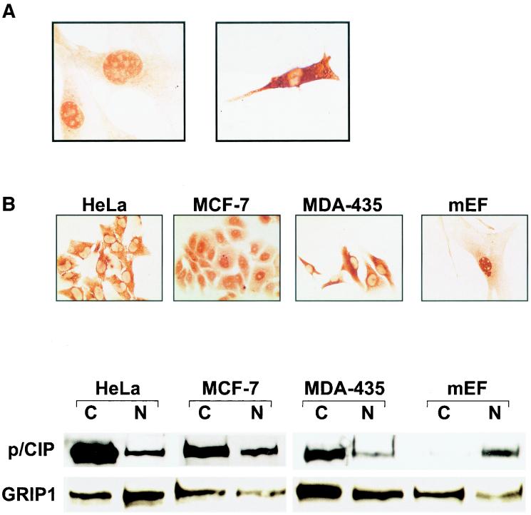
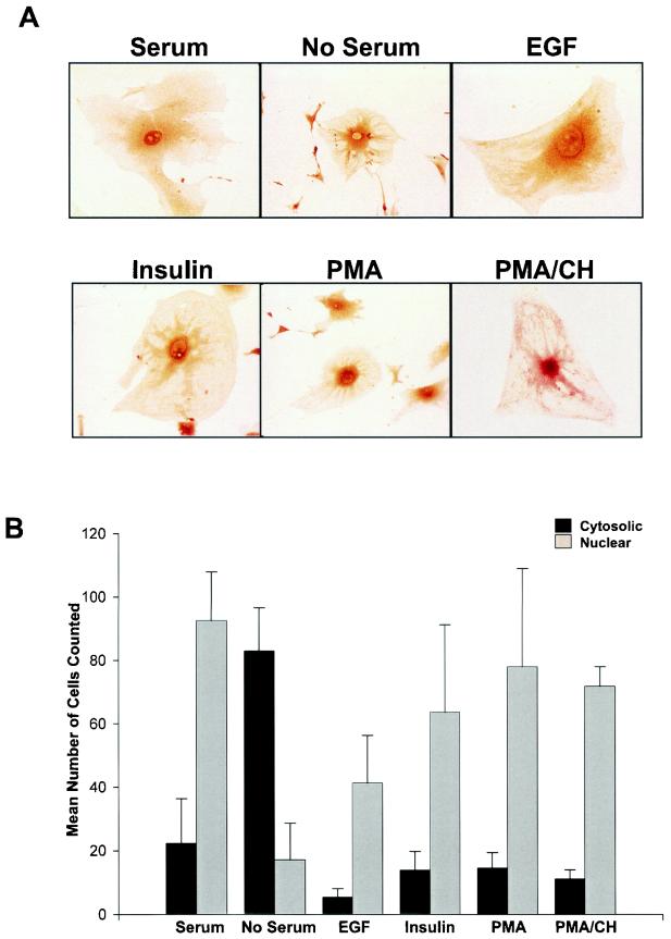
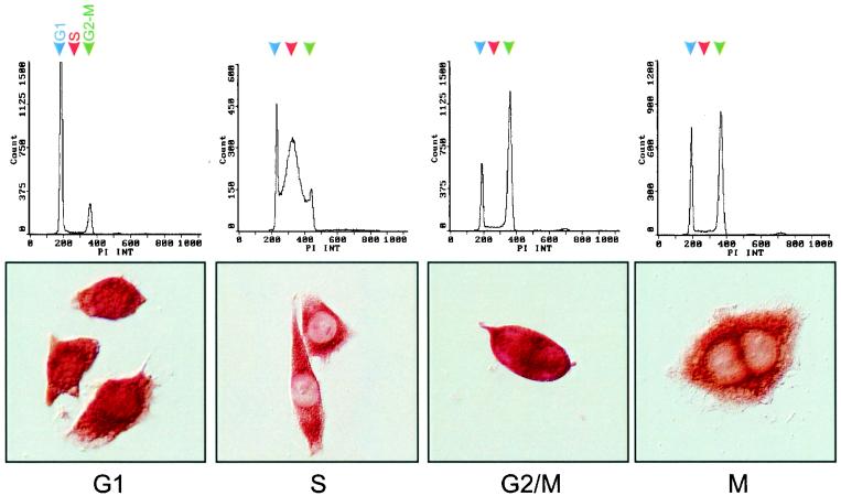
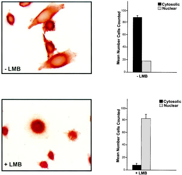
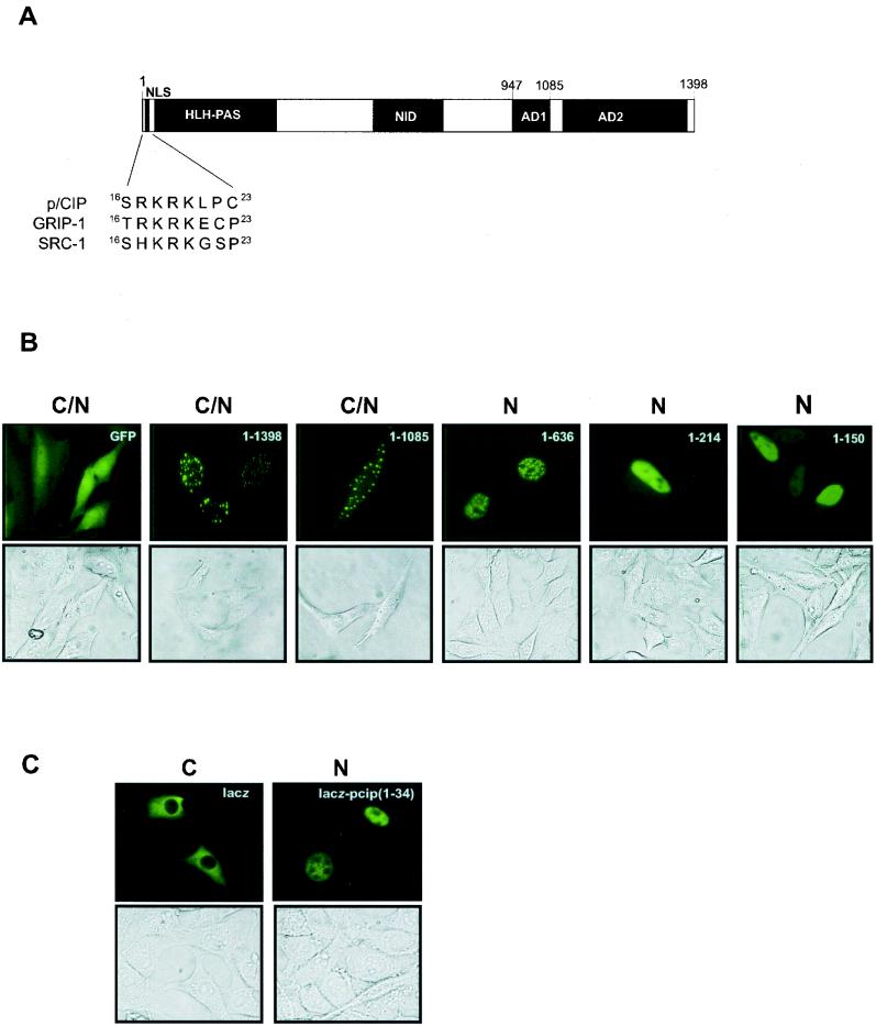
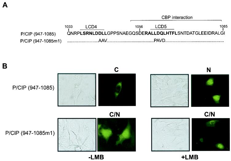
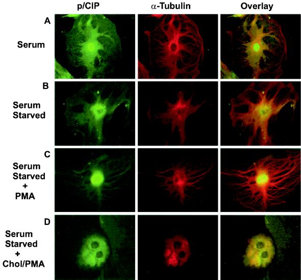
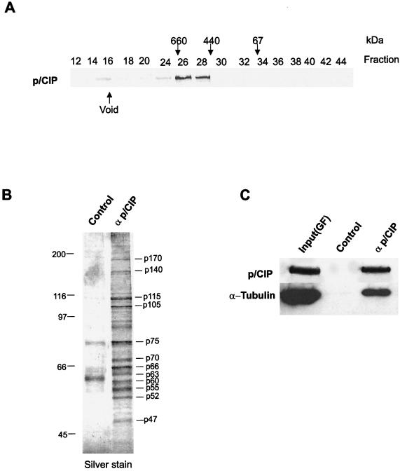
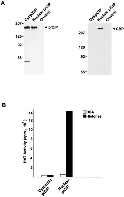
The transcriptional coactivator p/CIP is a member of a family of nuclear receptor coactivator/steroid receptor coactivator (NCoA/SRC) proteins that mediate the transcriptional activities of nuclear hormone receptors. We have found that p/CIP is predominantly cytoplasmic in a large proportion of cells in various tissues of the developing mouse and in a number of established cell lines. In mouse embryonic fibroblasts, serum deprivation results in the redistribution of p/CIP to the cytoplasmic compartment and stimulation with growth factors or tumor-promoting phorbol esters promotes p/CIP shuttling into the nucleus. Cytoplasmic accumulation of p/CIP is also cell cycle dependent, occurring predominantly during the S and late M phases. Leptomycin B (LMB) treatment results in a marked nuclear accumulation, suggesting that p/CIP undergoes dynamic nuclear export as well as import. We have identified a strong nuclear import signal in the N terminus of p/CIP and two leucine-rich motifs in the C terminus that resemble CRM-1-dependent nuclear export sequences. When fused to green fluorescent protein, the nuclear export sequence region is cytoplasmic and is retained in the nucleus in an LMB-dependent manner. Disruption of the leucine-rich motifs prevents cytoplasmic accumulation. Furthermore, we demonstrate that cytoplasmic p/CIP associates with tubulin and that an intact microtubule network is required for intracellular shuttling of p/CIP. Immunoaffinity purification of p/CIP from nuclear and cytosolic extracts revealed that only nuclear p/CIP complexes possess histone acetyltransferase activity. Collectively, these results suggest that cellular compartmentalization of NCoA/SRC proteins could potentially regulate nuclear hormone receptor-mediated events as well as integrating signals in response to different environmental cues.


Figures

FIG. 1.
FIG. 1.
Immunohistological analysis of p/CIP expression in mouse female reproductive organs. (A and B) Low magnification (A) and high magnification (B) of the same section of an ovary taken from a 6-week-old mouse. Predominantly nuclear staining was observed within the granulosa cells (Gc) of the tertiary follicle, and diffuse p/CIP staining was also observed within the cytoplasm. In the surrounding theca interna (Ti) and corpora lutea (Cl), mainly cytoplasmic staining was observed. The cubiodal cells (Cb) enveloping the entire ovary also displayed diffuse cytoplasmic staining. (C) Cross section of a uterine horn taken from an adult virgin mouse. Intense nuclear and cytoplasmic staining can be observed within the uterina epithelial cells (Ep), whereas in the surrounding stroma (St) and smooth muscle myometrium (Mm), cytoplasmic staining predominates. (D) Sagittal section of a uterus taken 7.5 d.p.c. Mature nonproliferating decidua (Md), close to the embryo proper, demonstrates little to no staining for p/CIP. Decidual cells in between the myometrium and the embryo show mainly diffuse cytoplasmic staining for p/CIP. In contrast, the outer layer of highly proliferative deciduas (Pd) demonstrates intense nuclear staining. Each of these tissues were isolated from approximately the same age and strain of mice. Control experiments were performed using nonimmune IgG or p/CIP IgG preincubated with antigen. In both cases, no staining was observed (data not shown). Oo, oocyte; Bl, blood; Am, antimesometrium; Ms, mesometrium.
FIG. 2.
FIG. 2.
Intracellular localization of p/CIP varies between cell types. (A) Differentiation of BC3H-1 cells stimulates cytoplasmic accumulation of p/CIP. Cells were placed in differentiation medium for 4 days (right panel) prior to immunostaining using anti-p/CIP antibody. (B) (Top) Immunohistochemical staining for p/CIP in various cell lines. Significant cytoplasmic staining was observed in the HeLa S3, MCF-7, and MDA-435 cell lines. In the MEFs, p/CIP was localized mainly to the nuclei of the cells. (Bottom) Western blot analysis of nuclear and cytosolic fractions isolated from the corresponding cell lines used in panel A. Approximately 20 μg of protein was loaded into each well prior to SDS-PAGE and Western blotting. The antibodies used are indicated on the left. C, cytosolic extract; N, nuclear extract;
FIG. 3.
FIG. 3.
p/CIP undergoes redistribution in MEFs. (A) Immunohistochemical analysis of p/CIP in MEFs grown in the presence or absence of serum, or in serum-starved cells treated with either 100 nM EGF, insulin, PMA, or PMA in the presence of cycloheximide (PMA/CH). (B) Graphical representation of the results of the experiment in panel A, showing the mean number of cells counted in 10 random fields of cells stained for p/CIP in the presence or absence of serum, EGF, insulin, PMA, or PMA with cycloheximide. Error bars indicate standard errors of the means.
FIG. 4.
FIG. 4.
Redistribution of p/CIP is cell cycle dependent. Subcellular compartmentalization of p/CIP was determined by immunohistochemistry of synchronized HeLa cells advancing through the cell cycle. HeLa cells were arrested in S phase on treatment with 2.5 mM thymidine for 36 h. Cells were collected for flow cytometry at 30-min intervals after removal of the blocker, and the proportions of cells in G1 (77.4%), S (68.8%), G2/M (66.2%), and M (57.5%) were determined by flow cytometry (top) or by immunostaining for p/CIP (bottom).
FIG. 5.
FIG. 5.
p/CIP distribution is determined by nuclear export as well as nuclear import. (Bottom) Immunohistochemical analysis of HeLa cells treated with 50 nM LMB resulted in accumulation of p/CIP in the nucleus. (Top) In the absence of LBM, endogenous p/CIP expression was predominantly cytoplasmic with some nuclear staining. The corresponding graphs on the right represent the mean numbers of cells in six random fields of cells stained for p/CIP in the absence or presence of LMB. Data are representative of four separate experiments. Error bars indicate standard errors of the means.
FIG. 6.
FIG. 6.
p/CIP contains an NLS in its amino terminus. (A) Schematic representation highlighting the NLS in p/CIP and the homologous regions found in GRIP-1 and mouse SRC-1. (B) Representative green fluorescent images of living cells expressing GFP-p/CIP fusion constructs. HeLa cells were transfected with expression plasmids containing GFP fused to p/CIP deletion mutants as indicated in the upper right-hand corner. The lower images represent the corresponding bright-field images. (C) Representative green fluorescent images of living cells expressing lacZ fused to GFP (left) or lacZ-GFP fused to p/CIP (aa 1-34). C, predominantly cytosolic localization; N, predominantly nuclear localization; C/N, cytosolic and nuclear localization observed in more than 75% of the cells in a random field of view consisting of approximately 50 cells.
FIG. 7.
FIG. 7.
The CBP/p300 interaction domain contains an NES region. (A) Amino acid sequence of the CBP/p300 interaction domain containing the two leucine-rich regions. The NES consensus sites are highlighted in bold. (B) Representative green fluorescent images of living cells expressing GFP-p/CIP fusion constructs. HeLa cells were transfected with expression plasmids containing GFP fused to p/CIP (aa 947-1085) or GFP-p/CIP (aa 947-1085m1) in which the leucine-rich regions were mutated as indicated in panel A. The cells were then treated with vehicle (−LMB) or 50 nM LMB (+ LMB) for 4 h. The images on the left represent the corresponding bright-field images. C, predominantly cytosolic localization; N, predominantly nuclear localization; C/N, cytosolic and nuclear localization observed in more than 75% of the cells in a random field of view consisting of approximately 50 cells.
FIG. 8.
FIG. 8.
Colocalization of p/CIP and α-tubulin in MEFs. p/CIP staining (green) and α-tubulin (red) are colocalized in the cytoplasm, as indicated by the yellow color in the overlay images. (A) Cells grown in the presence of serum. (B) Immunofluorescence of serum-starved cells. (C) Immunofluorescence of serum-starved cells which have been stimulated for 4 h with 100 nM PMA. (D) Immunofluorescence of serum-starved cells which have been stimulated for 4 h with 100 nM PMA after colchicine (Chol) treatment. No p/CIP shuttling was observed, in response to PMA, when microtubules were disrupted.
FIG. 9.
FIG. 9.
α-Tubulin is present in the affinity-purified cytosolic p/CIP complex. (A) Gel filtration chromatography of HeLa cell cytosolic extracts. (B) Silver stain SDS-PAGE gel of affinity-purified p/CIP-associated proteins isolated from HeLa cell cytosolic extracts. (C) Western blot of affinity- purified p/CIP isolated from HeLa cell cytosolic extract. p/CIP and its associated proteins were isolated from cytosolic extract by affinity chromatography using an anti-p/CIP affinity column or control affinity column as indicated above the lanes. Affinity-purified proteins were Western blotted and probed for either α-tubulin or p/CIP as indicated on the left. Input(GF) indicates an aliquot of the pooled p/CIP-containing fractions obtained from the gel filtration chromatography.
FIG. 10.
FIG. 10.
Only nuclear p/CIP possesses HAT activity. (A) Western blots of p/CIP isolated from either cytosolic (Cyt/p/CIP) or nuclear (Nuclear p/CIP) extracts using an anti-p/CIP affinity column or an anti-IgG column as a control. Aliquots of the affinity-purified nuclear and cytoplasmic p/CIP complexes were separated by SDS-PAGE, transferred to nitrocellulose, and probed with anti-p/CIP or anti-CBP antibodies indicated on the right of each blot. (B) HAT activity of affinity-purified complexes. P/CIP was affinity purified from the cytosolic and nuclear HeLa cell extracts, and aliquots of each were incubated with either BSA or free histones in a buffer containing [3H]acetyl-CoA. Histones were isolated, and the incorporation of [3H]acetyl-CoA was determined by liquid scintillation counting. The data are representative of two separate purifications.

Similar articles

Cited by

References

    1. Anafi, M., Y. F. Yang, N. A. Barlev, M. V. Govindan, S. L. Berger, T. R. Butt, and P. G. Walfish. 2000. GCN5 and ADA adaptor proteins regulate triiodothyronine/GRIP1 and SRC-1 coactivator-dependent gene activation by the human thyroid hormone receptor. Mol. Endocrinol. 14:718-732. - PubMed
    1. Anzick, S. L., J. Kononen, R. L. Walker, D. O. Azorsa, M. M. Tanner, X. Y. Guan, G. Sauter, O. P. Kallioniemi, J. M. Trent, and P. S. Meltzer. 1997. AIB1, a steroid receptor coactivator amplified in breast and ovarian cancer. Science 277:965-968. - PubMed
    1. Braiman, L., L. Sheffi-Friedman, A. Bak, T. Tennenbaum, and S. R. Sampson. 1999. Tyrosine phosphorylation of specific protein kinase C isoenzymes participates in insulin stimulation of glucose transport in primary cultures of rat skeletal muscle. Diabetes 48:1922-1929. - PubMed
    1. Brownell, J. E., and C. D. Allis. 1995. An activity gel assay detects a single, catalytically active histone acetyltransferase subunit in Tetrahymena macronuclei. Proc. Natl. Acad. Sci. USA 92:6364-6368. - PMC - PubMed
    1. Chen, D., H. Ma, H. Hong, S. S. Koh, S. M. Huang, B. T. Schurter, D. W. Aswad, and M. R. Stallcup. 1999. Regulation of transcription by a protein methyltransferase. Science 284:2174-2177. - PubMed

Publication types

MeSH terms

LinkOut - more resources