. 2002 Sep;22(18):6648-60.
doi: 10.1128/MCB.22.18.6648-6660.2002.

3' end processing of Drosophila melanogaster histone pre-mRNAs: requirement for phosphorylated Drosophila stem-loop binding protein and coevolution of the histone pre-mRNA processing system

Affiliations

3' end processing of Drosophila melanogaster histone pre-mRNAs: requirement for phosphorylated Drosophila stem-loop binding protein and coevolution of the histone pre-mRNA processing system

Zbigniew Dominski et al. Mol Cell Biol. 2002 Sep.

Abstract

Synthetic pre-mRNAs containing the processing signals encoded by Drosophila melanogaster histone genes undergo efficient and faithful endonucleolytic cleavage in nuclear extracts prepared from Drosophila cultured cells and 0- to 13-h-old embryos. Biochemical requirements for the in vitro cleavage are similar to those previously described for the 3' end processing of mammalian histone pre-mRNAs. Drosophila 3' end processing does not require ATP and occurs in the presence of EDTA. However, in contrast to mammalian processing, Drosophila processing generates the final product ending four nucleotides after the stem-loop. Cleavage of the Drosophila substrates is abolished by depleting the extract of the Drosophila stem-loop binding protein (dSLBP), indicating that both dSLBP and the stem-loop structure in histone pre-mRNA are essential components of the processing machinery. Recombinant dSLBP expressed in insect cells by using the baculovirus system efficiently complements the depleted extract. Only the RNA-binding domain plus the 17 amino acids at the C terminus of dSLBP are required for processing. The full-length dSLBP expressed in insect cells is quantitatively phosphorylated on four residues in the C-terminal region. Dephosphorylation of the recombinant dSLBP reduces processing activity. Human and Drosophila SLBPs are not interchangeable and strongly inhibit processing in the heterologous extracts. The RNA-binding domain of the dSLBP does not substitute for the RNA-binding domain of the human SLBP in histone pre-mRNA processing in mammalian extracts. In addition to the stem-loop structure and dSLBP, 3' processing in Drosophila nuclear extracts depends on the presence of a short stretch of purines located ca. 20 nucleotides downstream from the stem, and an Sm-reactive factor, most likely the Drosophila counterpart of vertebrate U7 snRNP.


Figures

FIG. 1.
FIG. 1.
dH3 pre-mRNA is efficiently processed in nuclear extracts from Drosophila cells. (A) Sequence alignment of the 3′ end region of all five Drosophila histone pre-mRNAs and the mouse H2a-614 pre-mRNA. The stem-loop structure is indicated with the line, and the U7 binding site in the mouse H2a pre-mRNA is boxed. The known cleavage site used in mammalian processing and the deduced cleavage site used in Drosophila processing (see below) are indicated by the arrow. The nucleotides are numbered beginning from the first nucleotide after the stem-loop. (B) In vitro processing of radiolabeled dH3 pre-mRNA in dNE from S-2 cells (lane 1) and 0- to 13-h-old embryos (lane 2). The reaction was carried out for 2 h at 22°C, and RNA was resolved by electrophoresis in a 6% denaturing gel. “Unproc” and “Proc” correspond to the input pre-mRNA and the upstream cleavage product (mRNA), respectively. The band indicated as the “Cut-off” corresponds to the 3′ cleavage product. (C) Time course of Drosophila 3′ end processing. The dH3 pre-mRNA was incubated for the indicated times in nuclear extract from S-2 cells. (D) The dH3 pre-mRNA was incubated in the S-2 nuclear extract in the presence of excess of 26-nucleotide stem-loop RNA (SL, lane 2), reverse stem RNA (RS, lane 3), or a 2′ O-methyl oligonucleotide complementary to the 5′ end of human U7 snRNA (A-hU7, lane 4). The control processing reaction is shown in lane 1.
FIG. 2.
FIG. 2.
dSLBP is essential for 3′ end processing of pre-mRNAs encoded by all Drosophila histone genes. (A) The downstream sequence of the Drosophila H1, H2a, H2b, H3, and H4 histone pre-mRNAs was fused at EcoRI site to the stem-loop and the cleavage site of the mouse H2a pre-mRNA, giving rise to a set of hybrid substrates denoted by an asterisk. The nucleotides are numbered beginning from the first nucleotide after the stem-loop. (B) In vitro processing of dH3∗ (left panel) and mouse H2a (right panel) pre-mRNAs in Drosophila S-2 and mouse nuclear extracts, respectively. The reaction was carried out in the absence (dNE or mNE) or in the presence (+SL) of excess 26-nucleotide RNA containing the stem-loop. (C) In vitro processing of the hybrid pre-mRNAs in S-2 nuclear extract in the absence (dNE) or in the presence of the stem-loop RNA (+SL).
FIG. 3.
FIG. 3.
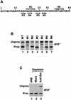
A sequence element downstream of the stem-loop is required for Drosophila histone pre-mRNA processing. (A) Sequence of the dH3∗ pre-mRNA starting first nucleotide after the stem-loop. The six mutations M1 to M6 are shown with the nucleotide substitutions indicated above and below the wild-type sequence. (B) In vitro processing of the wild-type (WT) and mutant dH3∗ pre-mRNAs (indicated above each lane) in the nuclear extract from S-2 cells. The processing of M1 and M2 and of M3 to M6 mutant pre-mRNAs was analyzed on two gels (lanes 1 to 3 and 4 to 7), accounting for the differences in separation of unprocessed and processed RNAs across the panel. (C) dNE from S-2 cells was incubated with a control monoclonal antibody (mock, lane 2) or anti-Sm antibody (lane 3), and the antibody complexes were removed from the extract with protein G beads. The processing activity of the depleted nuclear extract was assayed with dH3∗ pre-mRNA. Lane 1 represents untreated nuclear extract.
FIG. 4.
FIG. 4.
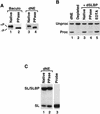
Drosophila and mammalian nuclear extracts cleave pre-mRNA at different sites. (A) dH3∗ pre-mRNA was processed in S-2 dNE (lane 1) and mouse H2a pre-mRNA was processed in either mouse myeloma (mNE, lane 2) or HeLa (hNE, lane 3) nuclear extracts. The length of the processing products (Proc) was compared after electrophoresis in an 8% low-resolution denaturing gel. The dH3∗ pre-mRNA (Unproc) substrate is longer from mouse H2a pre-mRNA at the 3′ end by 19 nucleotides, but the sequence of the 5′ end is identical. (B) The processing product of mouse H2a pre-mRNA generated in mNE (lane 2) was analyzed in high-resolution, 40-cm 12% polyacrylamide gels, next to a 48-nucleotide synthetic RNA ending precisely at the ACCCA sequence (lane 1). (C) Mammalian (lanes 1 and 3) and Drosophila (lane 2) processing products, generated as described for panel A, were analyzed side by side in a high-resolution gel, as described for panel B.
FIG. 5.
FIG. 5.
Processing of histone pre-mRNA in the heterologous nuclear extracts. (A) The mouse H2a pre-mRNA is processed with low efficiency in S-2 dNE (lane 4). Generation of the processing product, indicated with the arrow, is inhibited by excess RNA containing the wild-type stem-loop (SL, lane 6) but not the reverse stem mutation (RS, lane 5). Processing of dH3∗ pre-mRNA in a limited amount of S-2 dNE (lane 1) and mouse H2a pre-mRNA in mNE at 32 and 22°C (lanes 2 and 3) are shown for comparison. Processing of dH3∗ pre-mRNA was carried out under suboptimal conditions to reduce amount of the final product and better assess its migration mobility. (B) mNE does not process dH3∗ pre-mRNA. Processing of dH3∗ pre-mRNA was tested in nuclear extract from mouse myeloma cells (mNE, lane 2). Processing of the same substrate in Drosophila S-2 nuclear extract (dNE, lane 1) is shown for comparison.
FIG. 6.
FIG. 6.
Recombinant dSLBP restores processing activity to dSLBP-depleted nuclear extract. (A) Amino acid sequence of dSLBP. The RBD is underlined, and the arrows indicate the starting point of a 103-amino-acid deletion variant. (B) Diagram of the full-length dSLBP (FL) consisting of the N-terminal domain (dN), the Drosophila RBD (dRBD), and the 17-amino-acid end (dC). Regions included in three deletion mutants are shown below. The 103-amino-acid mutant starts 15 amino acids upstream from the Drosophila RBD, as shown in panel A, and contains the remainder of the protein, whereas the 88-amino-acid protein begins with the Drosophila RBD. The 71-amino-acid protein consists only of Drosophila RBD. (C) The S-2 nuclear extract was analyzed by Western blotting before (lane 1) and after depletion with dSLBP antibody (lane 2). The asterisk indicates a cross-reacting protein not removed from the extract during depletion. (D) The dH3∗ histone pre-mRNA was incubated in S-2 nuclear extract (lane 1), in dSLBP-depleted nuclear extract alone (lane 2), or in the presence of the baculovirus-expressed variants, as indicated (lanes 2 to 7). The H-D-H is a hybrid protein consisting of Drosophila RBD and two flanking domains from human SLBP (see Fig. 9A for details).
FIG. 7.
FIG. 7.
Phosphorylation of dSLBP is required for processing. (A) Western blotting of baculovirus-expressed dSLBP (left panel) and the dSLBP in the S-2 nuclear extract (right panel) before (lanes 1 and 3) and after (lanes 2 and 4) treatment with PPase. (B) Processing of the dH3∗ pre-mRNA was analyzed in dNE (lane 1), in the dSLBP-depleted dNE (lane 2), after the addition of the untreated baculovirus-expressed dSLBP (lane 3), in dSLBP treated with PPase in the presence of Mn2+ (lane 4), or in dSLBP treated with PPase in the presence of Mn2+ and 20 mM EDTA (lane 5). (C) Equal amounts of the dNE (lane 1) or the dNE treated with PPase (lane 2) were analyzed by a mobility shift assay for the ability to bind to a 26-nucleotide stem-loop RNA labeled with [32P]ATP at the 5′ end. Lane 3 contains only the free probe.
FIG. 8.
FIG. 8.
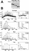
dSLBP contains four phosphates on the C-terminal region. (A) The deletion mutants of dSLBP shown in Fig. 5B were expressed by using the baculovirus system and purified. Aliquots of each variant were treated with PPase, and the untreated (lanes 1, 3, and 5) and dephosphorylated (lanes 2, 4, and 6) proteins were separated by gel electrophoresis and then detected by Western blotting. The mobility of the 103 (lanes 1 and 2)- and 88 (lanes 3 and 4)-amino-acid dSLBPs was increased after PPase treatment, whereas the mobility of the 71-amino-acid protein containing only the RBD was unchanged (lanes 5 and 6). (B) The full-length dSLBP and the 103-, 88-, and 71-amino-acid dSLBPs expressed in insects cells were analyzed by mass spectrometry as described in Materials and Methods. The charge/mass (m/z) spectrum is shown, and the charges on the individual clusters of peaks are indicated. The insets show a blowup of the most abundant ion. The various phosphorylated forms and proteins associated with sodium or potassium ions are indicated. The major peaks in the full-length, 103- and 88-amino-acid dSLBPs correspond to a molecular mass of 320 Da higher than that predicted from the amino acid sequence, corresponding to the four phosphate groups. The two satellite peaks indicated by the circle and square in the full-length dSLBP correspond to additional partially phosphorylated molecules containing five and six phosphates, respectively, whereas the peak indicated by the triangle corresponds to a protein containing three phosphates. The major peak in the 71-amino-acid SLBP corresponds to the molecular weight predicted from the amino acid sequence, indicating that the RBD is not phosphorylated.
FIG. 9.
FIG. 9.
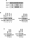
Drosophila and human SLBPs are not interchangeable. (A) Diagram of dSLBP and human SLBP (hSLBP) and a hybrid protein H-D-H composed of the RBD from dSLBP (dRBD) and both flanking domains from human SLBP (hN and hC). (B) The mouse H1t (left panel) and dH3∗ (right panel) pre-mRNAs were processed in the species-specific SLBP-depleted nuclear extracts alone (lane 2 in each panel) or in the presence of the baculovirus-expressed proteins, as indicated. Lane 1 in each panel shows processing in the control, undepleted nuclear extract. (C) Drosophila and human SLBPs inhibit processing in the heterologous nuclear extract. The Drosophila dH3∗ (left panel) and mouse H2a (right panel) pre-mRNAs were incubated in the Drosophila and mouse nuclear extracts, respectively (lane 1 in each panel) and in the same extracts in the presence of the indicated baculovirus-expressed SLBPs (lanes 2 and 3 in each panel).

Similar articles

Cited by

References

    1. Albig, W., and D. Doenecke. 1997. The human histone gene cluster at the D6S105 locus. Hum. Genet. 101:284-294. - PubMed
    1. Albig, W., P. Kioschis, A. Poustka, K. Meergans, and D. Doenecke. 1997. Human histone gene organization: nonregular arrangement within a large cluster. Genomics 40:314-322. - PubMed
    1. Battle, D. J., and J. A. Doudna. 2001. The stem-loop binding protein forms a highly stable and specific complex with the 3′ stem-loop of histone mRNAs. RNA 7:123-132. - PMC - PubMed
    1. Birnstiel, M. L., and F. J. Schaufele. 1988. Structure and function of minor snRNPs, p. 155-182. In M. L. Birnstiel (ed.), Structure and function of major and minor small ribonucleoprotein particles. Springer-Verlag, Berlin, Germany.
    1. Bond, U. M., T. A. Yario, and J. A. Steitz. 1991. Multiple processing-defective mutations in a mammalian histone premessenger RNA are suppressed by compensatory changes in U7 RNA both in vivo and in vitro. Genes Dev. 5:1709-1722. - PubMed

Publication types

MeSH terms

LinkOut - more resources