

The role of disulfide bond in the amyloidogenic state of beta(2)-microglobulin studied by heteronuclear NMR
- PMID: 12192077
- PMCID: PMC2373597
- DOI: 10.1110/ps.0213202
The role of disulfide bond in the amyloidogenic state of beta(2)-microglobulin studied by heteronuclear NMR
Abstract
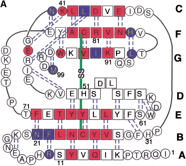
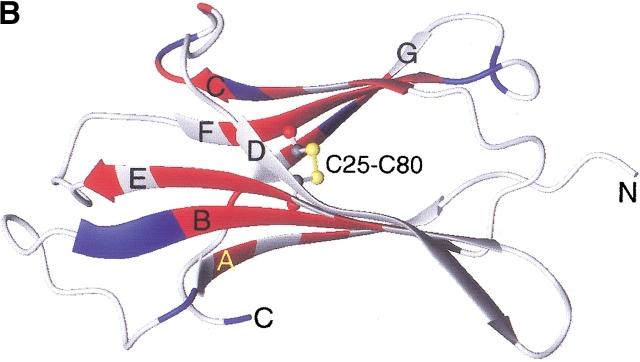
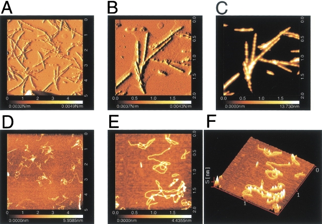
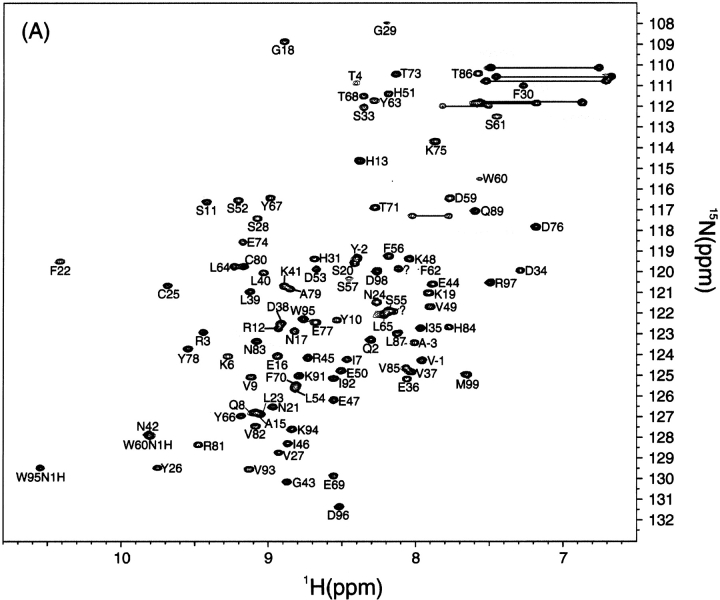
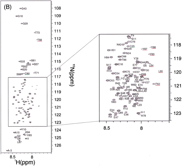
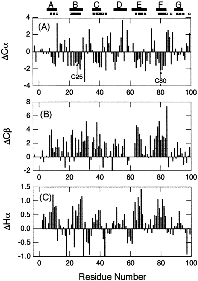
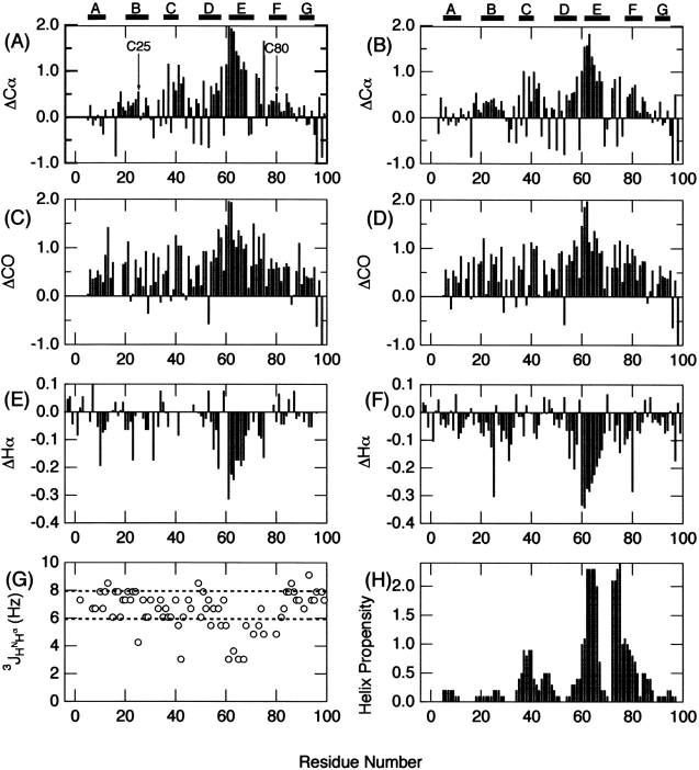
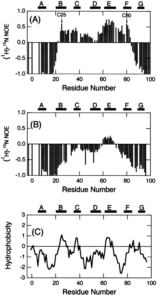
beta(2)-Microglobulin (beta2-m) is a major component of dialysis-related amyloid fibrils. Although recombinant beta2-m forms needle-like fibrils by in vitro extension reaction at pH 2.5, reduced beta2-m, in which the intrachain disulfide bond is reduced, cannot form typical fibrils. Instead, thinner and flexible filaments are formed, as shown by atomic force microscopy images. To clarify the role of the disulfide bond in amyloid fibril formation, we characterized the conformations of the oxidized (intact) and reduced forms of beta2-m in the acid-denatured state at pH 2.5, as well as the native state at pH 6.5, by heteronuclear NMR. [(1)H]-(15)N NOE at the regions between the two cysteine residues (Cys25-Cys80) revealed a marked difference in the pico- and nanosecond time scale dynamics between that the acid-denatured oxidized and reduced states, with the former showing reduced mobility. Intriguingly, the secondary chemical shifts, DeltaCalpha, DeltaCO, and DeltaHalpha, and (3)J(HNHalpha) coupling constants indicated that both the oxidized and reduced beta2-m at pH 2.5 have marginal alpha-helical propensity at regions close to the C-terminal cysteine, although it is a beta-sheet protein in the native state. The results suggest that the reduced mobility of the denatured state is an important factor for the amylodogenic potential of beta2-m, and that the marginal helical propensity at the C-terminal regions might play a role in modifying this potential.
Figures
Similar articles
-
A single disulfide bond differentiates aggregation pathways of beta2-microglobulin.J Mol Biol. 2005 Nov 25;354(2):473-82. doi: 10.1016/j.jmb.2005.09.075. Epub 2005 Oct 7. J Mol Biol. 2005. PMID: 16242719
-
The intrachain disulfide bond of beta(2)-microglobulin is not essential for the immunoglobulin fold at neutral pH, but is essential for amyloid fibril formation at acidic pH.J Biochem. 2002 Jan;131(1):45-52. doi: 10.1093/oxfordjournals.jbchem.a003076. J Biochem. 2002. PMID: 11754734
-
Conformational dynamics of beta(2)-microglobulin analyzed by reduction and reoxidation of the disulfide bond.J Biochem. 2003 Jun;133(6):731-6. doi: 10.1093/jb/mvg094. J Biochem. 2003. PMID: 12869529
-
Structural stability of amyloid fibrils of beta(2)-microglobulin in comparison with its native fold.Biochim Biophys Acta. 2005 Nov 10;1753(1):64-75. doi: 10.1016/j.bbapap.2005.08.002. Epub 2005 Aug 24. Biochim Biophys Acta. 2005. PMID: 16213801 Review.
-
Limited proteolysis in the investigation of beta2-microglobulin amyloidogenic and fibrillar states.Biochim Biophys Acta. 2005 Nov 10;1753(1):44-50. doi: 10.1016/j.bbapap.2005.09.004. Epub 2005 Sep 23. Biochim Biophys Acta. 2005. PMID: 16213198 Review.
Cited by
-
Fibril growth kinetics reveal a region of beta2-microglobulin important for nucleation and elongation of aggregation.J Mol Biol. 2008 Apr 18;378(1):251-63. doi: 10.1016/j.jmb.2008.01.092. Epub 2008 Feb 12. J Mol Biol. 2008. PMID: 18342332 Free PMC article.
-
Extremely Amyloidogenic Single-Chain Analogues of Insulin's H-Fragment: Structural Adaptability of an Amyloid Stretch.Langmuir. 2020 Oct 20;36(41):12150-12159. doi: 10.1021/acs.langmuir.0c01747. Epub 2020 Oct 7. Langmuir. 2020. PMID: 32988199 Free PMC article.
-
Disulfide bridges remain intact while native insulin converts into amyloid fibrils.PLoS One. 2012;7(6):e36989. doi: 10.1371/journal.pone.0036989. Epub 2012 Jun 1. PLoS One. 2012. PMID: 22675475 Free PMC article.
-
Competition between intramolecular and intermolecular interactions in an amyloid-forming protein.J Mol Biol. 2009 Jun 19;389(4):776-86. doi: 10.1016/j.jmb.2009.04.042. Epub 2009 Apr 23. J Mol Biol. 2009. PMID: 19393661 Free PMC article.
-
Small molecule-mediated inhibition of β-2-microglobulin-based amyloid fibril formation.J Biol Chem. 2017 Jun 23;292(25):10630-10638. doi: 10.1074/jbc.M116.774083. Epub 2017 May 3. J Biol Chem. 2017. PMID: 28468825 Free PMC article.
References
-
- Alexandrescu, A. 2001. An NMR-based quenched hydrogen exchange investigation of model amyloid fibrils formed by cold shock protein A. Pac. Symp. Biocomput. 67–78. - PubMed
-
- Bjorkman, P.J., Saper, M.A., Samraoui, B., Bennett, W.S., Strominger, J.L., and Wiley, D.C. 1987. Structure of the human class I histocompatibility antigen, HLA-A2. Nature 329 506–512. - PubMed
-
- Brange, J., Andersen, L., Laursen, E.D., Meyn, G., and Rasmussen, E. 1997. Toward understanding insulin fibrillation. J. Pharm. Sci. 86 517–525. - PubMed
-
- Bucciantini, M., Giannoni, E., Chiti, F., Baroni, F., Formigli, L., Zurdo, J., Taddei, N., Ramponi, G., Dobson, C.M., and Stefani, M. 2002. Inherent toxicity of aggregates implies a common mechanism for protein misfolding diseases. Nature 416 507–511. - PubMed
Publication types
MeSH terms
Substances
LinkOut - more resources
Full Text Sources
Molecular Biology Databases
Research Materials
