. 2002 Sep 3;99(18):11957-62.
doi: 10.1073/pnas.182412699. Epub 2002 Aug 21.

PDZ-domain interactions and apical expression of type IIa Na/P(i) cotransporters

Affiliations

PDZ-domain interactions and apical expression of type IIa Na/P(i) cotransporters

Nati Hernando et al. Proc Natl Acad Sci U S A. .

Abstract

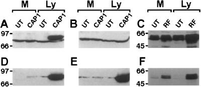
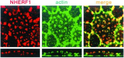
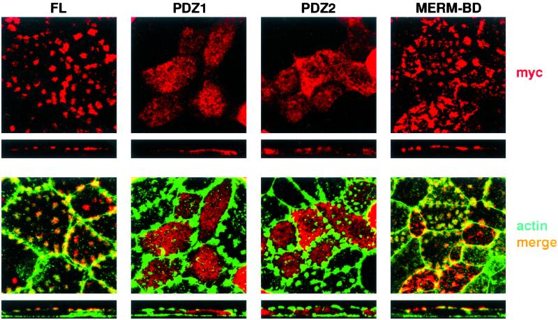
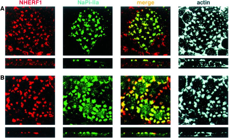
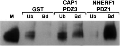
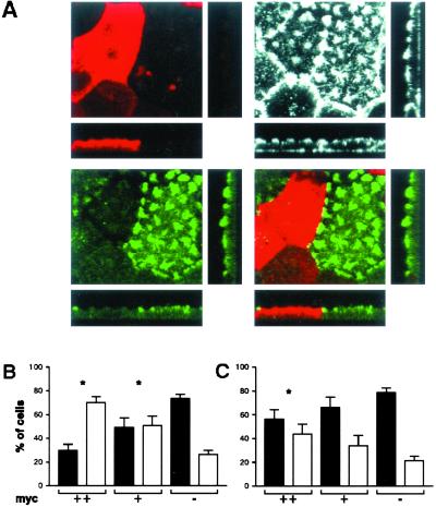
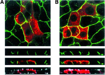
Type IIa Na/P(i) cotransporters are expressed in renal proximal brush border and are the major determinants of inorganic phosphate (P(i)) reabsorption. Their carboxyl-terminal tail contains information for apical expression, and interacts by means of its three terminal amino acids with several PSD95/DglA/ZO-1-like domain (PDZ)-containing proteins. Two of these proteins, NaPi-Cap1 and Na/H exchanger-regulatory factor 1 (NHERF1), colocalize with the cotransporter in the proximal brush border. We used opossum kidney cells to test the hypothesis of a potential role of PDZ-interactions on the apical expression of the cotransporter. We found that opossum kidney cells contain NaPi-Cap1 and NHERF1 mRNAs. For NHERF1, an apical location of the protein could be documented; this location probably reflects interaction with the cytoskeleton by means of the MERM-binding domain. Overexpression of PDZ domains involved in interaction with the cotransporter (PDZ-1/NHERF1 and PDZ-3/NaPi-Cap1) had a dominant-negative effect, disturbing the apical expression of the cotransporter without affecting the actin cytoskeleton or the basolateral expression of Na/K-ATPase. These data suggest an involvement of PDZ-interactions on the apical expression of type IIa cotransporters.


Figures

Figure 1
Figure 1
OK cells express endogenous NaPi-Cap1 and NHERF-1 mRNAs. Samples of 10 μg of polyA RNA extracted from mouse kidney cortex (K) and OK cells (OK) were separated in a 10% agarose denaturing gel, transferred to nitrocellulose membranes, and incubated overnight with [32P]dCTP-labeled NaPi-Cap1 (A) or NHERF1 (B) probes.
Figure 2
Figure 2
Endogenous NHERF-1 (C) but not NaPi-Cap1 (A and B) is recognized by available antibodies. Samples of Ly (100 μg) or Ms (50 μg) from untransfected (UT) or transfected (CAP1/RF) OK cells were separated on an SDS/10% PAGE and transferred to nitrocellulose membranes. Membranes were incubated with either a NaPi-Cap1 (Diphor1) antibody in the absence (A) or presence (B) of 50 μg/ml of the antigenic peptide, or with a NHERF antibody (C). The same membranes were subsequently incubated with a myc monoclonal antibody (DF).
Figure 3
Figure 3
Endogenous NHERF-1 is expressed in actin-containing apical patches. Confluent OK cultures plated on coverslips were stained with the NHERF antibody (in red) and with phalloidin-AlexaFluor to detect actin signal (in green). Samples were analyzed by confocal microscopy: squares represent apical focal planes and rectangles represent confocal cross sections. There was a total overlap between the NHERF1 and actin stainings, as reflected by the yellow signal of the merge composite, indicating that NHERF1 is accumulated in actin-containing apical patches.
Figure 4
Figure 4
The MERM-binding domain of NHERF1 retains apical expression. OK cultures were transfected with myc constructs containing either the full-length NHERF1 (FL) or only the PDZ-1, PDZ-2, or MERM-binding domain. Samples were stained with myc antibody (red) and with phalloidin-AlexaFluor (green) and were analyzed by confocal microscopy. (Upper) myc staining (NHERF-related signals). (Lower) myc/actin merge. The transfected FL protein as well as the construct containing only the MERM-binding domain reproduced the pattern of expression of the endogenous NHERF1; i.e., they appear in apical patches.
Figure 5
Figure 5
NaPi4 and NHERF1 colocalize in actin-containing apical patches. OK cells were transfected with either EGFP-NaPi (A) or with myc-NHERF1 (B). Cultures then were stained with antibodies against NHERF and actin (A) or against myc, NaPi4, and actin (B), respectively. Samples were analyzed by confocal microscopy. NaPi4 and NHERF1 colocalize in actin patches, as reflected by the yellow signal of the merge composite.
Figure 6
Figure 6
NaPi-Cap1 and NHERF1 pull down the endogenous (NaPi4) cotransporter. Samples of 200 μg of OK Ly were incubated with GST alone or with GST-PDZ3/Cap1 or GST-PDZ1/NHERF1, as indicated in Materials and Methods. Unbound (Ub; supernatants) and bound (Bd; pellets) samples as well as 50 μg of Ms were separated in an SDS/PAGE gel, transferred to a nitrocellulose membrane, and incubated with a NaPi4 antibody. PDZ3/CAP1 and PDZ1/NHERF1 led to the pull-down of NaPi4, indicating that NaPi4 is able to interact with both proteins.
Figure 7
Figure 7
Expression of PDZ1/NHERF1 and PDZ3/NaPi-Cap1 impairs apical expression of the endogenous NaPi4. OK cells were transfected with myc-fused PDZ1/NHERF (A, B) or PDZ3/Cap1 (C), and stained for myc (A, red), actin (A, white), and NaPi4 (A, green). In every transfection experiment, we consistently detected the presence of two types of cells: one with a strong myc signal (myc++) and a second one with a clear but weaker myc fluorescence (myc+). Discrimination between both groups was easy and consistently clear within single experiments. (A) Squares show apical focal planes and rectangles show confocal cross sections of cultures transfected with myc-fused PDZ1/NHERF. Untransfected (myc−) or transfected cells (myc++, myc+) showing a normal actin cytoskeleton were analyzed for the expression of strong (black bars) or weak (white bars) NaPi4 patches. (B and C) Bars = mean ± SE of nine independent experiments. *, P < 0.05.
Figure 8
Figure 8
Expression of PDZ1/NHERF1 and PDZ3/NaPi-Cap1 does not affect basolateral expression of the endogenous Na/K-ATPase. OK cells were transfected with myc-fused PDZ1/NHERF (A) or PDZ3/Cap1 (B) and stained for myc (red), actin (white), and Na/K-ATPase (green). Samples were analyzed by confocal microscopy: squares represent basal focal planes and rectangles represent confocal cross sections.

References

    1. Murer H, Kaissling B, Forster I, Biber J. In: The Kidney: Physiology and Pathophysiology. 3rd Ed. Seldin D W, Giebish G, editors. Philadelphia: Lippincott; 2000.
    1. Murer H, Hernando N, Forster I, Biber J. Physiol Rev. 2000;80:1373–1409. - PubMed
    1. Beck L, Karaplis A C, Amizuka N, Hewson A S, Ozawa H, Tenenhouse H S. Proc Natl Acad Sci USA. 1998;95:5372–5377. - PMC - PubMed
    1. Custer M, Lötscher M, Biber J, Murer H, Kaissling B. Am J Physiol. 1994;35:F767–F774. - PubMed
    1. Ritthaler T, Traebert M, Lötscher M, Biber J, Murer H, Kaissling B. Kidney Int. 1999;55:976–983. - PubMed

Publication types

Substances