Comparative Study
. 2002 Sep-Oct;4(5):388-99.
doi: 10.1038/sj.neo.7900234.

IL-13Ralpha2 is a glioma-restricted receptor for interleukin-13

Affiliations
Comparative Study

IL-13Ralpha2 is a glioma-restricted receptor for interleukin-13

Akiva Mintz et al. Neoplasia. 2002 Sep-Oct.

Abstract

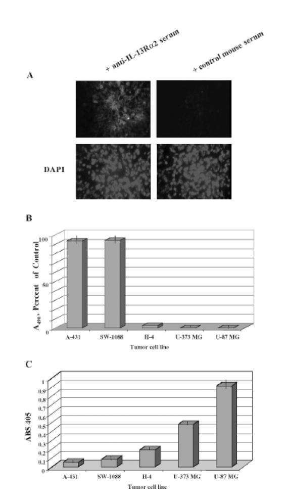
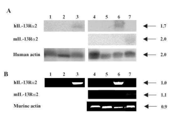
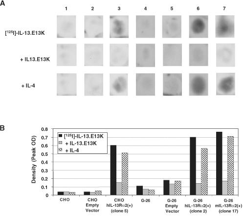
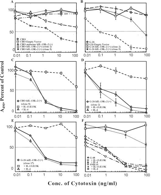
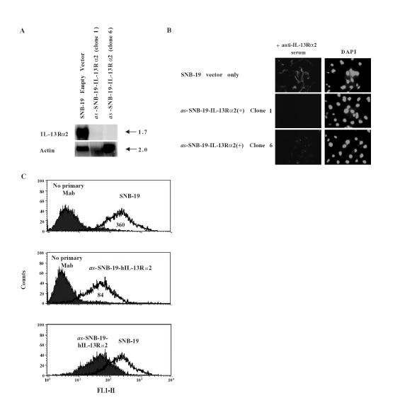
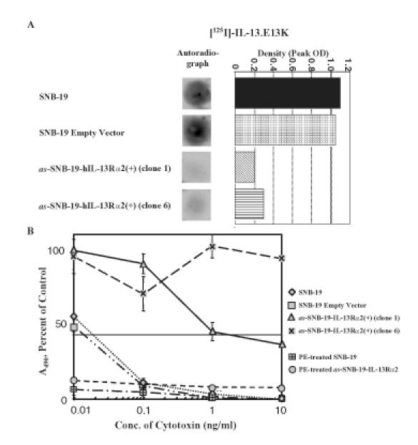
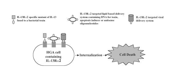
We have found that binding sites for interleukin-13 (IL-13) are overexpressed in a vast majority of high-grade astrocytomas (HGAs). These binding sites for IL-13 are distinct from the physiological receptor in that it does not bind IL-4. We also demonstrated that IL-13 receptor alpha 2 protein chain (IL-13Ralpha2), an IL-4-independent receptor for IL-13, is abundant among HGAs, but not in normal organs. To examine if IL-13Ralpha2 is the tumor-associated site for IL-13, we stably transfected normal Chinese hamster ovary (CHO) cells and glioma G-26 cells to express either human (h) or murine (m) IL-13Ralpha2. CHO-hIL-13Ralpha2(+) cells and G-26-h/mIL-13Ralpha2(+) cells, and not CHO and G-26 parental or mock-transfected cells, specifically bound IL-13 in an IL-4-independent manner. The IL-13Ralpha2(+) cells also became highly susceptible to the killing by an IL-13-based cytotoxic fusion protein. In loss of function studies, a HGA cell line, SNB-19, was transfected with antisense (as) hIL-13Ralpha2. as-SNB-19-hIL-13Ralpha2(+) cells lost their natural affinity towards IL-13 and became resistant to IL-13-based cytotoxins. The fact, that IL-13Ralpha2-positive cells bind IL-13 independent of IL-4, become susceptible to IL-13 cytotoxins, and cells deprived of IL-13Ralpha2 receptor lose these features, demonstrates that IL-13Ralpha2 is the brain tumor-associated receptor for IL-13.


Figures

Figure 1
Figure 1
(A) hIL-13Rα2 immunoreactivity in a GBM tumor specimen. Nonimmunized serum was used on a contiguous section of the same tumor as a negative control. Tissues were also stained for nuclear presence using DAPI. (B) Cytotoxicity of high-grade astrocytoma cells (SW-1088, U-373 MG, U-87 MG), low-grade astrocytoma cells (H-4), and epidermoid tumor cells (A-431) using 100 ng/ml IL-13.E13K-PE38QQR determined in a colorimetric cell proliferation assay. Vertical lines represent SD. (C) Reactivity of high-grade astrocytoma cells (U-87 MG, U-373 MG, SW-1088), low-grade astrocytoma cells (H-4), and epidermoid tumor cell (A-431) to anti-hIL-13Rα2 serum in a cell ELISA assay. Vertical lines represent SD.
Figure 2
Figure 2
(A) Northern blot of CHO and G-26 cells transfected with hIL-13Rα2 or mIL-13Rα2 and controls. Parental CHO cells (lane 1), mock-transfected CHO (lane 2), CHO-hIL-13Rα2(+) clone 5 (lane 3), parental G-26 cells (lane 4), mock-transfected G-26 (lane 5), G-26-hIL-13Rα2(+) clone 2 (lane 6), and G-26-mIL-13Rα2(+) clone 17 (lane 7) are shown. The size of transcripts is given in kilobases (kb). (B) RT-PCR of hIL-13α2 or mIL-13Rα2 in G-26 and CHO controls and transfected cells. The order of analyzed cells is the same as in (A). The size of PCR-amplified fragments is given in kilobases (kb).
Figure 3
Figure 3
(A) Autoradiography of CHO-hIL-13Rα2(+) clone 5, G-26-hIL-13Rα2(+) clone 2, G-26-mIL-13Rα2(+) clone 17, and control cells using labeled IL-13.E13K. Parental CHO cells (column 1), mock-transfected CHO (column 2), CHO-hIL-13Rα2(+) clone 5 (column 3), parental G-26 cells (column 4), mock-transfected G-26 (column 5), G-26-hIL-13Rα2(+) clone 2 (column 6), and G-26-mIL-13Rα2(+) clone 17 (column 7). The assay was performed in the absence of blocker or in the presence of an excess of unlabeled IL-13.E13K or IL-4. Each experiment was repeated at least four times and the results shown are a representative sample of one of the experiments. (B) Histogram of peak densities of 125I IL-13.E13K binding in the absence or presence of IL-13.E13K and IL-4 in CHO-hIL-13Rα2(+) clone 5, G-26-hIL-13Rα2(+) clone 2, G-26-mIL-13Rα2(+) clone 17, and control cells. Column designations are the same as in (A). Measurements were taken from scanned X-rays shown in (A).
Figure 4
Figure 4
Cytotoxicity using IL-13.E13K-PE38QQR determined in a colorimetric cell proliferation assay. Vertical bars represent SD. (A) CHO parental cells, CHO mock-transfected, and CHO-hIL-13Rα2(+) clones 2 and 5. (B) G-26 parental cells, G-26 mock-transfected, and G-26-hIL-13Rα2(+) clones 2 and 5. (C-E) Neutralization assays in which the cell killing by IL-13.E13K-PE38QQR in IL-13Rα2(+) clones is blocked in the presence of an excess of IL-13.E13K, but not IL-4, in CHO and G-26 cells. (F) G-48a and SNB-19 HGA cells and neutralization of IL-13.E13K-PE38QQR cytotoxicity in the presence of an excess of IL-13.E13K, but not IL-4.
Figure 5
Figure 5
(A) Northern blot of IL-13Rα2 and actin in SNB-19 mock-transfected, and as-SNB-19-hIL-13Rα2(+) clones 1 and 6. (B) IL-13Rα2 immunoreactivity in SNB-19 cells transfected with as-IL-13Rα2 (clones 1 and 6) and with vector only. DAPI nuclear staining was also performed. (C) Flow cytometry performed on SNB-19 parental cells and as-SNB-19-IL-13Rα2(+) clone 6 cells. The bottom graph juxtaposes the distribution of the two experiments. Mean values of each distribution are listed in the area under the peaks. Experiment was repeated three times.
Figure 6
Figure 6
(A) Autoradiography and histogram of peak densities of IL-13 binding sites in SNB-19 parental cells, SNB-19 mock-transfected, and in as-SNB-19-hIL-13Rα2(+) clones treated with labeled IL-13.E13K. Each experiment was repeated at least four times and the results shown are a representative sample of the experiments. (B) Cytotoxicity assay using IL-13.E13K-PE38QQR cytotoxin or wild-type PE in SNB-19 parental cells, SNB-19 mock-transfected, and in as-SNB-19-hIL-13Rα2(+) clones 1 and 6. Vertical bars represent SD.
Figure 7
Figure 7
Schemata of possible therapeutic interventions by using IL-13Rα2 as a molecularly defined target.

References

    1. Debinski W, Gibo DM, Hulet SW, Connor JR, Gillespie GY. Receptor for interleukin 13 is a marker and therapeutic target for human high-grade gliomas. Clin Cancer Res. 1999;5:985–990. - PubMed
    1. Debinski W, Gibo DM, Slagle B, Powers SK, Gillespie GY. Receptor for interleukin 13 is abundantly and specifically over-expressed in patients with glioblastoma multiforme. Int J Oncol. 1999;15:481–486. - PubMed
    1. Debinski W, Obiri NI, Powers SK, Pastan I, Puri RK. Human glioma cells over-express receptor for interleukin 13 and are extremely sensitive to a novel chimeric protein composed of interleukin 13 and Pseudomonas exotoxin. Clin Cancer Res. 1995;1:1253–1258. - PubMed
    1. Debinski W. An immune regulatory cytokine and high-grade gliomas: an unexpected link. Crit Rev Oncog. 1998;9:256–268. - PubMed
    1. Minty A, Chalon P, Derocq JM, Dumont X, Guillemot JC, Kaghad M, Labit C, Leplatois P, Liauzun P, Miloux B, Minty C, Casellas P, Loison G, Lupker J, Shire D, Ferrara P, Caput D. Interleukin-13 is a new human lymphokine regulating inflammatory and immune responses. Nature. 1993;36:248–251. - PubMed

Publication types

MeSH terms

LinkOut - more resources