. 2002 Aug 22:3:10.
doi: 10.1186/1471-2172-3-10.

The kappa B transcriptional enhancer motif and signal sequences of V(D)J recombination are targets for the zinc finger protein HIVEP3/KRC: a site selection amplification binding study

Affiliations

The kappa B transcriptional enhancer motif and signal sequences of V(D)J recombination are targets for the zinc finger protein HIVEP3/KRC: a site selection amplification binding study

Carl E Allen et al. BMC Immunol. .

Abstract

Background: The ZAS family is composed of proteins that regulate transcription via specific gene regulatory elements. The amino-DNA binding domain (ZAS-N) and the carboxyl-DNA binding domain (ZAS-C) of a representative family member, named kappaB DNA binding and recognition component (KRC), were expressed as fusion proteins and their target DNA sequences were elucidated by site selection amplification binding assays, followed by cloning and DNA sequencing. The fusion proteins-selected DNA sequences were analyzed by the MEME and MAST computer programs to obtain consensus motifs and DNA elements bound by the ZAS domains.

Results: Both fusion proteins selected sequences that were similar to the kappaB motif or the canonical elements of the V(D)J recombination signal sequences (RSS) from a pool of degenerate oligonucleotides. Specifically, the ZAS-N domain selected sequences similar to the canonical RSS nonamer, while ZAS-C domain selected sequences similar to the canonical RSS heptamer. In addition, both KRC fusion proteins selected oligonucleoties with sequences identical to heptamer and nonamer sequences within endogenous RSS.

Conclusions: The RSS are cis-acting DNA motifs which are essential for V(D)J recombination of antigen receptor genes. Due to its specific binding affinity for RSS and kappaB-like transcription enhancer motifs, we hypothesize that KRC may be involved in the regulation of V(D)J recombination.


Figures

Figure 1
Figure 1
KRC fusion proteins and site-selection EMSA Figure 1A. KRC fusion proteins. (Top) The full-length KRC protein is described schematically. In the ZAS-N and ZAS-C DNA-binding domains the zinc-fingers, acidic regions, and serine-threonine-rich regions highlighted. ZASN, ZAS-N domain; ZF3, zinc finger 3; NLS, nuclear localization signal; GTP, GTPase motif; ZASC, ZAS-C motif. (Bottom) KRC fusion proteins, KRC/ZAS-N and KRC/ZAS-C are described schematically. KRC/ZAS-N is a S-tag fusion protein containing the ZAS-N DNA-binding domain (nt 949–2167) KRC/ZAS-C is an Mbp fusion protein containing the ZAS-C DNA-binding domain (nt 5544–7015). These are the fusion proteins used in the site-selection assay described in this paper. Figure 1B and 1C. Electrophoretic mobility shift assays of the site selection procedures. (Bottom) A portion of the oligonucleotides (~0.2 ng and 5000 cpm) recovered from each round of site selection was 32P-labeled and incubated with KRC fusion proteins (~0.5 μg), (B) KRC/ZAS-N and (C) KRC/ZAS-C, in the presence of 10 μg poly(dI-dC). DNA-protein complexes and free probes were resolved in 6% polyacrylamide gels and visualized by exposing dried gels to X-ray films. The probes used in lanes 1 through 5 were derived from aliquots of DNA recovered from round one through five of site selection, respectively. C, DNA-protein complexes; and F, free probes.
Figure 2
Figure 2
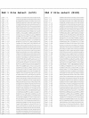
KRC-bound sequences recovered from site-selection experiments. The sequences were isolated from the pool of oligonucleotides remaining after five rounds of site selection with either the N-terminal KRC/ZAS-N or the C-terminal KRC/ZAS-C fusion proteins. 53 sequences were obtained from the KRC/ZAS-N-bound pool, and 49 sequences were obtained from the KRC/ZAS-C-bound pool. Sequences are shown as sequenced with a 5' BSS1 primer in the forward orientation: BSS1(N25)BSS2.
Figure 3
Figure 3
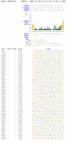
ZAS-N-selected sequence: MEME motif search and alignment, n = 25.Simplified position-specific probability matrix represents the probability of each possible letter appear a each possible position in an occurrence of the motif. Numbers are described as n/10 (eg 50 is represented as 5), with "0" described as ":". Information Content Diagram provides information of which positions in the motif are most (and least) highly conserved. Columns in the information content diagram are shaded according to the majority category of the letters occurring in that column of alignment. If no letter category has a frequency >0.5, the column is black. A = red, C = blue, G = orange, T = green. Summing of information content for each position gives the total information content of the motif, which is approximately equal to the log likelihood ratio divided by the number of occurrences times ln(2). Multilevel Consensus Sequence is calculated from the motif position-specific matrix where the most likely nucleotide is printed at the top of a column. Only letters with probabilities of 0.2 or higher are included. Summary Information is printed below the multilevel consensus sequence. "Width" describes the length of the motif. "Sites" describes the number of sequences in the dataset which contributed to the consensus sequence. The "log likelihood ratio" is the logarithm of the ratio of the probability of the occurrences of the motif given the motif model versus their probability given the null model. The "E-value" is an estimate of the expected number of motifs with the given log likelihood ratio (or higher), and with the same width and number of occurrences, that one would find in a similarly sized set of random sequences. Motif Alignment displays the occurrences of the motif in the dataset. Each site is identified by the name of the sequence where it occurs, the strand (+ or -), and the position in the sequence where the site begins. The p-value of a site is computed from the match score of the site with the position specific scoring matrix for the motif, and gives the probability of a random string having the same match score or higher. (Above is described in detail at MEME [30]).
Figure 4
Figure 4
ZAS-C-selected sequence: MEME motif search and alignment, n = 25 Figure legend as above (Figure 3).
Figure 5
Figure 5
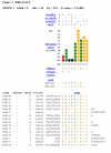
ZAS-N-selected sequence: MEME motif search and alignment, n = 9. Motif #1. Figure legend as above (Figure 3).
Figure 6
Figure 6
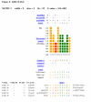
ZAS-N-selected sequence: MEME motif search and alignment, n = 9. Motif #2. Figure legend as above (Figure 3).
Figure 7
Figure 7
ZAS-C-selected sequence: MEME motif search and alignment, n = 9. Motif #1. Figure legend as above (Figure 3).
Figure 8
Figure 8
ZAS-C-selected sequence: MEME motif search and alignment, n = 9. Motif #3. Figure legend as above (Figure 3).
Figure 9
Figure 9
ZAS-N-selected sequence: MEME motif search and alignment, n = 9. Motif #1. Figure legend as above (Figure 3).
Figure 10
Figure 10
ZAS-N-selected sequence: MEME motif search and alignment, n = 5. Motif #1. Figure legend as above (Figure 3).
Figure 11
Figure 11
ZAS-N-selected sequence: MEME motif search and alignment, n = 5. Motif #2. Figure legend as above (Figure 3).
Figure 12
Figure 12
ZAS-C-selected sequence: MEME motif search and alignment, n = 5. Motif #1. Figure legend as above (Figure 3).
Figure 13
Figure 13
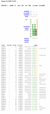
KRC/ZAS-N site selection consensus motif search: MAST results Human and murine genomic DNA identified by MAST by searching with the KRC/ZAS-N are listed. Features of DNA sequence from GenBank/NCBI are delineated by bar and footnote as follows: a. H. Sapiens COL4A6 gene, nt 16–60: COL4A6 exon 15 b. H. Sapiens IgH gene primer, DJ segment, 7–27: leukemia-specific primer c. H. Sapiens IgH gene primer, DJ segment, 30–46: D segment (Dxp1) d. H. Sapiens IgH gene primer, DJ segment, 47–59: J segment (J5) e. M. Musculus DNA for desmin-binding fragment DesF7, 1–107: LINE element The first line describes the GenBank-designated name of the DNA sequence. The next line lists the accession number and any publications associated with the sequence. The third line indicates the target sequence nucleotide positions of overlap as well as orientation. The combined p-value is defined as the probability of a randomly generated sequence of the same length having sequence p-values whose product is at least as small as the product of the sequence p-values of the matches of the motifs to the given sequence. The E-value of the match of a sequence in a database to a group of motifs is defined as the expected number of sequences in a random database of the same size that would match the motifs as well as the sequence does and is equal to the combined p-value of the sequence times the number of sequences in the database. (Above is described in detail at MAST [34]).
Figure 14
Figure 14
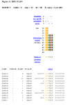
KRC/ZAS-C site selection consensus motif search: MAST results Human and murine genomic DNA identified by MAST by searching with the KRC/ZAS-C are listed. Nomenclature is described above (Figure 13). Features of DNA sequence from GenBank/NCBI are delineated by bar and footnote as follows: f. H. Sapiens ATP7B pseudogene, 1–54: ATP7B exon 6 g. H. Sapiens ATP7B pseudogene, > = 55: ATP7B exon 7 h. H. Sapiens clone 19r DH2-2/bcl-1 gene fusion reciprocal breakpoint, 1–54: IgHD (14q32) i. H. Sapiens clone 19r DH2-2/bcl-1 gene fusion reciprocal breakpoint, 55: N-nucleotide j. H. Sapiens clone 19r DH2-2/bcl-1 gene fusion reciprocal breakpoint, > = 56: bcl-1 (11q13) k. M. Musculus DNA for GFAP-binding fragment GFAPA10, 1–82: LINE element k. M. Musculus DNA for GFAP-binding fragment GFAPC13, 1–83: LINE element l. H. Sapiens ENL translocation, 1–51: ENL gene (partial) m. H. Sapiens ENL translocation, 55–84: 3' ENL 11q23 n. H. Sapiens IgH D1-4, 1–9: nanomer o. H. Sapiens IgH D1-4, 22–28: heptamer p. H. Sapiens IgH D1-4, 29–59: D region q. H. Sapiens IgH D1-4, 60–66: heptamer r. M. Musculus aldose reductase gene, 5–75: exon 4
Figure 15
Figure 15
Comparison of KRC-selected sequence with IgVκ and TCRAJ RSS elements. The KRC-bound sequences were compared to known RSS sequence in the murine TCR-alpha genes and the human immunoglobulin heavy chain genes, the loci of which have been fully sequenced [45,46]. Individual sequences in the training sets were compared manually with (A) RSS nonamers or (B) RSS hepamers defined in of the human Ig variable region gene segments of κ light chain (Table 1 of [45]) and the mouse TCRAJ gene segments (Fig. 2 of [46]). The nucleotide sequence of a RSS nonamer or heptamer was listed in the first column, followed by the name of the site selected sequence(s), and then by the gene segments with matching RSS elements. Sequence in the KRC fusion protein-bound oligonucleotides overlapped with heptamer, nonamer, or both in the RSS of 43.3% of human IgVκ genes and in 33.9% of murine TCRAJ genes. These calculations include only the IgVκ [45] and TCRAJ genes [46] which contain a nonamer, a heptamer, or both.
Figure 16
Figure 16
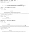
Comparison of KRC protein zinc finger domains. The zinc finger amino acid sequences of the KRC ZAS domains are compared. Mouse and human KRC ZAS-N amino acid sequences are aligned above the corresponding mouse and human KRC ZAS-C sequence. Above the sequences, the C and H represent the canonical Cys and His residues of the C2H2 domains, and the numbers represent the position within the alpha helix. The consensus sequence is derived from sequence analysis of all the known ZAS protein sequences [25]. The zinc finger 1 and zinc finger 2 domains within the ZAS domains are designated in green and blue font, respectively, in the consensus sequence. Amino acid residues that are common to all ZAS family members are capitalized. The critical amino acid residues within the α helical regions of the zinc finger regions are identical at all corresponding positions between the first and second zinc fingers of the ZAS-N and ZAS-C domains except at position 3 of the first zinc finger (outlined in gray) and at position 2 of the second zinc finger (outlined in gray). The linker region between the two zinc fingers is outlined in red.

References

    1. Seeler JS, Muchardt C, Suessle A, Gaynor RB. Transcription factor PRDII-BF1 activates human immunodeficiency virus type 1 gene expression. J Virol. 1994;68:1002–1009. - PMC - PubMed
    1. Brady JP, Kantorow M, Sax CM, Donovan DM, Piatigorsky J. Murine transcription factor α-A crystalline binding protein I. J Biol Chem. 1995;270:1221–1229. doi: 10.1074/jbc.270.8.3642. - DOI - PubMed
    1. Dörflinger U, Pscherer A, Moser M, Rümmele P, Schüle R, Buettner R. Activation of somatostatin receptor II expression by transcription factors MIBP1 and SEF-2 in the murine brain. Mol Cell Biol. 1999;19:3736–3747. - PMC - PubMed
    1. Hjelmsoe I, Allen CE, Cohn MA, Tulchinsky EM, Wu LC. The κB and V(D)J recombination signal sequence binding protein KRC regulates transcription of the mouse metastasis associated gene S100A4/mts1. J Biol Chem. 2000;275:913–920. doi: 10.1074/jbc.275.2.913. - DOI - PubMed
    1. Tanaka K, Matsumoto Y, Nakatani F, Iwamoto Y, Yamada Y. A zinc finger transcription factor, alpha A-crystallin binding protein 1, is a negative regulator of the chondrocyte-specific enhancer of the alpha 1 (II) collagen gene. Mol Cell Biol. 2000;20:4428–4435. doi: 10.1128/MCB.20.12.4428-4435.2000. - DOI - PMC - PubMed

Publication types

MeSH terms

LinkOut - more resources