

Case Reports
doi: 10.1136/fn.87.2.f125.
Portal vein thrombosis causing neonatal cerebral infarction
Affiliations
- PMID: 12193520
- PMCID: PMC1721458
- DOI: 10.1136/fn.87.2.f125
Item in Clipboard
Case Reports
Portal vein thrombosis causing neonatal cerebral infarction
Arch Dis Child Fetal Neonatal Ed.
2002 Sep.
Abstract
Neonatal cerebral infarction often occurs in the absence of known risk factors. Two such cases are described in which portal vein thrombosis was documented during two dimensional echocardiography. In both cases, infarcts were consistent with embolic events. A novel mechanism is proposed, which may explain some cases of "idiopathic" neonatal cerebral infarction.
Figures

Magnetic resonance diffusion weighted image showing acute infarcts in the right parieto-occipital and right posterior frontal lobes involving cortex and subcortical white matter. T1 and T2 acquisition sequences showed very subtle abnormality only.

Additional magnetic resonance diffusion weighted images from case 1 demonstrating acute cerebral infarcts.

Computed tomography showing right mesial frontoparietal high signal with low signal along cortex. Findings consistent with acute infarct of the right pericallosal artery.
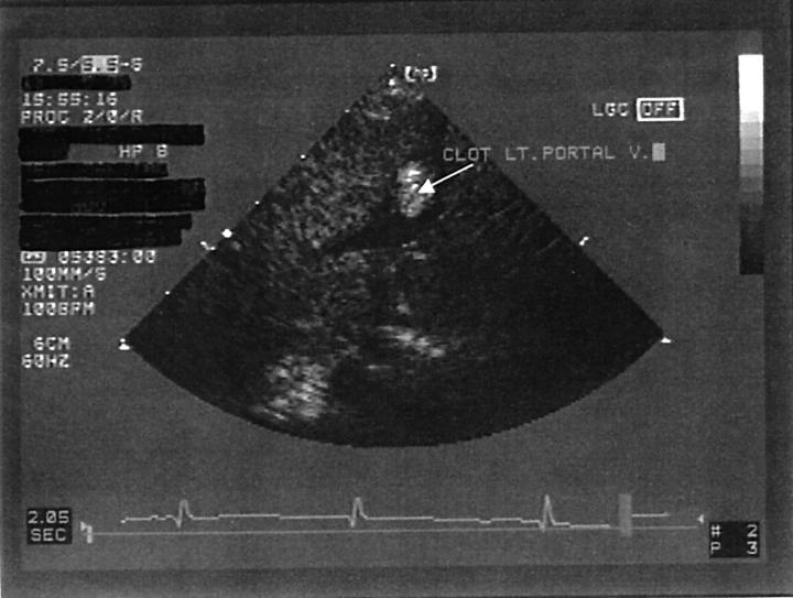
Two dimensional echocardiogram from the subcostal view showing thrombosis of the left portal vein.

Normal fetal circulation. Paradoxical thromboemboli could travel from the portal vein to the systemic circulation as shown by arrows. LA, Left atrium; LV, left ventricle; RA, right atrium; RV, right ventricle; SVC, superior vena cava; IVC, inferior vena cava.
Publication types
MeSH terms
LinkOut - more resources
Full Text Sources
Medical