. 2002 Sep 3;99(18):11872-7.
doi: 10.1073/pnas.172384399. Epub 2002 Aug 22.

Prospective isolation of human clonogenic common myeloid progenitors

Affiliations

Prospective isolation of human clonogenic common myeloid progenitors

Markus G Manz et al. Proc Natl Acad Sci U S A. .

Abstract

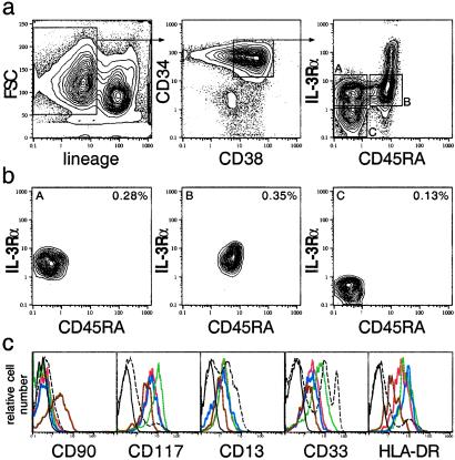
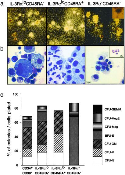
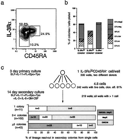
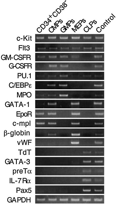
The hierarchical development from hematopoietic stem cells to mature cells of the hematolymphoid system involves progressive loss of self-renewal capacity, proliferation ability, and lineage potentials. Here we show the prospective isolation of early developmental intermediates, the human clonogenic common myeloid progenitors and their downstream progeny, the granulocyte/macrophage and megakaryocyte/erythrocyte progenitors. All three populations reside in the lineage-negative (lin(-)) CD34(+)CD38(+) fraction of adult bone marrow as well as in cord blood. They are distinguishable by the expression of the IL-3R alpha chain, the receptor of an early-acting hematopoietic cytokine, and CD45RA, an isoform of a phosphotyrosine phosphatase involved in negative regulation of cytokine signaling. Multipotent progenitors, early lymphoid progenitors, and the here-defined myeloid progenitors express distinct profiles of hematopoiesis-affiliated genes. The isolation of highly purified hematopoietic intermediates provides tools to better understand developmental programs underlying normal and leukemic hematopoiesis.


Figures

Figure 1
Figure 1
Prospective isolation of myeloid progenitors in adult human bone marrow. (a) The linCD34+CD38+ fraction was subdivided according to IL-3Rα and CD45RA expression (ac). (b) Reanalysis of the sorted populations. Percentages relative to mononuclear bone marrow cells are shown. (c) Additional antigen expression profile on CD34+CD38 (brown), IL-3RαloCD45RA (red), IL-3RαloCD45RA+ (green), and IL-3RαCD45RA (blue) populations. Isotype control gated on total linCD34+CD38+ cells (black) and marker expression on CD34 bone marrow cells (dashed line) are shown.
Figure 2
Figure 2
Colony formation in methylcellulose. (a) Morphology of day-14 colonies derived from sorted progenitors (inverted light microscope, ×4). (Right Insert) CFU-megakaryocyte/erythroid (×10). (b) Cellular morphology form single-picked colonies: CFU-granulocyte/erythroid/macrophage/megakaryocyte (GEMM, Left), CFU-granulocyte/macrophage (Center), burst-forming units/erythroid (Right), and CFU-megakaryocyte (Right Insert) (Giemsa, ×100 oil). (c) Clonogenic myeloid colony formation in methylcellulose. Scored were 288 wells receiving linCD34+CD38 cells (three donors), 336 wells receiving linCD34+CD38+IL-3RαloCD45RA cells, and 384 wells each receiving linCD34+CD38+IL-3RαloCD45RA+ and linCD34+CD38+IL-3RαCD45RA cells (four donors each). CFU-MegE, CFU-megakaryocyte/erythroid; CFU-GM, CFU-granulocyte/macrophage; BFU-E, burst-forming units/erythroid; CFU-Meg, CFU-megakaryocyte; CFU-M, CFU-macrophage; CFU-G, CFU-granulocyte.
Figure 3
Figure 3
LinCD34+CD38+IL-3RαloCD45RA cells are progenitors of IL-3RαloCD45RA+ and IL-3RαCD45RA cells and have clonal bipotent granulocyte/macrophage and erythrocyte/megakaryocyte potential. (a) IL-3RαloCD45RA cells give rise to both IL-3RαloCD45RA+ and IL-3RαCD45RA cells after 72-h culture on Sys-1stromal cells in the presence of SCF, IL-11, FL, Epo, and Tpo. (b) Colony readout of sorted cells from primary cultures (gates in a) in methylcellulose. CFU-MegE, CFU-megakaryocyte/erythroid; CFU-Meg, CFU-megakaryocyte; BFU-E, burst-forming units erythroid; CFU-GM, CFU-granulocyte/macrophage; CFU-M, CFU-macrophage; CFU-G, CFU-granulocyte. (c) Myeloid differentiation potential of single IL-3RαloCD45RA cells. Single cells were expanded in primary cultures and then transferred to methylcellulose (see Materials and Methods).
Figure 4
Figure 4
IL-3RαloCD45RA cells contain no lymphoid-committed progenitor cells. (a) Development of CD19+ and CD56+ cells from 33 lin CD34+CD38+IL-7Rα+CD10+ cells after 4 weeks of culture. (b) No CD19+ or CD56+ cells develop from 5,000 IL-3RαloCD45RA cells under the same conditions. Plots show cell fractions gated on human CD45+ cells.
Figure 5
Figure 5
Differential mRNA expression of hematopoiesis-affiliated genes in primitive progenitors (CD34+CD38), myeloid-committed progenitors, and lymphoid-committed progenitors. Total CD34+ bone marrow cells were used as controls. GM-CSFR, granulocyte/macrophage colony-stimulating factor receptor; G-CSFR, granulocyte colony-stimulating factor receptor; GAPDH, glyceraldehyde-3-phosphate dehydrogenase; vWG, von Willebrand factor; C/EBPɛ, CCAAT-enhancer-binding protein ɛ; MPO, myeloperoxidase; EpoR, erythropoietin receptor; TdT, terminal deoxynucleotidyl transferase.

References

    1. Kondo M, Weissman I L, Akashi K. Cell. 1997;91:661–672. - PubMed
    1. Akashi K, Traver D, Miyamoto T, Weissman I L. Nature (London) 2000;404:193–197. - PubMed
    1. de Wynter E A, Heyworth C M, Mukaida N, Jaworska E, Weffort-Santos A, Matushima K, Testa N G. J Leukocyte Biol. 2001;70:455–460. - PubMed
    1. Debili N, Coulombel L, Croisille L, Katz A, Guichard J, Breton-Gorius J, Vainchenker W. Blood. 1996;88:1284–1296. - PubMed
    1. Debili N, Robin C, Schiavon V, Letestu R, Pflumio F, Mitjavila-Garcia M T, Coulombel L, Vainchenker W. Blood. 2001;97:2023–2030. - PubMed

Publication types