. 2002 Sep 3;99(18):11908-13.
doi: 10.1073/pnas.172398899. Epub 2002 Aug 22.

Direct evidence that sulfhydryl groups of Keap1 are the sensors regulating induction of phase 2 enzymes that protect against carcinogens and oxidants

Affiliations

Direct evidence that sulfhydryl groups of Keap1 are the sensors regulating induction of phase 2 enzymes that protect against carcinogens and oxidants

Albena T Dinkova-Kostova et al. Proc Natl Acad Sci U S A. .

Abstract

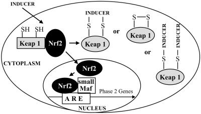
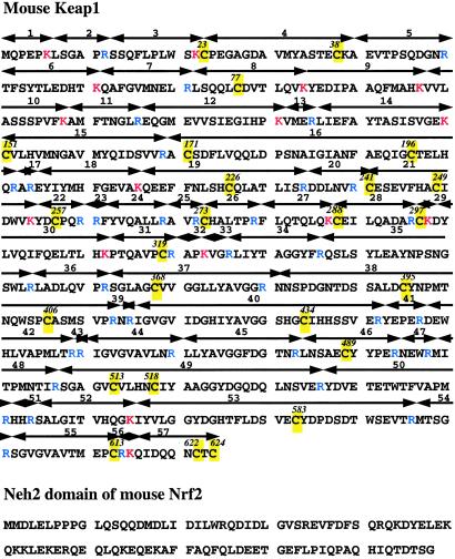
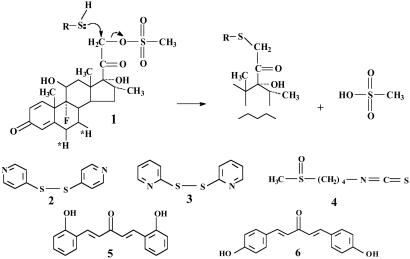
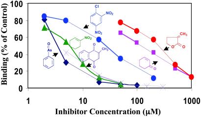
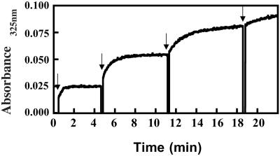
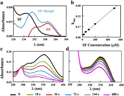
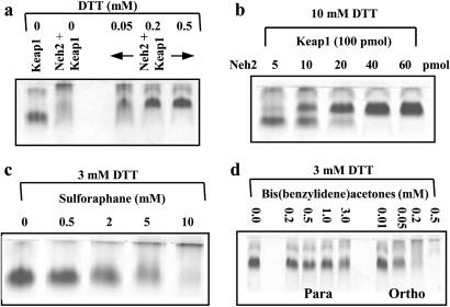
Coordinate induction of phase 2 proteins and elevation of glutathione protect cells against the toxic and carcinogenic effects of electrophiles and oxidants. All inducers react covalently with thiols at rates that are closely related to their potencies. Inducers disrupt the cytoplasmic complex between the actin-bound protein Keap1 and the transcription factor Nrf2, thereby releasing Nrf2 to migrate to the nucleus where it activates the antioxidant response element (ARE) of phase 2 genes and accelerates their transcription. We cloned, overexpressed, and purified murine Keap1 and demonstrated on native gels the formation of complexes of Keap1 with the Neh2 domain of Nrf2 and their concentration-dependent disruption by inducers such as sulforaphane and bis(2-hydroxybenzylidene)acetone. The kinetics, stoichiometry, and order of reactivities of the most reactive of the 25 cysteine thiol groups of Keap1 have been determined by tritium incorporation from [(3)H]dexamethasone mesylate (an inducer and irreversible modifier of thiols) and by UV spectroscopy with sulforaphane, 2,2'-dipyridyl disulfide and 4,4'-dipyridyl disulfide (titrants of thiol groups), and two closely related Michael reaction acceptors [bis(2- and 4-hydroxybenzylidene)acetones] that differ 100-fold in inducer potency and the UV spectra of which are bleached by thiol addition. With large excesses of these reagents nearly all thiols of Keap1 react, but sequential reaction with three successive single equivalents (per cysteine residue) of dipyridyl disulfides revealed excellent agreement with pseudo-first order kinetics, rapid successive declines in reaction velocity, and the stoichiometric formation of two equivalents of thiopyridone per reacted cysteine. This finding suggests that reaction of cysteine thiols is followed by rapid formation of protein disulfide linkages. The most reactive residues of Keap1 (C(257), C(273), C(288), and C(297)) were identified by mapping the dexamethasone-modified cysteines by mass spectrometry of tryptic peptides. These residues are located in the intervening region between BTB and Kelch repeat domains of Keap1 and probably are the direct sensors of inducers of the phase 2 system.


Figures

Figure 1
Figure 1
Mechanism of phase 2 response regulation. Nrf2 is anchored in the cytoplasm by binding to Keap1, which is attached to the actin cytoskeleton. Inducers disrupt the Keap1–Nrf2 complex, and Nrf2 migrates to the nucleus where it forms heterodimers with other transcription factors such as small Maf that bind to the ARE regions of phase 2 genes and accelerate their transcription. Several types of modifications of Keap1 by inducers are shown.
Figure 2
Figure 2
Primary structures of murine Keap1 (Upper) and Neh2 domain of Nrf2 (amino acid residues 1–98) (Lower). Tryptic peptides of Keap1 are numbered, and residues are highlighted: cysteine (yellow), arginine (blue), and lysine (red).
Figure 3
Figure 3
Irreversible reaction of Dex-mes (1) with a thiol group (Upper) and structures of the chemical probes used: 4,4′-dipyridyl disulfide (2), 2,2′-dipyridyl disulfide (3), sulforaphane (4), 2-HBA (5), and 4-HBA (6).
Figure 4
Figure 4
(a) SDS/PAGE of purified Keap1. (b) SDS/PAGE of Keap1 after labeling with [3H]Dex-mes: 5 μg of Keap1 was incubated with 50 pmol of [3H]Dex-mes for 60 min at 25°C, and unbound Dex-mes was removed by gel filtration. Labeled Keap1 was separated on a 10% gel and stained with Coomassie blue. The gel lane was cut into 2-mm slices, each digested with 30% H2O2, and radioactivity was counted.
Figure 5
Figure 5
Inhibition of [3H]Dex-mes binding to Keap1. Each reaction mixture contained 10 μl (3.5 μg, 50 pmol) of Keap1 and 80 μl of 25 mM Tris-Cl/2 mM EDTA/0.01% Tween 20, pH 8.0. A range of concentrations of competitors (10 μl in 50% acetonitrile/50% water) was added, and the mixture was incubated for 30 min at 25°C. Then 10 μl of [3H]Dex-mes (27 pmol) was added, and incubation was continued for an additional 60 min. [3H]Dex-mes bound to protein was separated from unbound steroid by gel filtration, and radioactivity in the protein fraction was determined.
Figure 6
Figure 6
Reaction of thiol groups of Keap1 with 4,4′-dipyridyl disulfide. To a solution of 0.5 μM Keap1 in 0.3 ml of 5 mM potassium phosphate buffer, pH 7.0, at 25°C, was added 0.5 μM (1 equivalent) 4,4′-dipyridyl disulfide in four sequential steps at times indicated with the arrows. Immediately after mixing, the increase in absorbance at 325 nm was monitored at 0.1-s intervals.
Figure 7
Figure 7
Reaction of Keap1 with sulforaphane. (a) Absorption spectra of 50 μM sulforaphane (SF), the reaction mixture of 50 μM sulforaphane after it has been incubated with 0.7 μM Keap1 for 90 min in 5 mM potassium phosphate buffer, pH 8.0, at 25°C against a Keap1 blank (SF+Keap1), and their difference spectrum (DF). (b) Plot of the reaction rates of sulforaphane with 0.7 μM Keap1 under the same reaction conditions as a function of the concentration of sulforaphane. (c and d) Absorption spectrum kinetics of the reaction of 2-HBA with Keap1 in the absence (c) or presence (d) of 20 mM sulforaphane.
Figure 8
Figure 8
Native gel electrophoresis showing complex formation between Keap1 (100 pmol) and the Neh2 domain (50 pmol) of Nrf2, and their dissociation. Concentration-dependent effects of DTT (a), Neh2 (b), sulforaphane (c), and 2-(ortho) and 4-(para) hydroxylated bis-(benzylidene)acetones (d) are shown. The binding reactions and native gel electrophoresis were carried out as described in Experimental Procedures. The 11.9-kDa Neh2 fragment migrates rapidly as a single band and is not shown in these gels.

References

    1. Kensler T W. Environ Health Perspect. 1997;105:965–970. - PMC - PubMed
    1. Talalay P. Biofactors. 2000;12:5–11. - PubMed
    1. Talalay P, Fahey J W, Holtzclaw W D, Prestera T, Zhang Y. Toxicol Lett. 1995;82/83:173–179. - PubMed
    1. Talalay P, De Long M J, Prochaska H J. Proc Natl Acad Sci USA. 1988;85:8261–8265. - PMC - PubMed
    1. Prestera T, Holtzclaw W D, Zhang Y, Talalay P. Proc Natl Acad Sci USA. 1993;90:2965–2969. - PMC - PubMed

Publication types

MeSH terms