. 2002 Sep 3;99(18):11634-9.
doi: 10.1073/pnas.182393899. Epub 2002 Aug 22.

A linear lattice model for polyglutamine in CAG-expansion diseases

Affiliations

A linear lattice model for polyglutamine in CAG-expansion diseases

Melanie J Bennett et al. Proc Natl Acad Sci U S A. .

Abstract

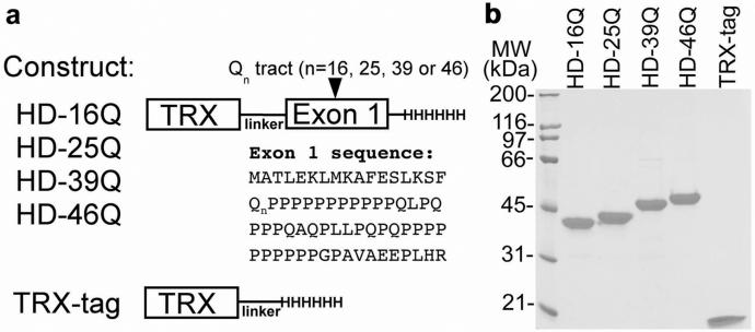
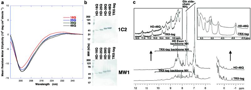
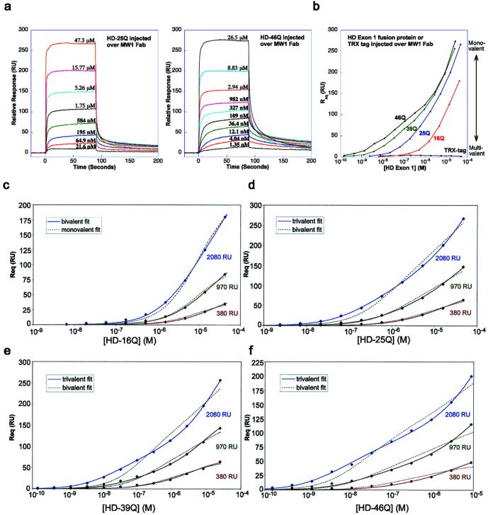
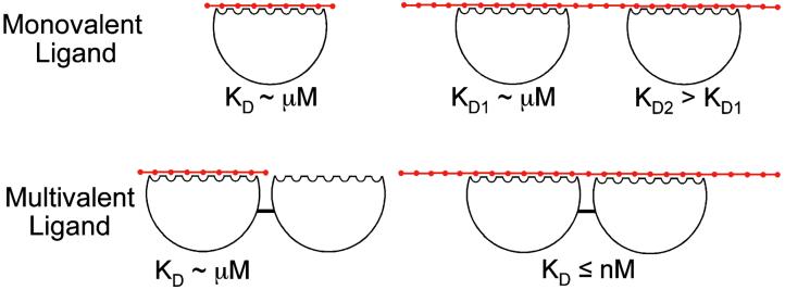
Huntington's disease and several other neurological diseases are caused by expanded polyglutamine [poly(Gln)] tracts in different proteins. Mechanisms for expanded (>36 Gln residues) poly(Gln) toxicity include the formation of aggregates that recruit and sequester essential cellular proteins [Preisinger, E., Jordan, B. M., Kazantsev, A. & Housman, D. (1999) Phil. Trans. R. Soc. London B 354, 1029-1034; Chen, S., Berthelier, V., Yang, W. & Wetzel, R. (2001) J. Mol. Biol. 311, 173-182] and functional alterations, such as improper interactions with other proteins [Cummings, C. J. & Zoghbi, H. Y. (2000) Hum. Mol. Genet. 9, 909-916]. Expansion above the "pathologic threshold" ( approximately 36 Gln) has been proposed to induce a conformational transition in poly(Gln) tracts, which has been suggested as a target for therapeutic intervention. Here we show that structural analyses of soluble huntingtin exon 1 fusion proteins with 16 to 46 glutamine residues reveal extended structures with random coil characteristics and no evidence for a global conformational change above 36 glutamines. An antibody (MW1) Fab fragment, which recognizes full-length huntingtin in mouse brain sections, binds specifically to exon 1 constructs containing normal and expanded poly(Gln) tracts, with affinity and stoichiometry that increase with poly(Gln) length. These data support a "linear lattice" model for poly(Gln), in which expanded poly(Gln) tracts have an increased number of ligand-binding sites as compared with normal poly(Gln). The linear lattice model provides a rationale for pathogenicity of expanded poly(Gln) tracts and a structural framework for drug design.


Figures

Figure 1
Figure 1
Expression and purification of HD exon 1 constructs. (a) HD exon 1 and TRX-tag constructs are represented schematically. (b) SDS/PAGE analysis of purified proteins (0.1 nmol; ≈2.5 μg of each protein) under reducing conditions.
Figure 2
Figure 2
Structural characterization of HD exon 1. (a) CD spectra. Each curve represents the difference between the spectrum of an HD exon 1 fusion protein and the spectrum of the TRX-tag control protein (i.e., the CD signal attributable to HD exon 1). (b) Western blots using 1C2 and MW1. Equimolar amounts of HD exon 1 fusion proteins or TRX-tag were loaded and probed with a 1:2000 dilution of ascites fluid containing 1C2 (Chemicon) or 200 nM purified MW1. (c) Comparison between 1H NMR spectra of TRX-tag and HD-46Q. Only the amide and aromatic proton region and the upfield-shifted methyl region are shown. (Insets) Expanded views.
Figure 3
Figure 3
Biosensor analyses of HD exon 1–MW1 Fab interactions. (a) Representative sensorgrams in which MW1 Fab is coupled at a density of 2,080 RU and HD-25Q (Left) or HD-46Q (Right) is injected at indicated concentrations. (b) Req versus the concentration (logarithmic scale) of injected analyte. Multivalent interactions are favored at low concentrations of each injected protein (low RU response region) and monovalent interactions are favored at high concentrations (high RU response region). The greatest differentiation between expanded poly(Gln) (e.g., HD-46Q) and normal poly(Gln) (e.g., HD-16Q) occurs in the low RU response region, because of multivalent binding of HD-46Q to MW1 Fab. (c–f) Global fits of biosensor data for HD exon 1 fusion proteins injected over MW1 Fab coupled to the chip at low, medium, or high density. Best-fit binding curves to the experimental data points are shown as continuous lines. Alternative models that do not adequately fit the data are shown as dashed lines.
Figure 4
Figure 4
Linear lattice model for poly(Gln). Poly(Gln) is a linear array of n repeating units (red dots). A monovalent ligand binds the lattice by interacting with several consecutive units. Binding of a first monovalent ligand occurs with an affinity that increases modestly with lattice length (e.g., 14 μM for HD-25Q and 1.5 μM for HD-46Q binding MW1 Fab, Table 2). A second ligand molecule can bind to a poly(Gln) tract that contains more than one binding site, but binds with weaker affinity than the first ligand. A multivalent ligand can bind with high avidity to a longer poly(Gln) tract by interacting with two binding sites simultaneously (the macroscopic KD is the product of the microscopic KD1 and KD2 values). Thus, normal and pathologic poly(Gln) can be distinguished if a ligand binds multivalently only to pathologic poly(Gln). The existence of such a multivalent ligand in vivo at concentrations below the KD for binding normal (≤36) poly(Gln), but above the KD for binding expanded (>36) poly(Gln) could explain the correlation between poly(Gln) length and disease incidence.

References

    1. Masino L, Pastore A. Brain Res Bull. 2001;56:183–189. - PubMed
    1. Altschuler E, Hud N V, Mazrimas J A, Rupp B. J Peptide Res. 1997;50:73–75. - PubMed
    1. Chen S, Berthelier V, Yang W, Wetzel R. J Mol Biol. 2001;311:173–182. - PubMed
    1. Masino L, Kelly G, Leonard K, Trottier Y, Pastore A. FEBS Lett. 2002;513:267–272. - PubMed
    1. Lescure A, Lutz Y, Eberhard D, Jacq X, Krol A, Grummt I, Davidson I, Chambon P, Tora L. EMBO J. 1994;13:1166–1175. - PMC - PubMed

Publication types