

Review
doi: 10.1136/jcp.55.9.641.
Changes produced in the urothelium by traditional and newer therapeutic procedures for bladder cancer
Affiliations
- PMID: 12194991
- PMCID: PMC1769754
- DOI: 10.1136/jcp.55.9.641
Item in Clipboard
Review
Changes produced in the urothelium by traditional and newer therapeutic procedures for bladder cancer
J Clin Pathol.
2002 Sep.
Abstract
A handful of traditional and newer therapeutic procedures, such as chemotherapy, immunotherapy, radiotherapy, photodynamic and laser treatment, and gene therapy, are used to treat epithelial malignancies of bladder origin. These treatment modalities, used either intravesically or systemically, produce morphological changes in the urothelial mucosa that can be mistaken for carcinoma. The pathologist must be able to separate toxic and drug related alterations from tumour related changes. The clinical history is usually invaluable in this assessment.
Figures
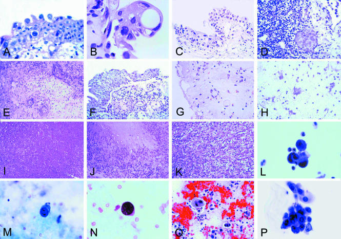
(A) Early histological changes in the urothelium after mitomycin C treatment. The superficial umbrella cells are large, vacuolated, and binucleated (original magnification, ×400; haematoxylin and eosin stained). (B) The effect of treatment with thiotepa. The changes are more pronounced than in (A) (see also text) (original magnification, ×400; haematoxylin and eosin stained). (C) The effect of treatment with cyclophosphamide. Large cells with large bizarre nuclei are easily identifiable (original magnification, ×250; haematoxylin and eosin stained). (D) The effect of treatment with BCG. Residual urothelial carcinoma. Denudation of the urothelium is present (upper right corner) (original magnification, ×400; haematoxylin and eosin stained). (E) The effect of treatment with interferon α. Oedema of the lamina propria, or subepithelial connective tissue, and perivascular collections of inflammatory cells are present (original magnification, ×400; haematoxylin and eosin stained). (F) Acute radiation cystitis. Partial detachment of the urothelium from the lamina propria where a mild inflammatory infiltrate is recognisable (original magnification, ×250; haematoxylin and eosin stained). (G) Acute radiation cystitis. Oedema of the lamina propria with atypical looking endothelial and stromal cells (original magnification, ×250; haematoxylin and eosin stained). (H) Atypical looking stromal cells (“radiation fibroblasts”) similar to those seen in giant cell cystitis (original magnification, ×400; haematoxylin and eosin stained). (I) Coagulation necrosis after laser treatment (original magnification, ×100; haematoxylin and eosin stained). (J) Postsurgical necrobiotic granuloma (original magnification, ×250; haematoxylin and eosin stained). (K) Postoperative spindle cell nodule (original magnification, ×250; haematoxylin and eosin stained). (L) Urinary cytology after topic mitomycin C treatment. Cluster of atypical looking cells with degenerative features (original magnification, ×400; Papanicolaou stain). (M) Urinary cytology after systemic cyclophosphamide treatment. Atypical looking cell with enlarged, eccentrically located, and extremely hyperchromatic nucleus (original magnification, ×400; Papanicolaou stain). (N) Urinary cytology after systemic cyclophosphamide treatment. Atypical looking cell in a patient with reactivation of polyomavirus infection (original magnification, ×400; Papanicolaou stain). (O) Urinary bladder cytology after external beam radiation. Degenerated urothelial cells and extensive background debris with histiocytes (original magnification, ×250; Papanicolaou stain). (P) Atypical epithelial cells in the urinary cytology after BCG treatment in a patient with urothelial carcinoma in situ (original magnification, ×400; Papanicolaou stain).
References
-
- Eble JN, Banks ER. Postsurgical necrobiotic granulomas of the urinary bladder. Urology 1990;35:454–7. - PubMed
-
- Belldegrun AS, Franklin JR, O'Donnell MA, et al. Superficial bladder cancer: the role of interferon-α. J Urol 1998;159:1793–801. - PubMed
-
- Antonakopoulous GN, Hicks RM, Berry RJ. The subcellular basis of damage to the urinary bladder induced by irradiation. J Pathol 1984;143:103–16. - PubMed
-
- Bostwick DG, Mikuz G. Urothelial papillary (exophytic) neoplasms. Virchows Arch [In press.] - PubMed
Publication types
MeSH terms
Substances
LinkOut - more resources
Full Text Sources
Other Literature Sources
Medical