. 2002 Aug;17(2):211-20.
doi: 10.1016/s1074-7613(02)00365-5.

In vivo depletion of CD11c+ dendritic cells abrogates priming of CD8+ T cells by exogenous cell-associated antigens

Affiliations

In vivo depletion of CD11c+ dendritic cells abrogates priming of CD8+ T cells by exogenous cell-associated antigens

Steffen Jung et al. Immunity. 2002 Aug.

Abstract

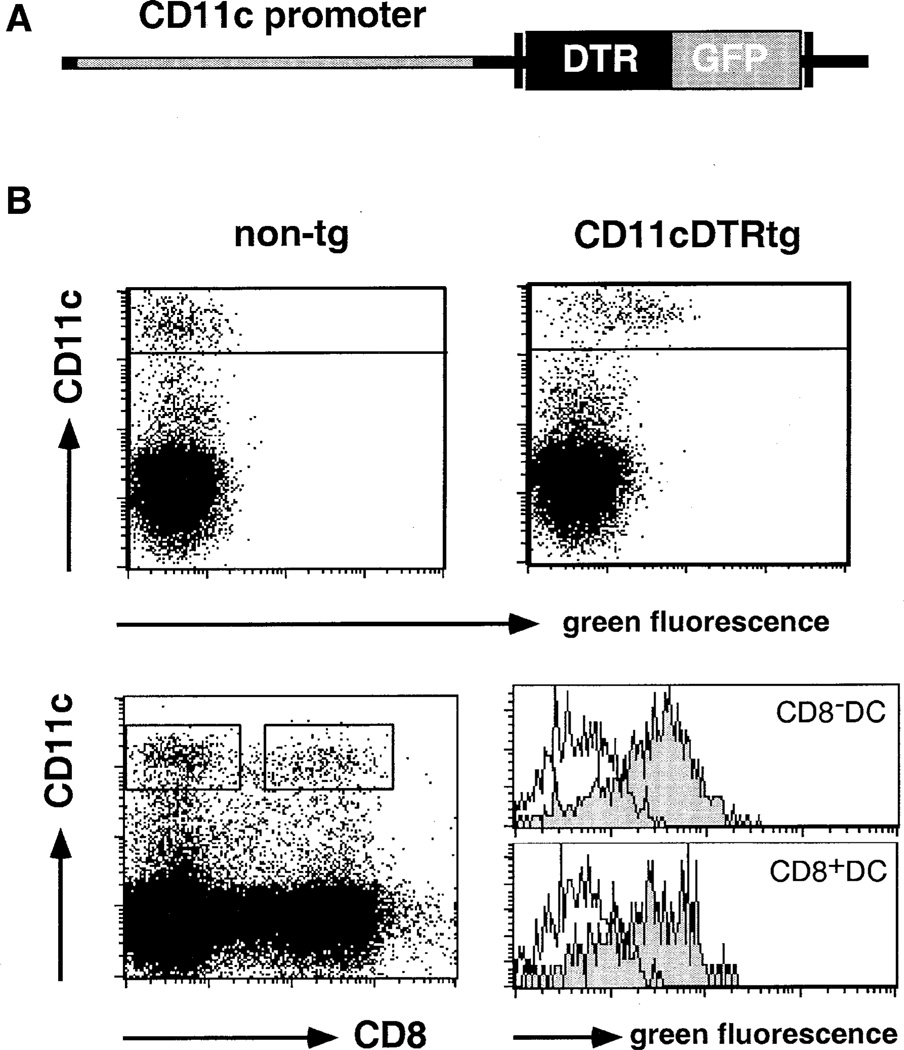
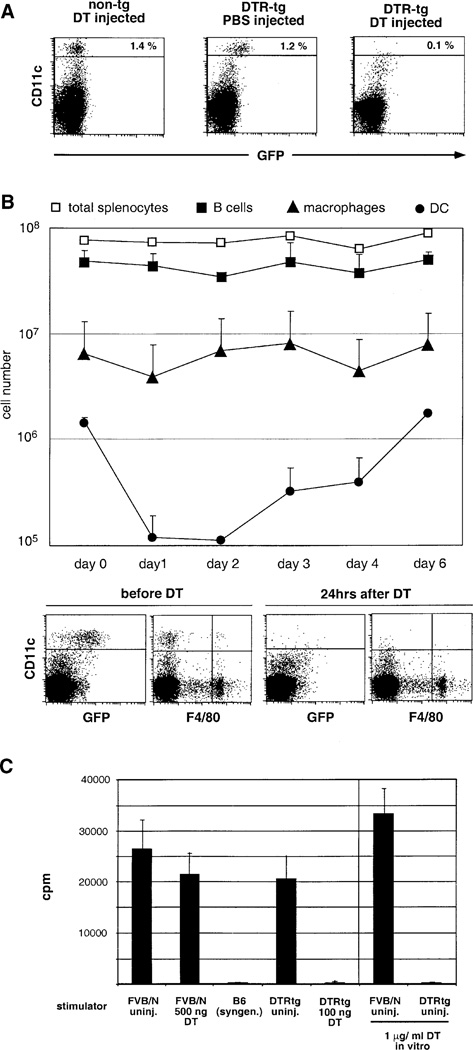
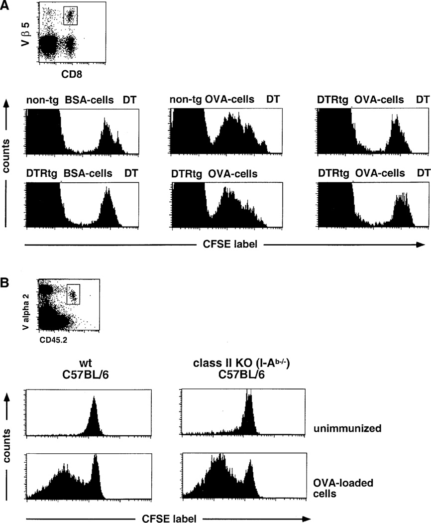
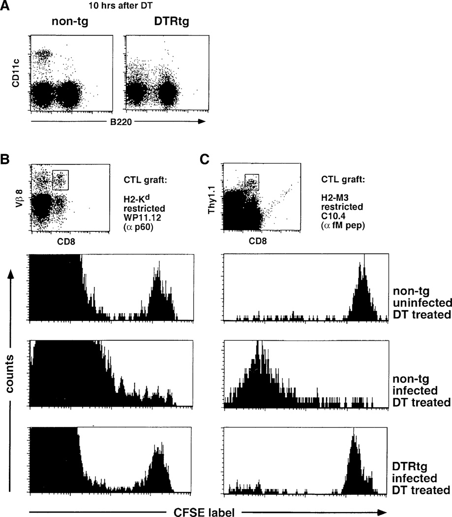
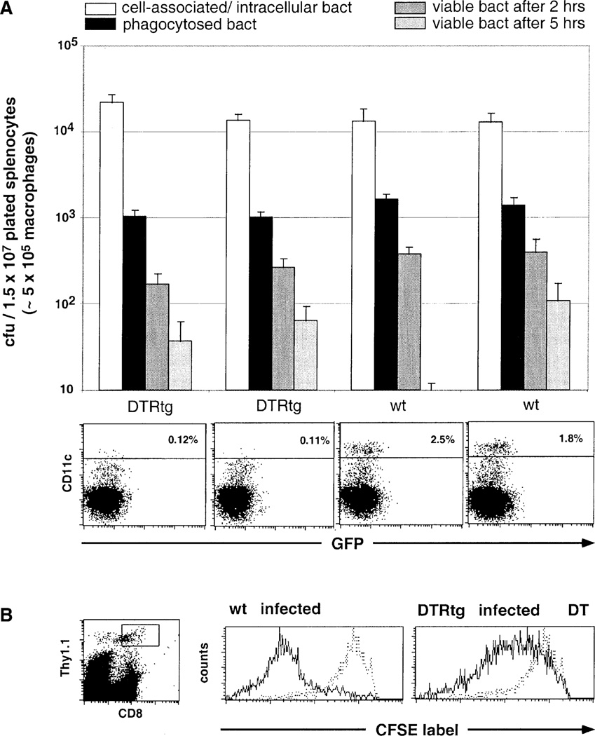
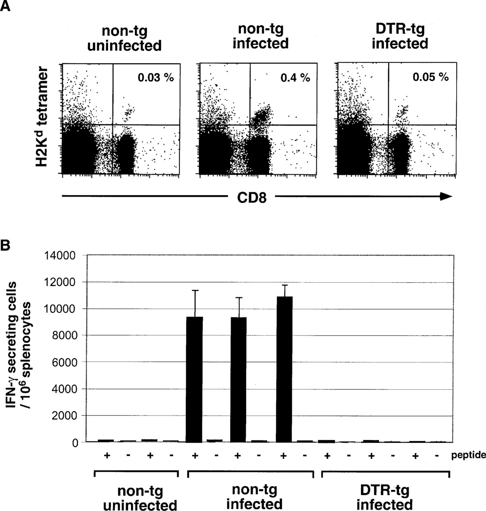
Cytotoxic T lymphocytes (CTL) respond to antigenic peptides presented on MHC class I molecules. On most cells, these peptides are exclusively of endogenous, cytosolic origin. Bone marrow-derived antigen-presenting cells, however, harbor a unique pathway for MHC I presentation of exogenous antigens. This mechanism permits cross-presentation of pathogen-infected cells and the priming of CTL responses against intracellular microbial infections. Here, we report a novel diphtheria toxin-based system that allows the inducible, short-term ablation of dendritic cells (DC) in vivo. We show that in vivo DC are required to cross-prime CTL precursors. Our results thus define a unique in vivo role of DC, i.e., the sensitization of the immune system for cell-associated antigens. DC-depleted mice fail to mount CTL responses to infection with the intracellular bacterium Listeria monocytogenes and the rodent malaria parasite Plasmodium yoelii.


Figures

Figure 1
Figure 1. Phenotypic Characterization of DTR Transgenic Mice
(A) Schematic representation of the DTR/GFP transgene. (B) Flow cytometric analysis of spleen cells of DTR transgenic (line 57) and nontransgenic FVB/N mice indicating GFP expression in CD11c+ DC. Histograms show GFP expression in CD8+ and CD8 DC subsets. Histogram gates are indicated in the dot plot. Open histograms, wild-type cells; filled histograms, DTR transgenic cells. Dead cells were excluded by propidium iodide (PI) staining.
Figure 2
Figure 2. DT Sensitivity of the DC Compartment in DTR Transgenic Mice
(A) Flow cytometric analysis of spleen cells of wt and DTR-transgenic (line 57) FVB/N mice 24 hr after injection of 100 ng DT or control vehicle. (B) Analysis of splenic APC populations of homozygous CD11c-DTR transgenic mice (line 57) after i.p. injection of 100 ng DT(day 0). Data represent mean of two mice per time point and are representative of two independent experiments. B cells are represented by squares and defined as being CD19+; macrophages are represented by triangles and defined as being F4/80+; DC are presented by circles and defined as being CD11c+. Open squares represent total splenocyte counts. Dot blots below the graph show a representative flow cytometric analysis of two time points. (C) Mixed leukocyte reaction with 8 × 10⁵ stimulator splenocytes (FVB/N; H2q) and 105 responder T cells (C57BL/6; H2b). Data are representative of three experiments with similar results. Note that the MLR was also abrogated by addition of DT during the culture resulting in in vitro DC depletion.
Figure 3
Figure 3. DC Dependence of In Vivo CTL Cross-Priming with Cell-Associated Antigen
(A) Flow cytometric analysis of CFSE-labeled OT-I CD8+ T cell graft (106 cells) in DT-treated nontransgenic and DTR transgenic (line 11) C57Bl/6 mice 2 days after immunization with OVA-loaded β2m−/− splenocytes or control splenocytes loaded with BSA. Histograms represent cells gated according to scatter, CD8, and Vβ5 expression as indicated in dot plots. Note that the large CFSE-negative peak represents host Vβ 5+ CD8 T cells. (B) Analysis of CFSE-labeled OT-I T cells (CD45.2, 106 cells) 3 days after immunization of wt C57Bl/6 (B6.SJL, CD45.1) and MHC class II-deficient mice (I-Ab −/−, CD45.1) with OVA-loaded β2m−/− splenocytes. Histograms represent cells gated according to scatter, expression of CD8, and the allotypic marker CD45.2 as indicated in dot plots. Dead cells were excluded by PI staining.
Figure 4
Figure 4. DC Dependence of Classical and Nonclassical MHC-Restricted Anti-Listeria CTL Responses
(A) Flow cytometric analysis of splenocyte of nontransgenic and DTR transgenic (line 57) littermate BALB/c mice 10 hr after DT injection (4 ng/g body weight). (B) Flow cytometric analysis of CFSE-labeled WP11.12 CD8+ T cell graft (106 cells) in DT-treated nontransgenic and DTR transgenic (line 57) BALB/c mice 4 days after i.v. injection of 2000 live L. monocytogenes. Histograms represent cells gated according to scatter, CD8, and Vβ 8 expression as indicated in dot plots. Note that the large CFSE peak represents host Vβ 8+ CD8 T cells. Dead cells were excluded by PI staining. (C) Flow cytometric analysis of CFSE-labeled C10.4 CD8+ T cell graft (Thy1.1, 106 cells) in spleens of DT-treated DTR transgenic (line 57) and nontransgenic littermate CB6F1 mice (Thy1.2) 4 days after i.v. injection of 2000 live L. monocytogenes. Histograms represent cells gated according to scatter, CD8, and Thy1.1 expression as indicated in dot plots. Dead cells were excluded by PI staining.
Figure 5
Figure 5. Functionality of Macrophage Compartment in DC-Depleted Spleens
(A) In vitro analysis of phagocytic and bacteriocidal activity of macrophages isolated from spleens of two DT-treated DTR transgenic (line 57) and two nontransgenic littermate CB6F1 mice. Macrophages isolated by plastic adherence were incubated with the noninvasive Shigella mutant BS176 (at an moi of 1:10). Open bars indicate counts of cell-associated and intracellular BS176; black bars indicate counts of phagocytosed BS176; and dark and light gray bars indicate counts of BS176 surviving 2 hr and 5 hr inside the macrophages, respectively. Standard deviations are based on triplicate cultures. FACS blots below bar diagram show respective splenocyte analysis. Data are representative of two experiments with similar results. (B) Flow cytometric analysis of a CFSE-labeled WP11.12 CD8+ T cell line graft (Thy1.1, 106 cells) in DT-treated nontransgenic and DTR transgenic (line 57) BALB/c mice (Thy1.2) 4 days after i.v. injection of 2000 live L. monocytogenes. Histograms represent cells gated according to scatter, CD8, and Thy1.1 expression, as indicated in dot plot. Stippled histograms represent CFSE-profile of uninfected wt control. Dead cells were excluded by PI staining.
Figure 6
Figure 6. DC Dependence of the CTL Response to Malaria Sporozoites
(A) Flow cytometric analysis of spleens wt and DTRtg recipients of anti-sporozoite CTL precursor grafts 4 days after infection with Plasmodium yoelii sporozoites. Data are representative of three mice per group. All mice received a single DT injection (4 ng/g body weight) 10 hr prior to infection. Cells are gated according to scatter. (B) ELISPOT assay of splenocytes of non-transgenic and DTR transgenic (line 57) BALB/c CTL-precursor recipients 4 days after infection with Plasmodium yoelii sporozoites. Black bars, number of sporozoite-peptide (SYVPSAEQI)-specific IFNγ-producing cells; open bars, number of IFNγ producers in absence of peptide.

References

    1. Albert ML, Sauter B, Bhardwaj N. Dendritic cells acquire antigen from apoptotic cells and induce class I-restricted CTLs. Nature. 1998;392:86–89. - PubMed
    1. Banchereau J, Steinman RM. Dendritic cells and the control of immunity. Nature. 1998;392:245–252. - PubMed
    1. Bellone M, Iezzi G, Rovere P, Galati G, Ronchetti A, Protti MP, Davoust J, Rugarli C, Manfredi AA. Processing of engulfed apoptotic bodies yields T cell epitopes. J. Immunol. 1997;159:5391–5399. - PubMed
    1. Bennett SR, Carbone FR, Karamalis F, Flavell RA, Miller JF, Heath WR. Help for cytotoxic-T-cell responses is mediated by CD40 signalling. Nature. 1998;393:478–480. - PubMed
    1. Berg RE, Princiotta MF, Irion S, Moticka JA, Dahl KR, Staerz UD. Positive selection of an H2–M3 restricted T cell receptor. Immunity. 1999;11:33–43. - PubMed

Publication types

MeSH terms

Substances