. 2002 Sep 1;22(17):7373-9.
doi: 10.1523/JNEUROSCI.22-17-07373.2002.

Selective electrical silencing of mammalian neurons in vitro by the use of invertebrate ligand-gated chloride channels

Affiliations

Selective electrical silencing of mammalian neurons in vitro by the use of invertebrate ligand-gated chloride channels

Eric M Slimko et al. J Neurosci. .

Abstract

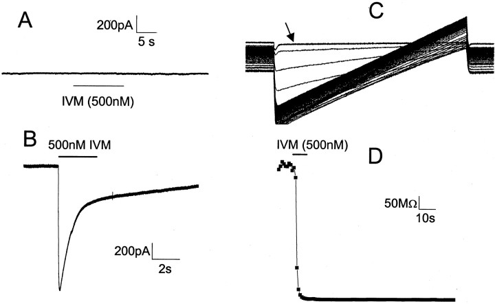
Selectively reducing the excitability of specific neurons will (1) allow for the creation of animal models of human neurological disorders and (2) provide insight into the global function of specific sets of neurons. We focus on a combined genetic and pharmacological approach to silence neurons electrically. We express invertebrate ivermectin (IVM)-sensitive chloride channels (Caenorhabditis elegans GluCl alpha and beta) with a Sindbis virus and then activate these channels with IVM to produce inhibition via a Cl- conductance. We constructed a three-cistron Sindbis virus that expresses the alpha and beta subunits of a glutamate-gated chloride channel (GluCl) along with the green fluorescent protein (EGFP) marker. Expression of the C. elegans channel does not affect the normal spike activity or GABA/glutamate postsynaptic currents of cultured embryonic day 18 hippocampal neurons. At concentrations as low as 5 nm, IVM activates a Cl- current large enough to silence infected neurons effectively. This conductance reverses in 8 hr. These low concentrations of IVM do not potentiate GABA responses. Comparable results are observed with plasmid transfection of yellow fluorescent protein-tagged (EYFP) GluCl alpha and cyan fluorescent protein-tagged (ECFP) GluCl beta. The present study provides an in vitro model mimicking conditions that can be obtained in transgenic mice and in viral-mediated gene therapy. These experiments demonstrate the feasibility of using invertebrate ligand-activated Cl- channels as an approach to modulate excitability.


Figures

Fig. 1.
Fig. 1.
Modified pSinRep5 construct, pSinRep5tsgGluClαβEGFP, designed to express three genes with three subgenomic promoters: the GluCl α subunit, the GluCl β subunit, and the reporter EGFP. The resulting Sindbis virus is vSinGluClαβEGFP.
Fig. 2.
Fig. 2.
Both GluCl α and β are required for functional channels in HEK 293 cells. A, Voltage-clamp record of a HEK 293 cell transfected with GluCl α and EGFP shows that this subunit alone does not respond to applications of 500 nmIVM. Five of five cells showed no response to this subunit alone.B, Voltage-clamp record of a HEK 293 cell transfected with GluCl α and β subunits shows a transient IVM-induced current. Five of five cells transfected in this manner showed a robust response to IVM. C, Voltage ramps from –90 to –40 mV before (arrow), during, and after the application of 500 nm IVM. The increasing slopes show the IVM-induced conductance. The waveforms also show a clear change in reversal potential during the development of the conductance. Ramps 100 msec in duration were delivered at 1 sec intervals. D, Input resistance of the cell in C. A conductance develops and then remains relatively constant after the application of IVM.
Fig. 3.
Fig. 3.
Activation of GluCl conductance by IVM over a 100-fold concentration range (A, 500 nm;B, 50 nm; C, 5 nm). Top panels, Current-clamp data from cells that displayed spontaneous activity. Bottom panels, Input conductance measured with voltage-clamp ramps from –80 to –50 mV for 200 msec duration at intervals of 1 sec. Eachpanel represents data from a different cell. Uninfected and GFP-infected neurons had no response to IVM applied similarly.
Fig. 4.
Fig. 4.
IVM silences GluCl-expressing neurons.A, Silenced cells no longer respond to glutamate. Shown are current-clamp traces from a GluCl-infected cell.Dots indicate 10 msec applications of 100 μm glutamate. A, Left, Before activation with IVM the cell responds to glutamate with action potentials.A, Right, After activation with 5 nm IVM the cell no longer responds to the same glutamate application. The 1 sec applications of glutamate could produce a slight depolarization, but no action potentials (data not shown). B, GluCl-infected cells in current clamp, showing responses to depolarizing current pulses (0–30 pA, 5 pA increments). In control solutions the cells responded with action potentials, but the IVM-induced (5 nm) Cl conductance prevented action potentials (bottom panel). Cells remained silenced even in responses to current pulses as high as 200 pA (data not shown). C, An uninfected cell showing no reduction in excitability by 5 nm IVM.
Fig. 5.
Fig. 5.
There is a large variation in expression levels that seems to be culture dependent. Each pointrepresents the average of five cells in a culture dish. Approximately one-half of the cultures that were surveyed have no response to 500 nm IVM, and these cultures were excluded from the plot. Thepoints plotted here represent the cultures that do respond to 500 nm IVM. Note that this graph represents data taken at 0 and 5 nm IVM; the points have been spread out to enhance visualization. The arrowrepresents average cell conductance in 0 nm IVM, 100 μm muscimol for comparison.
Fig. 6.
Fig. 6.
Neurons cotransfected with separate plasmids encoding EYFP-tagged GluCl α and ECFP-tagged GluCl β show fluorescence. A, Diagram indicating that the fluorescent protein was inserted in the M3–M4 loop of each subunit.B, Measured input conductance of neurons transfected with the GluCl α–EYFP fusion and GluCl β–ECFP fusion.NT, Neurons that have not been transfected;AB, neurons transfected with both subunits;A, neurons transfected with only the GluCl α–EYFP subunit. All bars represent data from three independent cultures, 10 cells per culture. C, A bright-field image at 40× showing several neurons, witharrows indicating three neurons that have been transfected. D, The field of view inC imaged with an ECFP filter set.E, The field of view in C imaged with an EYFP filter set.
Fig. 7.
Fig. 7.
IVM-induced chloride conductance deactivates several hours after IVM washout. The input conductance of cells that were infected with vSinαβEGFP was measured first without IVM and then in the presence of 5 nm IVM. After 1 hr of washing there is little recovery in cell conductance, whereas after 8 hr the conductance returns to uninfected levels. The last barshows that IVM-induced conductance can be reactivated. Eachbar represents 10 cells in two cultures.
Fig. 8.
Fig. 8.
Expression of GluCl does not alter glutamate- or GABA-evoked currents. A, Evoked currents were measured in GluCl-infected and uninfected neurons with 10 msec agonist puffs (100 μm GABA, 1 mm glutamate). The amplitude, rise time, and decay time were measured. The plot shows these three parameters relative to the measurements in uninfected cells. GABA responses are shown on the left and glutamate responses on the right. For each of the two ligands thebars represent data pooled from 10 cells from two different cultures. B, C, Analysis of GABA mIPSCs in the absence (B) and presence (C) of 5 nm IVM. The peak amplitude histogram (top) and decay time histogram (bottom) are shown. The amplitude data are shown in 10 pA bins, and the decay time data are shown in 10 msec bins. Note that there is no apparent difference in histogram shape between the 0 and 5 nm IVM case for either measurement. In B, the data are pooled from 20 events each from five neurons in one culture; in C, the data are pooled from 30 events each from five neurons in one culture.

References

    1. Baines RA, Uhler JP, Thompson A, Sweeney ST, Bate M. Altered electrical properties in Drosophila neurons developing without synaptic transmission. J Neurosci. 2001;21:1523–1531. - PMC - PubMed
    1. Crunelli V, Forda S, Kelly JS. Blockade of amino acid-induced depolarizations and inhibition of excitatory postsynaptic potentials in rat dentate gyrus. J Physiol (Lond) 1983;341:627–640. - PMC - PubMed
    1. Cully DF, Paress PS. Solubilization and characterization of a high-affinity ivermectin binding site from Caenorhabditis elegans. Mol Pharmacol. 1991;40:326–332. - PubMed
    1. Cully DF, Vassilatis DK, Liu KK, Paress PS, Van der Ploeg LH, Schaeffer JM, Arena JP. Cloning of an avermectin-sensitive glutamate-gated chloride channel from Caenorhabditis elegans. Nature. 1994;371:707–711. - PubMed
    1. Ehrengruber MU, Doupnik CA, Xu Y, Garvey J, Jasek MC, Lester HA, Davidson N. Activation of heteromeric G-protein-gated inward rectifier K+ channels overexpressed by adenovirus gene transfer inhibits the excitability of hippocampal neurons. Proc Natl Acad Sci USA. 1997;94:7070–7075. - PMC - PubMed

Publication types

MeSH terms