. 2002 Sep 1;22(17):7380-8.
doi: 10.1523/JNEUROSCI.22-17-07380.2002.

Lack of neurodegeneration in transgenic mice overexpressing mutant amyloid precursor protein is associated with increased levels of transthyretin and the activation of cell survival pathways

Affiliations

Lack of neurodegeneration in transgenic mice overexpressing mutant amyloid precursor protein is associated with increased levels of transthyretin and the activation of cell survival pathways

Thor D Stein et al. J Neurosci. .

Abstract

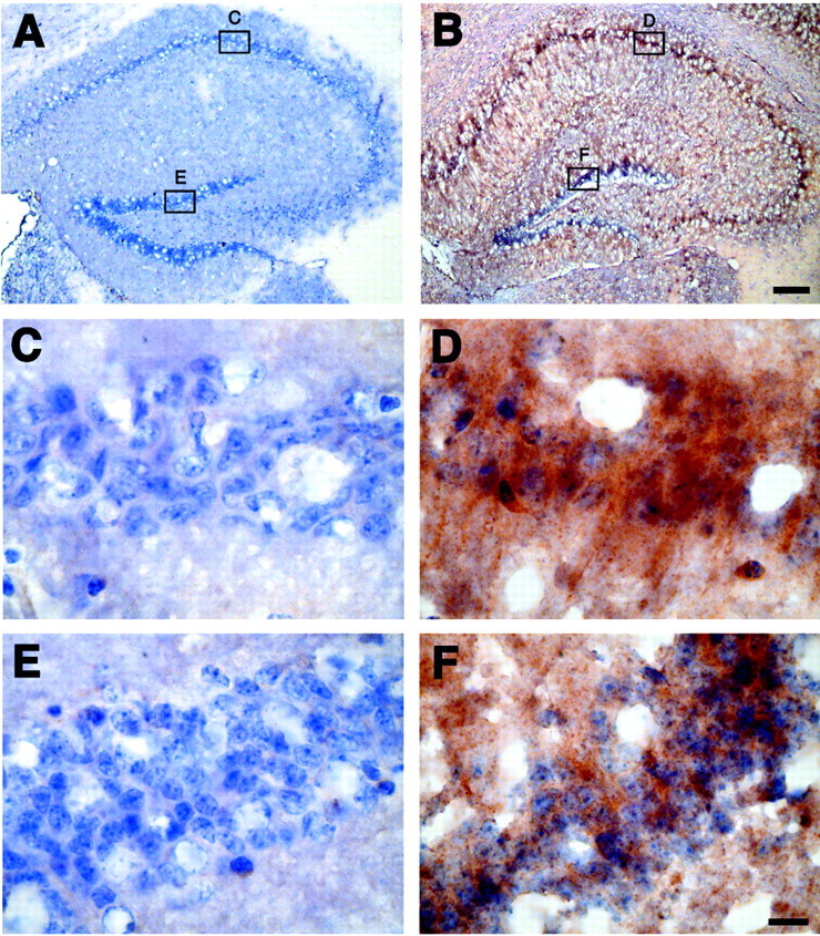
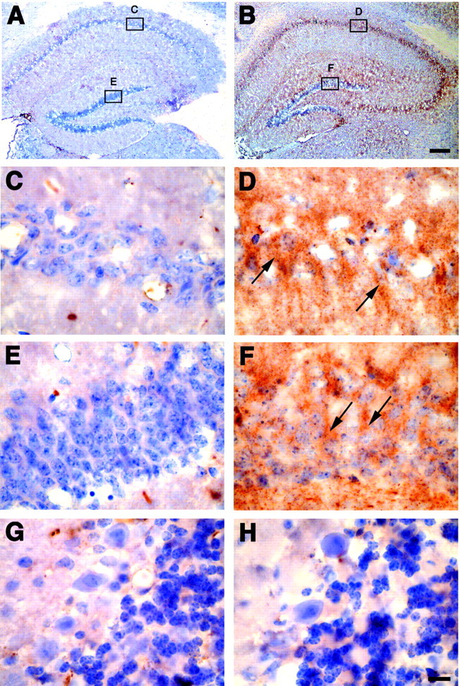
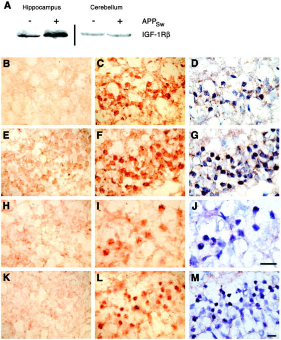
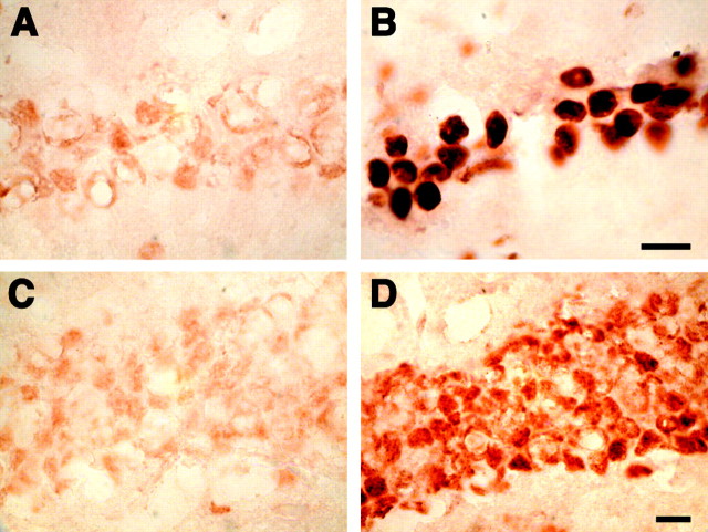
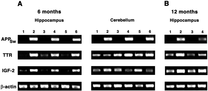
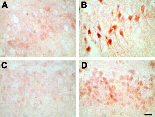
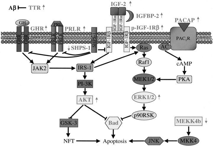
Tg2576 mice overexpress a mutant form of human amyloid precursor protein with the Swedish mutation (APP(Sw)), resulting in high beta-amyloid (Abeta) levels in the brain. Despite this, amyloid plaques do not develop until 12 months of age, and there is no neuronal loss in mice as old as 16 months. Gene expression profiles in the hippocampus and cerebellum of 6-month-old APP(Sw) mice were compared with age-matched controls. The expression of transthyretin, a protein shown to sequester Abeta and prevent amyloid fibril formation in vitro, and several genes in the insulin-signaling pathway, e.g., insulin-like growth factor-2, were increased selectively in the hippocampus of APP(Sw) mice. Concomitant activation of the insulin-like growth factor-1 receptor, Akt, and extracellular signal-regulated protein kinase 1 and 2 as well as increased phosphorylation of Bad also were unique to the hippocampus of APP(Sw) mice. In addition, the increased expression of transthyretin and insulin-like growth factor-2 and the increased phosphorylation of Bad in hippocampal neurons were maintained in 12-month-old APP(Sw) mice when compared with age-matched controls. These results suggest that the slow progression and lack of full-fledged Alzheimer's disease pathology in the hippocampal neurons of APP(Sw) mice result from the genetic reprogramming of neural cells to cope with increased levels of Abeta.


Figures

Fig. 1.
Fig. 1.
Immunohistochemistry for TTR in the hippocampus of control and APPSw overexpressing mice. Sections from nontransgenic mice (A, C, E) and APPSw mice (B, D, F) were immunostained for TTR and counterstained with hematoxylin. TTR is increased throughout the hippocampus in APPSw mice (B) compared with control mice, which contain little to no TTR (A). TTR levels in APPSw mice are largest around the neurons in CA1 (D) and the dentate gyrus (F). There is virtually no immunostaining for TTR in CA1 (C) and the dentate gyrus (E) of nontransgenic mice. Scale bars: (inB) A, B, 150 μm; (inF) C–F, 10 μm.
Fig. 2.
Fig. 2.
Immunohistochemistry for IGF-2 in control and APPSw overexpressing mice. Sections from nontransgenic mice (A, C, E, G) and APPSwmice (B, D, F, H) were immunostained for IGF-2 and counterstained with hematoxylin. IGF-2 is increased throughout the hippocampus in APPSw mice (B) compared with control mice, which contain little to no IGF-2 (A). IGF-2 levels in APPSw mice are largest around the neurons in CA1 (D) and the dentate gyrus (F). The arrows highlight examples of IGF-2-positive neurons in APPSw mice. There is little immunostaining for IGF-2 in CA1 (C) and the dentate gyrus (E) of nontransgenic mice. IGF-2 expression is unchanged in the cerebellum in which there is little immunostaining in either nontransgenic mice (G) or APPSw mice (H). Scale bars: (in B)A, B, 150 μm; (in H)C–H, 10 μm.
Fig. 3.
Fig. 3.
Activation of the IGF-1 receptor, Akt, and Erk1/2 in the hippocampus of APPSw mice. A, Immunoprecipitation for phosphorylated tyrosine and subsequent immunoblotting for the β-subunit of the insulin-like growth factor-1 receptor (IGF-1Rβ) revealed an increase in tyrosine-phosphorylated IGF-1Rβ in APPSw mice (+APPSw) when compared with nontransgenic controls (−APPSw) in the hippocampus but not in the cerebellum. B–G, Hippocampal neurons in CA1 stain faintly or not at all for Akt phosphorylated at Thr308 in nontransgenic control animals (B), but neurons in CA1 in APPSw mice are phospho-Akt positive (C, D). Hippocampal neurons in the dentate gyrus (DG) are faintly positive for phospho-Akt in control mice (E), but this staining is increased in the DG of APPSw mice (F, G). A hematoxylin counterstain of C andF reveals the laminar organization of the CA1 and DG neurons and the nuclear localization of phospho-Akt within these neurons in APPSw mice (D, G).H–M, Hippocampal neurons in CA1 (H) or DG (K) in control mice do not show significant staining for Erk1 phosphorylated at Thr202 and Tyr204 or Erk2 phosphorylated at Thr183 and Tyr185. However, APPSw mice do show positive staining for phospho-Erk1/2 in CA1 (I, J) and DG (L, M). A hematoxylin counterstain of I andL reveals the laminar organization of the CA1 and DG neurons and the nuclear localization of phopsho-Erk1/2 within these neurons in APPSw mice (J, M). Scale bars: J (for H–J), M (for B–G, K, L), 10 μm.
Fig. 4.
Fig. 4.
The 6-month-old APPSw mice have increased levels of phosphorylated Bad in hippocampal neurons. Nontransgenic mice have low levels of Bad phosphorylated at Ser112 in CA1 (A) and dentate gyrus (C) neurons, but levels of phospho-Bad are increased in CA1 (B) and the dentate gyrus (D) of APPSw mice. Scale bars:B (for A, B), D (forC, D), 10 μm.
Fig. 5.
Fig. 5.
RT-PCR from the hippocampus and cerebellum of APPSw and nontransgenic mice. A, The 6-month-old mice that contain and express the human APPSwtransgene (lanes 2, 4, 6) have increased levels of transthyretin (TTR) and insulin-like growth factor-2 (IGF-2) in the hippocampus when compared with nontransgenic littermates (lanes 1, 3, 5). In the cerebellum the increase in TTR expression is reduced and the increase in IGF-2 is eliminated when comparing APPSw mice (lanes 2, 4, 6) with nontransgenic littermates (lanes 1, 3, 5). B, At 12 months the increased expression of TTR and IGF-2 in the hippocampus of APPSw mice remains (lanes 2, 4) when compared with nontransgenic controls (lanes 1, 3).
Fig. 6.
Fig. 6.
The 12-month-old APPSw mice have increased levels of phosphorylated Bad in hippocampal neurons. Nontransgenic mice have low levels of Bad phosphorylated at Ser112 in CA1 (A) and dentate gyrus (C) neurons, but levels of phospho-Bad are increased in CA1 (B) and the dentate gyrus (D) of APPSw mice. Scale bar: (inD) A–D, 10 μm.
Fig. 7.
Fig. 7.
Hypothetical schema showing integration of neuroprotective Akt and Erk1/2 pathways in APPSw mice. All differentially expressed genes or proteins (grayed text) that have been shown previously to have a role in the inhibition or activation of apoptosis or are involved in Aβ sequestration are shown. Also indicated is the increased (↑) or decreased (↓) gene expression or protein activation in APPSw mice. Between proteins the black arrows represent the activation of one protein by another, and the bars represent inhibition. AC, Adenylate cyclase; GHR, growth hormone receptor;IRS-1, insulin receptor substrate 1;MKK4, MAPK kinase 4; NFT, neurofibrillary tangles; p-IGF-1Rβ, tyrosine-phosphorylated IGF-1 receptor β; p90RSK, 90 kDa ribosomal S6 kinases;PAC1R, type I PACAP receptor;PKA, protein kinase A; PRLR, prolactin receptor.

References

    1. Beilharz EJ, Russo VC, Butler G, Baker NL, Connor B, Sirimanne ES, Dragunow M, Werther GA, Gluckman PD, Williams CE, Scheepens A. Co-ordinated and cellular-specific induction of the components of the IGF/IGFBP axis in the rat brain following hypoxic–ischemic injury. Brain Res Mol Brain Res. 1998;59:119–134. - PubMed
    1. Berlanga JJ, Gualillo O, Buteau H, Applanat M, Kelly PA, Edery M. Prolactin activates tyrosyl phosphorylation of insulin receptor substrate 1 and phosphatidylinositol-3-OH kinase. J Biol Chem. 1997;272:2050–2052. - PubMed
    1. Bonni A, Brunet A, West AE, Datta SR, Takasu MA, Greenberg ME. Cell survival promoted by the Ras-MAPK signaling pathway by transcription-dependent and -independent mechanisms. Science. 1999;286:1358–1362. - PubMed
    1. Calhoun ME, Wiederhold KH, Abramowski D, Phinney AL, Probst A, Sturchler-Pierrat C, Staufenbiel M, Sommer B, Jucker M. Neuron loss in APP transgenic mice. Nature. 1998;395:755–756. - PubMed
    1. Costoya JA, Finidori J, Moutoussamy S, Senaris R, Devesa J, Arce VM. Activation of growth hormone receptor delivers an antiapoptotic signal: evidence for a role of Akt in this pathway. Endocrinology. 1999;140:5937–5943. - PubMed

Publication types

MeSH terms