Case Reports
. 2002 Sep 1;22(17):7462-70.
doi: 10.1523/JNEUROSCI.22-17-07462.2002.

The myotonia congenita mutation A331T confers a novel hyperpolarization-activated gate to the muscle chloride channel ClC-1

Affiliations
Case Reports

The myotonia congenita mutation A331T confers a novel hyperpolarization-activated gate to the muscle chloride channel ClC-1

Maike Warnstedt et al. J Neurosci. .

Abstract

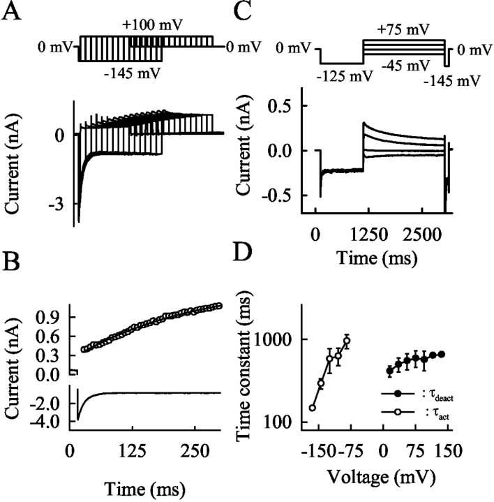
Mutations in the muscle chloride channel gene CLCN1 cause myotonia congenita, an inherited disorder of skeletal muscle excitability leading to a delayed relaxation after muscle contraction. Here, we examine the functional consequences of a novel disease-causing mutation that predicts the substitution of alanine by threonine at position 331 (A331T) by whole-cell patch-clamp recording of recombinant mutant channels. A331T hClC-1 channels exhibit a novel slow gate that activates during membrane hyperpolarization and closes at positive potentials. This novel gate acts in series with fast opening and closing transitions that are common to wild-type (WT) and mutant channels. Under conditions at which this novel gate is not activated, i.e., a holding potential of 0 mV, the typical depolarization-induced activation gating of WT hClC-1 was only slightly affected by the mutation. In contrast, A331T hClC-1 channels with an open slow gate display an altered voltage dependence of open probability. These novel gating features of mutant channels produce a decreased open probability at -85 mV, the normal muscle resting potential, leading to a reduced resting chloride conductance of affected muscle fibers. The A331T mutation causes an unprecedented alteration of ClC-1 gating and reveals novel processes defining transitions between open and closed states in ClC chloride channels.


Figures

Fig. 1.
Fig. 1.
Evolutionary conservation of the region of the A331T mutation. Location of residue 331 using the transmembrane topology of StClC from Salmonella typhimurium (Dutzler et al., 2002) and alignment of residues 327–341 of the A331T hClC-1 channels with ClC-0 from the electric organ from Torpedo marmorata, the WT human ClC isoforms ClC-1 to ClC-7, a plant ClC channel from Arabidopsis thaliana, and a prokaryotic ClC channel from Methanococcus jannaschii.
Fig. 2.
Fig. 2.
Electrophysiological characterization of WT and A331T hClC-1 channels. Whole-cell current recordings from tsA201 cells expressing WT (A) or A331T (B) hClC-1 channels. Cells were held at 0 mV, and voltage steps between −165 and +75 mV in 60 mV intervals were applied, each followed by a fixed pulse to −125 mV. C,D, Voltage dependence of the averaged normalized instantaneous (●) and late (▾) current amplitude. Current amplitudes were normalized by dividing by the peak current amplitude at −145 mV. Means ± SEM are shown from 10 (C) or 13 (D) cells.
Fig. 3.
Fig. 3.
Voltage-dependent gating in A331T mutant channels.AC, Voltage dependence of amplitudes of the three fractional currents [A1: fast deactivating (●) (A);A2: slow deactivating (▪) (B); C: nondeactivating (▾) (C)]. Corresponding WT data are given asopen symbols. D, Voltage dependence of the relative open probability from cells perfused with the standard internal solution expressing A331T (●) and WT (○) channels. Means ± SEM from 13 (A331T) and 10 (WT) cells. To construct these activation curves, normalized instantaneous current amplitudes at a fixed potential of −125 mV were measured after a 0.2 sec prepulse and plotted versus the preceding potential. Solid lines represent fits with single Boltzmann terms (A331T: V0.5 = −59.6 ± 5.6 mV, kV = 21.8 ± 1.5 mV, Pmin = 0.3,n = 13; WT: V0.5 = −79.9 ± 1.4 mV, kV = 20.5 ± 0.4 mV, Pmin = 0.1,n = 10). E, Voltage dependence of the relative open probability from cells perfused with an internal solution with low chloride concentration ([Cl]int = 4 mm) expressing A331T (▪, n = 6) and WT (■, n =5) channels. To prevent internal Cl accumulation, cells were clamped to a holding potential (HP) of −85 mV between two consecutive voltage steps. Solid linesrepresent fits with single (A331T) or double (WT) Boltzmann terms (A331T:V0.5 = −6.9 ± 6.2 mV,kV = 37.6 ± 3.6 mV,n = 6; WT: V0.5 = −105.1 ± 3.1 mV, kV = 16.2 ± 1.7 mV and V0.5 = −42.2 ± 19.2 mV, kv = 33.3 ± 4.5 mV,n = 5). Arrows demonstrate the difference between relative open probability of WT and mutant channels at the resting potential of the muscle (−85 mV).
Fig. 4.
Fig. 4.
Voltage dependence of the relative open probability of A331T hClC-1 channels determined after three distinct conditioning pulses. AC, Current–responses of one cell to a pulse protocol consisting of a fixed conditioning pulse, a variable prepulse (from −125 to +75 mV in 40 mV intervals), followed by a fixed test step to −125 mV. Measurements differ in the conditioning potential: 0 mV (A), +75 mV (B), and −85 mV (C). D, Plot of the instantaneous current amplitude at −125 mV versus the preceding potential, after a conditioning pulse to 0 mV (●), +75 mV (○), and −85 mV (▾). Data were obtained from four cells; for each cell all three conditioning pulses were tested, and amplitudes were normalized to the maximum current amplitude observed for each particular cell.
Fig. 5.
Fig. 5.
A novel hyperpolarization-induced gate in A331T mutant channels. A, B, Current–responses of cells expressing WT hClC-1 channels (A) or expressing A331T (B) hClC-1 channels to a three-step pulse protocol, consisting of a variable conditioning pulse (between +55 and −165 mV), a fixed prepulse (to +75 mV), and a fixed test step (to −125 mV). C, Voltage dependence of the normalized maximum current amplitude measured at +75 mV as obtained from current recordings shown in A and B, for WT channels (○, n = 4) and A331T hClC-1 (●,n = 6) under standard internal and external solutions. D, Voltage dependence of the opening of the slow gate of A331T hClC-1 channels, measured using standard external and internal solutions (●, n = 6), using standard external and low Cl internal solution (▪,n =5), or using standard internal solution and an external solution in which the NaCl was completely substituted with NaNO3 (▾, n = 3). Solid lines in C and D represent Boltzmann fits to the original data.
Fig. 6.
Fig. 6.
Time course of opening and closing of the novel hyperpolarization-induced gate. A, Current–response of a tsA201 cell expressing A331T mutant channels to a pulse protocol consisting of a pulse to −145 mV followed by a voltage step +100 mV. The duration of the first voltage step was increased stepwise. B, Time dependence of the maximum current amplitude at the +100 mV step (○) and of the current amplitude at −145 mV obtained from the recording shown inA. C, Current–response to voltage steps between −45 and +75 mV, each after a fixed prepulse to −125 mV. D, Voltage dependence of time constants of activation (○) and deactivation (●). Means ± SEM from between three and six cells.
Fig. 7.
Fig. 7.
Gating properties of WT-A331T heterodimeric channels. A, Whole-cell current recordings from tsA201 cells transfected with the WT-A331T concatameric construct. The cell was held at 0 mV, and voltage steps between −165 and +75 mV in 60 mV intervals were applied, each followed by a fixed pulse to −125 mV.B, Current–responses of cells expressing WT-A331T heterodimeric channels to a three-step pulse protocol, consisting of a variable conditioning pulse (between +55 and −165 mV), a fixed prepulse (to +75 mV), and a fixed test step (to −125 mV).C, Voltage dependence of the maximum current amplitude measured at +75 mV as obtained from current recordings shown inB, for heterodimeric WT-A331T channels (▾), homodimeric WT (○), and homodimeric A331T hClC-1 channels (●).D, Plot of the instantaneous current amplitude at −125 mV versus the preceding potential, after a conditioning pulse to 0 mV (●), +75 mV (○), and −85 mV (▾). Data were obtained from four cells; at each cell all three conditioning pulses were tested, and data were normalized to the maximum value observed at the particular cell.
None

References

    1. Adrian RH, Bryant SH. On the repetitive discharge in myotonic muscle fibres. J Physiol (Lond) 1974;240:505–515. - PMC - PubMed
    1. Barry PH. JPCalc, a software package for calculating liquid junction potential corrections in patch-clamp, intracellular, epithelial and bilayer measurements and for correcting junction potential measurements. J Neurosci Methods. 1994;51:107–116. - PubMed
    1. Beck CL, Fahlke C, George AL., Jr Molecular basis for decreased muscle chloride conductance in the myotonic goat. Proc Natl Acad Sci USA. 1996;93:11248–11252. - PMC - PubMed
    1. Bryant SH. Muscle membrane of normal and myotonic goats in normal and low external chloride. Fed Proc. 1962;21:312.
    1. Bryant SH, Morales-Aguilera A. Chloride conductance in normal and myotonic muscle fibres and the action of monocarboxylic aromatic acids. J Physiol (Lond) 1971;219:361–383. - PMC - PubMed

Publication types

MeSH terms

LinkOut - more resources