. 2002 Sep 1;22(17):7596-605.
doi: 10.1523/JNEUROSCI.22-17-07596.2002.

Heterotopic neurons with altered inhibitory synaptic function in an animal model of malformation-associated epilepsy

Affiliations

Heterotopic neurons with altered inhibitory synaptic function in an animal model of malformation-associated epilepsy

Maria Elisa Calcagnotto et al. J Neurosci. .

Abstract

Children with brain malformations often exhibit an intractable form of epilepsy. Although alterations in cellular physiology and abnormal histology associated with brain malformations has been studied extensively, synaptic function in malformed brain regions remains poorly understood. We used an animal model, rats exposed to methylazoxymethanol (MAM) in utero, featuring loss of lamination and distinct nodular heterotopia to examine inhibitory synaptic function in the malformed brain. Previous in vitro and in vivo studies demonstrated an enhanced susceptibility to seizure activity and neuronal hyperexcitability in these animals. Here we demonstrate that inhibitory synaptic function is enhanced in rats exposed to MAM in utero. Using in vitro hippocampal slices and whole-cell voltage-clamp recordings from visualized neurons, we observed a dramatic prolongation of GABAergic IPSCs onto heterotopic neurons. Spontaneous IPSC decay time constants were increased by 195% and evoked IPSC decay time constants by 220% compared with age-matched control CA1 pyramidal cells; no change in IPSC amplitude or rise time was observed. GABA transport inhibitors (tiagabine and NO-711) prolonged evoked IPSC decay kinetics of control CA1 pyramidal cells (or normotopic cells) but had no effect on heterotopic neurons. Immunohistochemical staining for GABA transporters (GAT-1 and GAT-3) revealed a low level of expression in heterotopic cell regions, suggesting a reduced ability for GABA reuptake at these synapses. Together, our data demonstrate that GABA-mediated synaptic function at heterotopic synapses is altered and suggests that inhibitory systems are enhanced in the malformed brain.


Figures
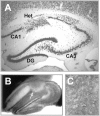
Fig. 1.
Fig. 1.
Nodular heterotopia in rats exposed to MAMin utero. A, Coronal hippocampal tissue section stained with the neuron-specific antibody neuronal-specific nuclear protein (Mullen et al., 1992). Note the presence of a cluster of displaced, pyramidal-like neurons in stratum CA1 pyramidale.B, Frame-grabber image of an acute hippocampal slice (300 μm) under direct IR-DIC visualization (10×). A nodular heterotopia in CA1 is indicated by the arrow.C, IR-DIC image of displaced, pyramidal-like neurons at higher magnification (40×). CA1, Stratum CA1 pyramidale; CA3, stratum CA3 pyramidale;DG, dentate gyrus; Het, nodular heterotopia.
Fig. 2.
Fig. 2.
Evoked IPSCs. A, Representative eIPSC recordings obtained in hippocampal slices from control animals (CA1 pyramidal and layer II/III supragranular neocortex) and MAM-exposed rats (normotopic, heterotopic, and interneuron). Eachtrace is an average of six sweeps. B, Normalized traces comparing a heterotopic neuron with a CA1 control cell (top) and a normotopic pyramidal cell (bottom). Note that the decay of the evoked IPSC is significantly prolonged for heterotopic pyramidal neurons compared with the two other cell types. C, Plot of the decay time constant for all cells. Data are presented as the mean ± SEM; *p < 0.001 using a one-way ANOVA. Eachbar represents 5–13 cells. normo, Normotopic; het; heterotopic; Int, interneuron; Ctx, cortex.
Fig. 3.
Fig. 3.
Spontaneous IPSCs. A, Representative sIPSC recordings in hippocampal slices from control and MAM-exposed rats. Individual events are shown at a faster time resolution at the right of each trace(asterisk in left trace).B–D, Plot of the cumulative data for all cells. sIPSC decay time constant, amplitude, and 10–90% rise time are shown. Data are presented as the mean ± SEM; *p < 0.001 using a one-way ANOVA. Each bar represents 14–21 cells.E, Representative event histograms for a control CA1 pyramidal neuron and an age-matched heterotopic cell at P17. Decay time constants are plotted for 100 individual sIPSC events;arrowhead indicates mean. Note the shift toward sIPSCs with longer decay time constants in the heterotopic cell.normo, Normotopic; het; heterotopic;Int, interneuron; Ctx, cortex.
Fig. 4.
Fig. 4.
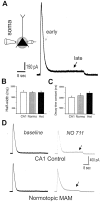
GABA responses in control and heterotopic neurons. A, Representativetraces of responses to picospritzer GABA application at the cell soma (soma) or cell dendrite (dendrite) at a concentration of 5 mmillustrating the similarity in evoked responses between different cell types. B, Schematic of the recording configuration and picospritzer location. C, Plots of the somatic and dendritic response to local GABA application; GABA-evoked current amplitude and 10–90% rise time are plotted. Data are presented as mean ± SEM; each bar represents five cells.Normo, Normotopic; het; heterotopic.
Fig. 5.
Fig. 5.
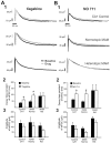
GABA transport inhibitors do not alter eIPSC responses on heterotopic neurons. A, Normalizedtraces of evoked IPSC responses before (Baseline) and ∼7 min after application of tiagabine (A1) or NO-711 (B2). Note the prolongation of eIPSC decay in the presence of a GABA transport inhibitor for CA1 control pyramidal and normotopic (Normo) neurons. These drugs did not alter the eIPSC recorded on heterotopic (Het) neurons. Cumulative data for all GABA transport inhibitor experiments are plotted for decay time constants (A2, tiagabine; B2, NO-711) and eIPSC amplitudes (A3, tiagabine; B3, NO-711). Data are presented as mean ± SEM; eachbar represents 8–16 cells. *p < 0.001 using a one-way ANOVA. The IPSC responses before and after the application of GABA transport inhibitor were scaled to the same peak amplitude (i.e., normalized).
Fig. 6.
Fig. 6.
Local GABA applications in the presence of a GABA transport inhibitor. A, Representativetrace from a heterotopic pyramidal neuron during somatic GABA application (5 mm). Note the presence of an outward current with early and late components. B, Plot of the half-width for GABA-evoked somatic currents. Data plotted represent the early outward current component. C, Plot of the decay time constant for GABA-evoked somatic currents. Data are presented as mean ± SEM. D, Traces showing a GABA-evoked somatic current before (baseline) and ∼7 min after bath application of NO-711. Note that a late outward current (arrow) appears during perfusion with the GABA transport inhibitor. Normo, Normotopic; Het, heterotopic.
Fig. 7.
Fig. 7.
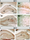
GABA transporter expression. A1, Coronal hippocampal section showing GAT-1 labeling around cell bodies in CA1–CA3 stratum pyramidale and granule cells of the dentate gyrus. This section is from a control, saline-treated rat. A2, A close-up section of CA1 showing GAT-1 staining at higher resolution (location indicated by asterisk in A1).B1, B2, GAT-1 labeling for a coronal hippocampal section from an MAM-exposed rat at low (B1)- and high (B2)-power magnification. Note the diffuse GAT-1 labeling around cell bodies in the nodular heterotopia (arrows). C, D, Same for GAT-3. Magnification (Zeiss stereoscope): A1,B1, C, D, 1.6×;A2, B2, 132×. Scale bars:A1, B1, 600 μm; A2,B2, 80 μm; C, D, 500 μm. CA1, Stratum CA1 pyramidale; CA3, stratum CA3 pyramidale; DG, dentate gyrus.

References

    1. Amano S, Ihara N, Uemura S, Yokoyama M, Ikeda M, Serikawa T, Sasahara M, Kataoka H, Hayase Y, Hazama F. Development of a novel rat mutant with spontaneous limbic-like seizures. Am J Pathol. 1996;149:329–336. - PMC - PubMed
    1. Andermann F. Cortical dysplasias and epilepsy: a review of the architectonic, clinical, and seizure patterns. Adv Neurol. 2000;84:479–496. - PubMed
    1. Andre V, Marescaux C, Nehlig A, Fritschy JM. Alterations of hippocampal GABAergic system contribute to development of spontaneous recurrent seizures in the rat lithium-pilocarpine model of temporal lobe epilepsy. Hippocampus. 2001;11:452–468. - PubMed
    1. Avoli M, Bernasconi A, Mattia D, Olivier A, Hwa GG. Epileptiform discharges in the human dysplastic neocortex: in vitro physiology and pharmacology. Ann Neurol. 1999;46:816–826. - PubMed
    1. Baraban SC, Schwartzkroin PA. Electrophysiology of CA1 pyramidal neurons in an animal model of neuronal migration disorders: prenatal methylazoxymethanol treatment. Epilepsy Res. 1995;22:145–156. - PubMed

Publication types

MeSH terms