. 2002 Sep 1;22(17):7627-38.
doi: 10.1523/JNEUROSCI.22-17-07627.2002.

Distinct actions of Emx1, Emx2, and Pax6 in regulating the specification of areas in the developing neocortex

Affiliations

Distinct actions of Emx1, Emx2, and Pax6 in regulating the specification of areas in the developing neocortex

Kathie M Bishop et al. J Neurosci. .

Abstract

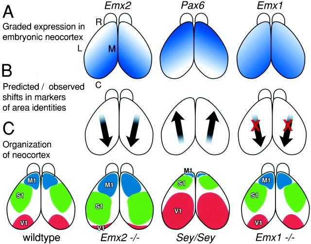
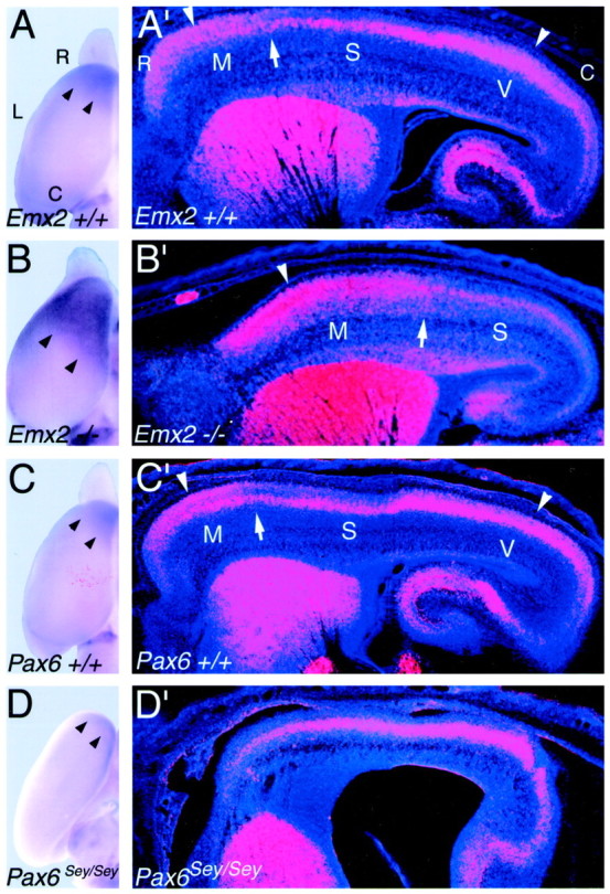
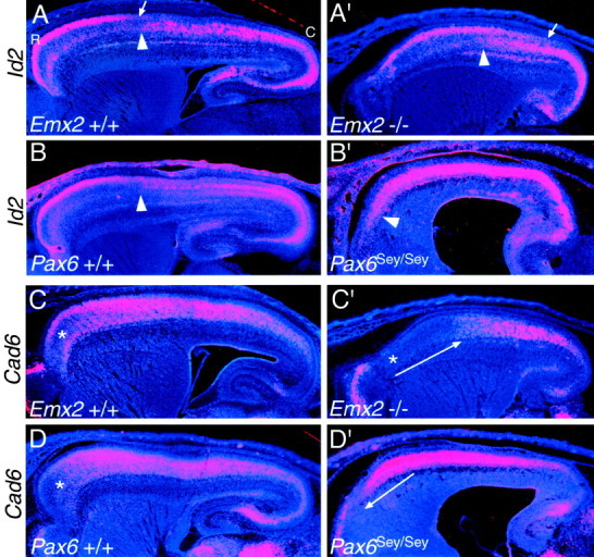
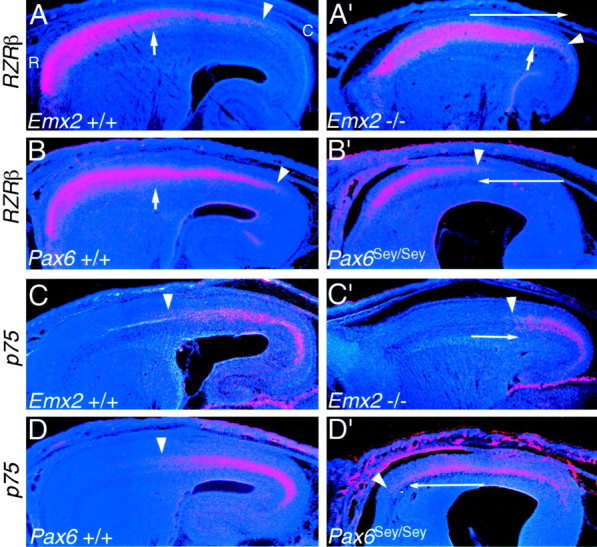
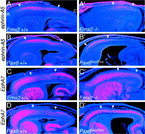
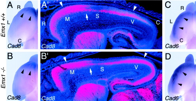
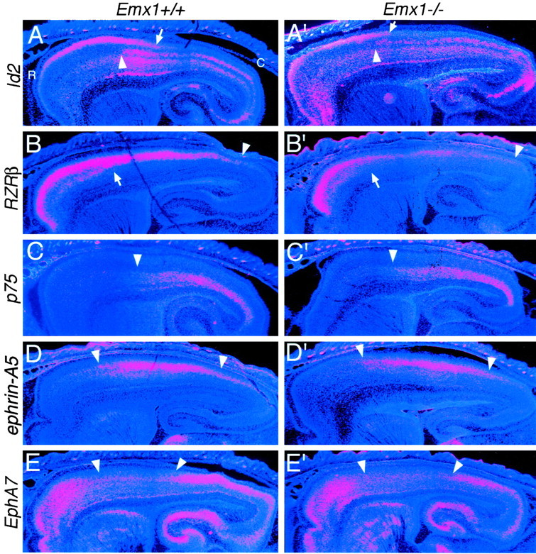
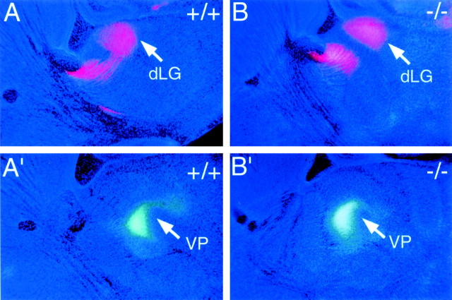
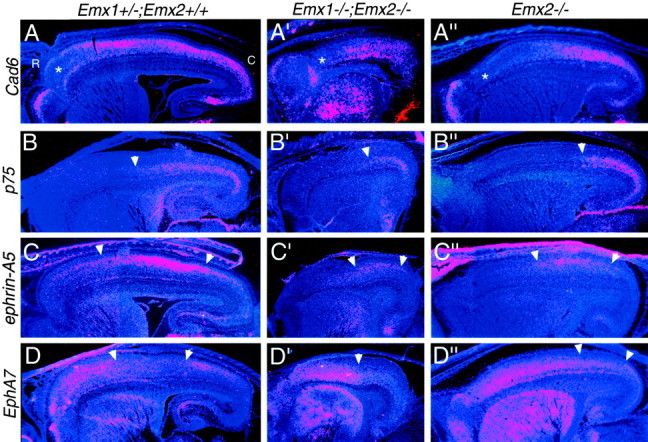
The mammalian neocortex is organized into subdivisions referred to as areas that are distinguished from one another by differences in architecture, axonal connections, and function. The transcription factors EMX1, EMX2, and PAX6 have been proposed to regulate arealization. Emx1 and Emx2 are expressed by progenitor cells in a low rostrolateral to high caudomedial gradient across the embryonic neocortex, and Pax6 is expressed in a high rostrolateral to low caudomedial gradient. Recent evidence has suggested that EMX2 and PAX6 have a role in the genetic regulation of arealization. Here we use a panel of seven genes (Cad6, Cad8, Id2, RZRbeta, p75, EphA7, and ephrin-A5) representative of a broad range of proteins as complementary markers of positional identity to obtain a more thorough assessment of the suggested roles for EMX2 and PAX6 in arealization, and in addition to assess the proposed but untested role for EMX1 in arealization. Orderly changes in the size and positioning of domains of marker expression in Emx2 and Pax6 mutants strongly imply that rostrolateral areas (motor and somatosensory) are expanded, whereas caudomedial areas (visual) are reduced in Emx2 mutants and that opposite effects occur in Pax6 mutants, consistent with their opposing gradients of expression. In contrast, patterns of marker expression, as well as the distribution of area-specific thalamocortical projections, appear normal in Emx1 mutants, indicating that they do not exhibit changes in arealization. This lack of a defined role for EMX1 in arealization is supported by our finding of similar shifts in patterns of marker expression in Emx1; Emx2 double mutants as in Emx2 mutants. Thus, our findings indicate that EMX2 and PAX6 regulate, in opposing manners, arealization of the neocortex and impart positional identity to cortical cells, whereas EMX1 appears not to have a role in this process.


Figures

Fig. 1.
Fig. 1.
Hypotheses, predicted results, and interpretations of analyses in this study. Diagrams are of dorsal views of the mouse neocortex. A, Graded expression patterns of the transcription factors Emx2, Pax6, andEmx1 across the embryonic neocortex. Emx2and Emx1 are expressed in a high caudomedial to low rostrolateral gradient, whereas Pax6 is expressed in an opposing gradient. B, Arrows indicate the direction of the predicted shifts in markers of area identity inEmx2, Pax6 (Sey/Sey), andEmx1 loss-of-function mutants, if these genes are involved in regulating arealization of the neocortex. The predicted shifts are observed in Emx2 and Pax6mutants but not in Emx1 mutants (indicated by red X marks). C, Organization of the mouse neocortex into areas predicted by our findings. These diagrams are not intended to show the exact sizes and shapes of the primary neocortical areas but rather to depict the disproportionate changes in area size and positioning, or no changes, in arealization in the different mutants. These predicted organizations suggested by our analyses of gene markers and area-specific thalamocortical projections are limited because the Emx2 and Pax6 mutants die on the day of birth, before areas become anatomically and functionally distinct, and thalamocortical projections do not develop inPax6 mutants. For simplicity, only the primary visual (V1), motor (M1), and somatosensory (S1) areas are shown. C, Caudal;L, lateral; M, medial; R, rostral; Sey, small eye mutant.
Fig. 2.
Fig. 2.
Opposing changes in the expression domains of the cadherin, Cad8 in Emx2, andPax6 (Sey/Sey) mutants.AD, Dorsal views of whole mounts of P0 cortical hemisphere of Emx2 wild-type (+/+) (A), Emx2 mutant (−/−) (B), Pax6 wild-type (+/+) (C), and Pax6(Sey/Sey) mutant (D) processed forin situ hybridization using digoxygenin-labeled riboprobes for Cad8. Arrowheads mark the caudal limit of the rostral expression domain ofCad8 in the superficial layers, which is characteristic of motor areas. A′–D′, Sagittal sections through E18.5 brains of mice of the corresponding genotypes as inAD, processed for in situ hybridization using S35-labeled riboprobes for Cad8. Sections were later counterstained with bisbenzimide. Sections are taken from similar mediolateral positions; rostral is to the left and dorsal is to thetop. Each panel is a montage of single-exposure photos using dark-field illumination with a red filter to view the silver grains and with UV fluorescence to view the counterstain. Marked are the approximate locations of the motor (M), somatosensory (S), and visual (V) areas in the wild-type cortex and their shifted locations in theEmx2−/− cortex suggested by the expansion and caudal shift in patterns of Cad8 expression, which are unique in each of these areas in wild-type mice. Arrowheads inA′–C′ mark rostral and caudal expression domains in the superficial layers characteristic of motor and visual areas; in comparison, expression is substantially diminished in the superficial layers of the intervening somatosensory area.Arrows in A′–C′ mark the presumed border between motor and somatosensory areas. TheCad8 expression rostral to these arrowsis the expression that is evident in the whole mounts shown inAC. This superficial rostral expression domain characteristic of motor areas is essentially absent inPax6 (Sey/Sey) mutants (D,D′). The wild type and mutants in each pair are age-matched littermates. See Results for details. C, Caudal; L, lateral; R, rostral.
Fig. 3.
Fig. 3.
Id2 and Cad6, used as markers of rostral neocortical areas, show opposing shifts inEmx2 and Pax6 mutants. In situ hybridizations on sagittal sections through the forebrain of E18.5 mice using S35-labeled riboprobes for the HLH transcription factor, Id2(A–B′) or the cadherin,Cad6 (C–D′), and counterstained with bisbenzimide are shown. Sections are fromEmx2 wild-type (+/+) and mutant (−/−) littermates orPax6 wild-type (+/+) and mutant (Sey/Sey) littermates and are taken from similar medial–lateral positions. Eachpanel is a montage of single-exposure photos using dark-field illumination with a red filter to view the silver grains and UV fluorescence to view the counterstain. Id2 exhibits a graded expression in superficial layers of rostral areas in wild-type mice; the arrows in A and A′ mark the position where the expression declines to very low levels. Thearrowheads in AB′ mark the transition from low to high expression reported in layer 5; this transition corresponds to the border between motor and somatosensory areas. The asterisks in C, C′, andD mark a domain of low Cad6 expression normally characteristic of far rostral neocortex. This domain of low expression expands and shifts caudally in Emx2 mutants, whereas inPax6 mutants it shifts rostrally and essentially disappears (C′, D′, long arrows). See Results for details. C, Caudal; R, rostral.
Fig. 4.
Fig. 4.
The complementary expression patterns ofRZRβ and p75 show opposing shifts inEmx2 and Pax6 mutants. In situ hybridizations on sagittal sections through the forebrain of E18.5 mice using S35-labeled riboprobes for either the nuclear receptor RZRβ (AB′) or the neurotrophin receptorp75 (CD′) and counterstained with bisbenzimide are shown. Sections are fromEmx2 wild-type (+/+) and mutant (−/−) littermates orPax6 wild-type (+/+) and mutant (Sey/Sey) littermates and are taken from similar medial–lateral positions. Eachpanel is a montage of single-exposure photos using dark-field illumination with a red filter to view the silver grains and UV fluorescence to view the counterstain. RZRβ shows two distinct high rostral to low caudal gradients across the wild-type neocortex (A, B), a gradient in superficial layers that extends farther caudally (marked byarrowheads) than a gradient within the deeper layers (marked by short arrows). Both gradients ofRZRβ expression expand caudally in Emx2mutants (A′) and constrict rostrally inPax6 mutants (B′). p75 is expressed in the deep layers in roughly the caudal half of the wild-type neocortex (C, D).p75 expression constricts caudally inEmx2 mutants (C′) and expands rostrally in Pax6 mutants (D′). Thearrowheads mark the rostral limit of expression. Thelong arrows in A′–D′ indicate the opposing shifts in expression in the mutants. See Results for details. C, Caudal; R, rostral.
Fig. 5.
Fig. 5.
The complementary expression patterns ofephrin-A5 and EphA7, used as markers of intermediate neocortical areas, show opposing shifts inEmx2 and Pax6 mutants. In situ hybridizations on sagittal sections through the forebrain of E18.5 mice using S35-labeled riboprobes for the axon guidance ligand, ephrin-A5, and one of its receptors, EphA7, and counterstained with bisbenzimide are shown. Sections are from Emx2 wild-type (+/+) and mutant (−/−) littermates (AB′) orPax6 wild-type (+/+) and mutant (Sey/Sey) littermates (CD′) and are taken from similar medial–lateral positions. Each panel is a montage of single-exposure photos using dark-field illumination with a red filter to view the silver grains and UV fluorescence to view the counterstain. In wild-type mice, ephrin-A5 has high expression centered on the somatosensory area (A,B), whereas EphA7 has low expression centered on the somatosensory area (C,D). These domains shift caudally in Emx2mutants (A′–B′) and rostrally inPax6 mutants (C′–D′).Arrowheads mark the domains of highephrin-A5 or low EphA7 expression. See Results for details. C, Caudal; R, rostral.
Fig. 6.
Fig. 6.
Patterned expression ofCad8 and Cad6 appears normal inEmx1 mutants. AD, Dorsal views of whole mounts of E18.5 cortical hemispheres ofEmx1 wild-type (+/+) and mutant (−/−) mice processed for in situ hybridization using digoxygenin-labeled riboprobes for Cad8 (A, B) or Cad6 (C, D).Arrowheads in A and B mark the caudal limit of the rostral Cad8 expression domain characteristic of motor areas and mark the lateral expression domain ofCad6 in C and D.A′, B′, Sagittal sections through E18.5 brains of Emx1 wild-type (+/+) and mutant (−/−) mice processed for in situ hybridization using S35-labeled riboprobes for Cad8.Sections were later counterstained with bisbenzimide. Sections are taken from similar medial–lateral positions; rostral is to theleft and dorsal is to the top. Eachpanel is a montage of single-exposure photos using dark-field illumination with a red filter to view the silver grains and UV fluorescence to view the counterstain. Marked are the approximate locations of the motor (M), somatosensory (S), and visual (V) areas suggested by the patterns ofCad8 expression, which are unique in each of these areas in wild-type mice. Arrowheads inA′–B′ mark rostral and caudal expression domains in the superficial layers characteristic of motor and visual areas; in comparison, expression is substantially diminished in the superficial layers of the intervening somatosensory area. Arrows inA′–B′ mark the presumed border between motor and somatosensory areas. The Cad8 expression rostral to these arrows is the expression that is evident in the whole mounts shown in A andB. The wild-type and mutants in each pair are age-matched littermates. See Results for details. C, Caudal; L, lateral; R, rostral.
Fig. 7.
Fig. 7.
The expression domains of genes that mark rostral, caudal, or intermediate areas of the neocortex appear normal in Emx1 mutants. In situ hybridizations on sagittal sections through the forebrain of E18.5 Emx1 wild-type (+/+) and mutant (−/−) littermates using S35-labeled riboprobes forId2, RZRβ, p75,ephrin-A5, and EphA7 and counterstained with bisbenzimide are shown. Sections are taken from similar medial–lateral positions. Each panel is a montage of single-exposure photos using dark-field illumination using a red filter to view the silver grains and UV fluorescence to view the counterstain.Id2 exhibits a graded expression in superficial layers of rostral areas in wild-type mice; the arrows inA and A′ mark the position where the expression declines to very low levels. The arrowheadsin AB′ mark the transition from low to high expression reported in layer 5; this transition corresponds to the border between motor and somatosensory areas. RZRβ is expressed in two distinct high rostral to low caudal gradients across the neocortex (B, B′); a gradient in superficial layers that extends farther caudally (marked byarrowheads) than a gradient within the deeper layers (marked by short arrows). p75 is expressed in the deep layers in roughly the caudal half of the neocortex (C, C′). Thearrowheads mark the rostral limit of expression.ephrin-A5 has high expression centered on the somatosensory area (D, D′), whereasEphA7 has low expression centered on the somatosensory area (E, E′). Arrowheadsmark the domains of high ephrin-A5 or lowEphA7 expression. Overall, the areal expression patterns of this panel of marker genes are similar in wild-type and mutant neocortex. See Results for details. C, Caudal;R, rostral.
Fig. 8.
Fig. 8.
Area-specific thalamocortical projections appear normal in Emx1 mutant mice. Sagittal sections through P0Emx1 wild type (+/+) and mutant (−/−) brains showing retrograde DiI (red) and DiA (green-bluish) labeling and bisbenzimide counterstain (dark blue). Rostral is to theleft and dorsal is to the top in allpanels. A, B, An injection of DiI into visual (occipital) cortex retrogradely labels neurons in the dorsal lateral geniculate nucleus (dLG) in bothEmx1+/+ and Emx1−/− mice.C, D, An injection of DiA into somatosensory (parietal) cortex of the same set of brains retrogradely labels neurons in the ventroposterior thalamic nucleus (VP) in both Emx1+/+ andEmx1−/− mice. Each pair of sections is from the same brain; the dLG-labeled sections are lateral to those with the VP labeling. See Results for details.
Fig. 9.
Fig. 9.
The relative positioning of expression domains of gene markers in Emx1; Emx2 double mutant neocortex resembles that in Emx2 single mutant neocortex. Shown are in situ hybridizations on sagittal sections through the forebrain of E18.5 (A–D) wild-type (i.e.,Emx1+/−; Emx2 +/+) (A′–D′) Emx double mutant (Emx1−/−; Emx2−/−), and (A′′–D′′) Emx2 single mutant (Emx2−/−) mice using S35-labeled riboprobes for Cad6,p75, ephrin-A5, or EphA7and later counterstained with bisbenzimide. Note that the neocortex of the Emx double mutant is reduced to approximately half of the wild-type area. Sections are taken from similar medial–lateral positions. Each panel is a montage of single-exposure photos using dark-field illumination and a red filter to view the silver grains and UV fluorescence to view the counterstain. Theasterisks in AA′′ mark a domain of low Cad6 expression normally characteristic of far rostral neocortex. This domain of low expression expands and shifts caudally in Emx double mutants and Emx2 mutants.p75 is expressed in the deep layers in roughly the caudal half of the wild-type neocortex (B); this expression domain constricts caudally in Emx double mutants andEmx2 mutants (B′, B′′). The arrowheads mark the rostral limit of expression. In wild-type mice, ephrin-A5 has high expression centered on the somatosensory area (A, B), whereasEphA7 has low expression centered on the somatosensory area (C, D). These domains shift caudally in Emx double mutants and Emx2 mutants (C′, D′, C′′,D′′). Arrowheads mark the domains of highephrin-A5 or low EphA7 expression. See Results for details. C, Caudal; R, rostral.

References

    1. Bishop KM, Goudreau G, O'Leary DDM. Regulation of area identity in the mammalian neocortex by Emx2 and Pax6. Science. 2000;288:344–349. - PubMed
    1. Caric D, Gooday D, Hill RE, McConnell SK, Price DJ. Determination of the migratory capacity of embryonic cortical cells lacking the transcription factor Pax-6. Development. 1997;124:5087–5096. - PubMed
    1. Cecchi C, Boncinelli E. Emx homeogenes and mouse brain development. Trends Neurosci. 2000;23:347–352. - PubMed
    1. Chenn A, Braisted JE, McConnell SK, O'Leary DDM. Development of the cerebral cortex: mechanisms controlling cell fate, laminar and areal patterning, and axonal connectivity. In: Dowan WM, Zipursky L, Jessell T, editors. Molecular and cellular approaches to neural development. Oxford UP; New York: 1997. pp. 440–473.
    1. Cohen-Tannoudji M, Babinet C, Wassef M. Early determination of a mouse somatosensory cortex marker. Nature. 1994;368:460–463. - PubMed

Publication types

MeSH terms

Substances